ObjectiveTo explore the effect of all-trans retinoic acid(ATRA) on inflammatory cytokines expression in human renal tubular epithelial cells under high glucose condition and its possible mechanisms. MethodsHK-2 cells were randomly divided into seven groups: blank control group, high glucose group, hypertonic group, ATRA intervention groups and Rho kinase inhibitor (Y27632) intervention group.All groups were treated for 48 hours.Reverse transcription polymerase chain reaction (RT-PCR) was used to evaluate RhoA and ROCK1 mRNA levels.ELISA was used to detect interleukin-6 (IL-6) and tumor necrosis factor-α (TNF- α) expression in HK-2 cells. ResultsRT-PCR showed that mRNA expressions of RhoA and ROCK1 were not different between the hypertonic group and the blank group (P>0.05), but were significantly increased in the high glucose group (P<0.05).In the ATRA treatment group,RhoA and ROCK1 mRNA expressions were significantly decreased compared with the high glucose group in a concentration-dependent manner.RhoA mRNA expressions showed no difference between the Y27632 intervention group and the high glucose group, but ROCK1 mRNA expression was significantly reduced (P<0.05).By Pearson correlation analysis, we found that there was a positive correlation between RhoA and ROCK1 mRNA expressions in the high glucose group and the ATRA treatment group.ELISA showed that the IL-6 and TNF-α protein levels were not different between the control and the hypertonic group (P>0.05), but were significantly increased in the high glucose group and decreased in the ATRA or Y27632 treatment groups (P<0.05). ConclusionATRA decreases IL-6 and TNF-α protein levels in HK-2 cells induced by high glucose.The underlying mechanism may be associated with the RhoA/ROCK signaling pathway.
Fig.1
mRNA expressions of RhoA and ROCK1 in seven groups of cells
M.Mark of relative molecular quality;1.blank control group;2.high glucose group;3.hyperosmolar group;4.high glucose and low concentration ATRA group;5.high glucose and middle concentration ATRA group;6.high glucose and high concentration ATRA group;7.Y27632 intervention group
表 2
Tab.2
表 2
表 2
7组细胞RhoA mRNA及ROCK1 mRNA表达相对水平
Tab.2
Relative mRNA expressions of RhoA and ROCK1 in seven groups of cells x¯±s,n=3
组别
RhoA
ROCK1
空白对照组
0.249±0.007*1
0.356±0.009*1
高糖组
0.548±0.019
0.936±0.011
高渗组
0.248±0.002*1
0.352±0.011*1
高糖+低浓度ATRA组
0.482±0.012*1
0.687±0.009*1
高糖+中浓度ATRA组
0.392±0.010*1*2
0.384±0.009*1*2
高糖+高浓度ATRA组
0.276±0.012*1*2*3
0.257±0.010*1*2*3
Y27632干预组
0.553±0.017*2*3
0.143±0.012*1*2*3
F
358.047
2 095.962
Compared with high glucose group,*1P<0.05; compared with high glucose+low concentration ATRA group,*2P<0.05; compared with high glucose+middle concentration ATRA group,*3P<0.05
WADAJ,MAKINOH.Inflammation and the pathogenesis of diabetic nephropathy[J].Clin Sci,2013,124(3):139-152.
The most problematic issue in clinical nephrology is the relentless and progressive increase in patients with ESRD (end-stage renal disease) worldwide. The impact of diabetic nephropathy on the increasing population with CKD (chronic kidney disease) and ESRD is enormous. Three major pathways showing abnormality of intracellular metabolism have been identified in the development of diabetic nephropathy: (i) the activation of polyol and PKC (protein kinase C) pathways; (ii) the formation of advanced glycation end-products; and (iii) intraglomerular hypertension induced by glomerular hyperfiltration. Upstream of these three major pathways, hyperglycaemia is the major driving force of the progression to ESRD from diabetic nephropathy. Downstream of the three pathways, microinflammation and subsequent extracellular matrix expansion are common pathways for the progression of diabetic nephropathy. In recent years, many researchers have been convinced that the inflammation pathways play central roles in the progression of diabetic nephropathy, and the identification of new inflammatory molecules may link to the development of new therapeutic strategies. Various molecules related to the inflammation pathways in diabetic nephropathy include transcription factors, pro-inflammatory cytokines, chemokines, adhesion molecules, Toll-like receptors, adipokines and nuclear receptors, which are candidates for the new molecular targets for the treatment of diabetic nephropathy. Understanding of these molecular pathways of inflammation would translate into the development of anti-inflammation therapeutic strategies.
NAPOLI JL.Physiological insights into all-trans-retinoic acid biosynthesis[J].Biochim Biophys Acta,2012,1821(1):152-167.
All-trans-retinoic acid (atRA) provides essential support to diverse biological systems and physiological processes. Epithelial differentiation and its relationship to cancer, and embryogenesis have typified intense areas of interest into atRA function. Recently, however, interest in atRA action in the nervous system, the immune system, energy balance and obesity has increased considerably, especially concerning postnatal function. atRA action depends on atRA biosynthesis: defects in retinoid-dependent processes increasingly relate to defects in atRA biogenesis. Considerable evidence indicates that physiological atRA biosynthesis occurs via a regulated process, consisting of a complex interaction of retinoid binding-proteins and retinoid recognizing enzymes. An accrual of biochemical, physiological and genetic data have identified specific functional outcomes for the retinol dehydrogenases, RDH1, RDH10, and DHRS9, as physiological catalysts of the first step in atRA biosynthesis, and for the retinal dehydrogenases RALDH1, RALDH2, and RALDH3, as catalysts of the second and irreversible step. Each of these enzymes associates with explicit biological processes mediated by atRA. Redundancy occurs, but seems limited. Cumulative data support a model of interactions among these enzymes with retinoid binding-proteins, with feedback regulation and/or control by atRA via modulating gene expression of multiple participants. The ratio apo-CRBP1/holo-CRBP1 participates by influencing retinol flux into and out of storage as retinyl esters, thereby modulating substrate to support atRA biosynthesis. atRA biosynthesis requires the presence of both an RDH and an RALDH: conversely, absence of one isozyme of either step does not indicate lack of atRA biosynthesis at the site. This article is part of a Special Issue entitled: Retinoid and Lipid Metabolism.
NORRIS EJ,PATELYC,REINHART MB,et al.Differ-entiation and apoptosis induced by all-trans retinoic acid is associated with downregulation of peroxiredoxin 1 in myeloid leukemia cells[J].Cancer Res,2014,74(19):4239.
All-trans retinoic acid (ATRA) based differentiation therapy with potentially curative outcome in acute promyelocytic leukemia (APL), has limited benefit in non-APL acute myeloid leukemia (AML). Thus, identifying key mediators of ATRA-induced differentiation/apoptosis in AML cells may reveal new putative therapeutic targets. Previously, we reported (Leukemia 20: 1809, 2006) that the therapeutic efficacy of ATRA is enhanced in topoisomerase 2β (TOP2β)-deficient AML cell lines, both APL (HL-60, AP-1060) and non-APL (KG-1), in part due to decreased expression of the antioxidant enzyme, peroxiredoxin (PRDX) 2 and increased accumulation of reactive oxygen species (ROS). Based on preliminary observations that (a) ATRA leads to down-regulation of a related antioxidant protein, PRDX1 (RNA array analysis), and (b) PRDX1 mRNA expression relative to PRDX2 mRNA is significantly higher in HL-60, KG-1, and leukemic blast cells from 20 AML patients (qRT-PCR analysis), we evaluated the role of PRDX1 in sensitizing the apoptotic effects of ATRA. Specifically, we tested the hypothesis that ATRA induced growth arrest/apoptosis is associated with decreased PRDX1 levels and increased ROS, which can be modulated by TOP2β. Accordingly, downregulation of TOP2β can enhance apoptosis by increasing ROS, whereas overexpression of TOP2β can inhibit PRDX1 downregulation by ATRA and attenuate ROS-induced apoptosis. Treatment of HL-60, KG-1 and patient AML blast cells with 1 08M ATRA in the absence or presence of 0.1 08M ICRF193 (TOP2β catalytic inhibitor) led to a 3 to 4-fold decrease in PRDX1 mRNA and protein. No effect of ICRF193, which leads to degradation of TOP2β, was observed on PRDX1 levels. Interestingly, while treatment with ATRA + ICRF 193 did not enhance PRDX1 down regulation compared to ATRA alone, it did lead to significantly (p<.05) more apoptosis and greater ROS than either treatment alone. Since, expression of TOP2β correlates with that of PRDX1 in blast cells of AML patients, both of which are expressed at relatively high levels, we tested whether overexpression of TOP2β affects ATRA-induced decrease in PRDX1, ROS accumulation and apoptosis. Transfection of TOP2β in an amsacrine-resistant HL-60 cell line (AR), which lacks TOP2β, leads to overexpression of TOP2β and abrogation of ATRA-induced downregulation of PRDX1, ROS accumulation and apoptosis. In summary, the present study demonstrates that PRDX1 is an ATRA regulated protein, which plays an important role in modulating ATRA-induced differentiation/apoptosis in myeloid leukemia cells. Furthermore, down regulation of PRDX1 levels by ATRA can be manipulated by overexpression of TOP2β. Since overexpression of TOP2β is observed in patient AML blasts, the increased PRDX1 levels may partly explain the resistance of non-APL acute myeloid leukemia cells to ATRA induced differentiation and ROS-mediated apoptosis.
ZHOU TB,QIN YH,LI ZY,et al.All-trans retinoic acid treatment is associated with prohibitin expression in renal interstitial fibrosis rats[J].Int J Mol Sci,2012,13(3):2769-2782.
This study was performed to investigate the association of prohibitin with renal interstitial fibrosis (RIF) lesion and to explore the association of all-trans retinoic acid (ATRA) treatment with prohibitin expression in RIF rats. Rats were divided into three groups: the sham operation group (SHO), the model group subjected to unilateral ureteral obstruction (UUO), and the model group treated with ATRA (GA). Renal tissues were collected at 14 and 28 days after surgery, and the relevant indicators were detected. In comparison with the SHO group, the RIF index in the UUO group was markedly elevated (p < 0.01), and the RIF index in the GA group was alleviated compared with that in the UUO group (p < 0.01). Compared with the SHO group, the expression of prohibitin (protein or mRNA) in the UUO group was significantly reduced (each p < 0.01). Prohibitin expression in the GA group was markedly increased when compared with that in the UUO (p < 0.01). The expression of TGF-尾1 (protein and mRNA), protein expressions of Col-IV, fibronectin, 伪-SMA and cleaved Caspase-3, ROS generation and cell apoptosis index in the UUO group were markedly higher than those in the SHO group (all p < 0.01), and their expressions in the GA group were markedly down-regulated compared to those in the UUO group (all p < 0.01, respectively). The protein expression of prohibitin was negatively correlated with the RIF index, protein expression of TGF-尾1, Col-IV, fibronectin, 伪-SMA or cleaved Caspase-3, ROS generation and the cell apoptosis index (each p < 0.01). In conclusion, lower expression of prohibitin is associated with the RIF, and ATRA treatment is associated with increased prohibitin, which can prevent the progression of RIF.
ZHOU TB,WU WF,QIN YH,et al.Association of all-trans retinoic acid treatment with the renin-angiotensin aldosterone system expression in glomerulosclerosis rats[J].J Renin Angiotensin Aldosterone Syst,2013,14(4):299-307.
Abstract BACKGROUND AND OBJECTIVE: All-trans retinoic acid (ATRA), a promising therapeutic agent, has been confirmed in animal experiments as playing a protective role against renal diseases. The renin-angiotensin aldosterone system (RAAS) plays a key role in the pathogenesis of renal diseases, and RAAS inhibitors can prevent the progression of kidney diseases. In our previous study, we found that ATRA could play a protective role against glomerulosclerosis (GS) lesions in rats, and its effect was similar to RAAS inhibitors. However, whether ATRA treatment was associated with RAAS expression was not clear. METHODS: Six-week-old male Wistar rats were divided into three groups: sham operation group (SHO), glomerulosclerosis model group without treatment (GS) and GS model group treated with ATRA (GA). At the end of 13 weeks, the relevant samples were collected and analyzed. RESULTS: The mRNA and protein expression of angiotensin-converting enzyme 1 (ACE1) in the GS group was notably higher when compared with the SHO group. However, mRNA and protein expression of ACE1 in the ATRA treatment group was markedly down-regulated when compared with the GS group. Angiotensin-converting enzyme 2 (ACE2) expression (mRNA or protein) in the GS group was reduced compared with that in the SHO group, and ATRA markedly increased the mRNA and protein expression of ACE2 compared with the GS group. The levels of protein expression of angiotensin I and angiotensin II were significantly up-regulated in the GS group compared with those in the SHO group, and ATRA reduced their expression in the GA group when compared with the GS group. CONCLUSION: ATRA is associated with RAAS expression in GS rats, but its detailed mechanism needs to be elucidated by further research.
KIM MK.Pathophysiology of diabetic nephropathy[J].Kor-ean J Dia,2013,14(1):15-18.
[本文引用:1]
[7]
JERUMSG,EKINCIE,PNNAGIOTOPOULOSS,et al.Ear-ly glomerular filtration rate loss as a marker of diabetic nephropathy[J].Eur Endocrin,2012,8(1):27-31.
In the early 1980s, studies in type 1 diabetes suggested that glomerular filtration rate (GFR) loss begins with the onset of macroalbuminuria. However, recent evidence indicates that up to one-quarter of subjects with diabetes reach a GFR of less than 60 ml/min/1.73 m2 (chronic kidney disease [CKD] stage 3) before developing micro- or macroalbuminuria. Furthermore, the prospective loss of GFR can be detected in early diabetic nephropathy (DN) well before CKD stage 3. Early GFR loss usually reflects DN in type 1 diabetes but, in older patients with type 2 diabetes, the assessment of early GFR loss needs to take into account the effects of aging. The assessment of GFR is now feasible at clinical level, using formulas based on serum creatinine, age, gender, and ethnicity. Overall, the estimation of early GFR loss is more accurate with the Chronic Kidney Disease Epidemiology (CKD-EPI) formula than with the Modification of Diet in Renal Disease (MDRD) study formula, but there is some evidence that the CKD-EPI formula does not exhibit better performance than the MDRD formula for estimating GFR in diabetes. Both formulas underestimate GFR in the hyperfiltration range. Formulas based on the reciprocal of cystatin C can also be used to estimate GFR, but their cost and lack of assay standardization have delayed their use at clinical level. In summary, early GFR loss is an important marker of DN as well as a potentially reversible target for interventions in DN.
ROY MS,JANAL MN,CROSBYJ,et al.Markers of endo-thelial dysfunction and inflammation predict progression of diabetic nephropathy in African Americans with type 1 diabetes[J].Kid Int,2014,87(2):427-433.
Kidney International aims to inform the renal researcher and practicing nephrologists on all aspects of renal research. Clinical and basic renal research, commentaries, The Renal Consult, Nephrology sans Frontieres, minireviews, reviews, Nephrology Images, Journal Club. Published weekly online and twice a month in print.
LIM A KH,TESCH GH.Inflammation in diabetic nephro-pathy[J].Mediators Inflam,2012,(2012):146-154.
[本文引用:1]
[10]
XIEX,PENGJ,CHANGX,et al.Activation of RhoA/ROCK regulates NF-κB signaling pathway in experimental diabetic nephropathy[J].Mol Cellular Endocrinol,2013,369(1):86-97.
Both /ROCK and NF-κB signaling pathways play important roles in the of (DN). However, it remains unknown whether and how /ROCK regulates NF-κB signaling in diabetic kidneys. In cultured glomerular mesangial cells (GMCs), the high -activated NF-κB nuclear translocation and activity were attenuated by ROCK inhibitor Y27632 or dominant-negative mutant, indicating that /ROCK signaling regulates high -activated NF-κB pathway. Furthermore, NF-κB-regulated inflammatory factors and TGF-β1 were markedly increased in high -treated GMCs, leading to accumulation of (FN), an important component of (), This effect was also effectively attenuated by Y27632 or dominant-negative mutant. In -induced diabetic , treatment with ROCK inhibitor fasudil suppressed the /ROCK activation and NF-κB nuclear translocation, and significantly reduced the renal FN, and TGF-β1 protein levels. Thus, the /ROCK pathway may regulate NF-κB to upregulate inflammatory genes and mediate the of DN.
HERMANDEZ-PEDRON,GRANADOS-SOTOV,ORDON-EZG,et al.Vitamin A increases nerve growth factor and retinoic acid receptor beta and improves diabetic neuropathy in rats[J].Transl Res,2014,164(3):196-201.
All-trans retinoic acid (ATRA) promotes the endogenous expression of both nerve growth factor (NGF) and retinoic acid receptor beta (RAR-beta). We have previously shown that the administration of ATRA partly reverts the damage induced by diabetic neuropathy (DN). In this investigation, we evaluated the effects of vitamin A, a commercial, inexpensive compound of retinoic acid, on the therapy of DN. A total of 70 rats were randomized into 4 groups. Group A was the control, and groups B, C, and D received a total dose of 60 mg/kg streptozotocin intraperitoneally. When signs of DN developed, groups C and D were treated either with vitamin A (20,000 IU) or with ATRA 25 mg/kg for 60 days. Plasma glucose, contents of NGF, thermal and nociceptive tests, and RAR-beta expression were evaluated. All diabetic rats developed neuropathy. The treatment with vitamin A and ATRA reverted similarly the sensorial disturbances, which was associated with increased contents of NGF and RAR-beta expression. Our results indicate that the administration of vitamin A has the same therapeutic effect as ATRA on peripheral neuropathy and suggest its potential therapeutic use in patients with diabetes.
WANX,LIX,BOH,et al.All-trans retinoic acid protects renal tubular epithelial cells against hypoxia induced injury in vitro[J].Transplant Proc,2013,45(2):497-502.
It has been reported that the all-trans retinoic acid (atRA)-mediated protective effects in various cells are related to the inhibition of nuclear factor (NF)-κB activities. There exists some evidence that an increase in vascular endothelial growth factor (VEGF), which is expressed by proximal tubular epithelial cells and regulated by NFκB, may play a critical role in maintaining peritubular capillary endothelium in renal disease. By stimulating the production of VEGF, hypoxia is involved in tubulointerstitial fibrosis processes in various renal diseases.NRK52E cells survival rate was proportional to absorbance in dimethyl-thiazol-diphenyltetrazoliumbromide tests. Quantitative real-time polymerase chain reaction and Western blot were performed to assay the expression of VEGF, p65, and Scpep1. The activation of NFκB was determined by electrophoretic mobility shift assay. Co-immunoprecipitation analysis demonstrates that whether the Scpep1 and NFκB protein interacted.We demonstrated that the hypoxia-mimicking agent CoCl2 triggered hypoxia injury of rat proximal tubular epithelial cells and significantly reduced cell viability. Addition of atRA increased the cell survival rate. Under CoCl(2)-mimicking hypoxic conditions, the expression of VEGF and p65 increased. The addition of atRA significantly attenuated the expression of VEGF and p65. There was a similar variation of NFκB/DNA binding activities. atRA not only activated distinct pathways to stimulate the expression of Scpep1, a retinoid-inducible gene, under normoxic conditions, but also acted as a CoCl(2)-mimicking hypoxia.The protective effects of atRA against hypoxia-induced injury might be involved in suppression of VEGF expression via stimulating Scpep1 distinct pathways and inhibiting the NFκB pathway.

 , 涂卫平
, 涂卫平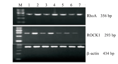
