Objective To investigate effect of Qindan particles on acute physiological change in rats under heatstroke, and to explore its mechanism. Methods Male anesthetized Sprague Dawley rats were randomly divided into normal control group, model control group, Qindan low-dose group, Qindan middle dose group and Qindan high-dose group. The model control group and Qindan groups were orally administered with vehicle (0.9% sodium chloride solution) or Qindan 10, 20 and 40 g·kg-1 for 30 days, respectively, followed by exposure to heat (42 ℃ for 75 min) before recovery at room temperature (RT, 24 ℃). The normal control group rats were treated with vehicle and were kept at room temperature. Core body temperature (Tc), heart rate (HR), mean arterial pressure (MAP) and systolic arterial blood pressure (SAP) were monitored. After the thermal damage, blood was collected immediately and the serum superoxide dismutase (SOD), malondialdehyde (MDA), total nitric oxide synthase (TNOS), induce nitric oxide synthase (iNOS) levels were detected. Part of the rats recovered at room temperature, and the time of death was observed. Observation of liver tissue pathological changes was carried out also. Results The Tc, MDA and iNOS in heatstroke model control group rats were (41.05±0.30) ℃, (11.66±2.25) μmol·L-1, (23.66±2.05) U·L-1, respectively, significantly higher than those of normal control group. The level of serum SOD was (291.22±51.17) U·mL-1, significantly lower than that of normal control group. After 60 min, the values of HR, MAP and SAP were maxed at (474.13±18.40) beat·min-1, (138.35±6.51) mmHg, and (187.12±7.85) mmHg, significantly higher than those of normal control group. After 75 min, the indexes fell rapidly to (309.58±22.47) beat·min-1, (104.11±4.26) mmHg, and (140.46±6.74) mmHg, respectively. The levels of Tc, MDA, iNOS fell to (39.94±0.17) ℃, (7.90±1.57) μmol·L-1, (17.20±1.57) U·L-1 and SOD rose to (373.51±38.78) U·mL-1 in Qindan particles high-dose group. After 75 min, the values of HR, MAP and SAP rose to (409.58±22.50) beat·min-1, (124.11±7.26) mmHg and (172.85±4.09) mmHg. ConclusionQindan particles can delay the onset of heatstroke and reduce the thermal damage, playing a protective role in rats under heat stress. This protective effect may be related to relieving oxidative stress reactions.
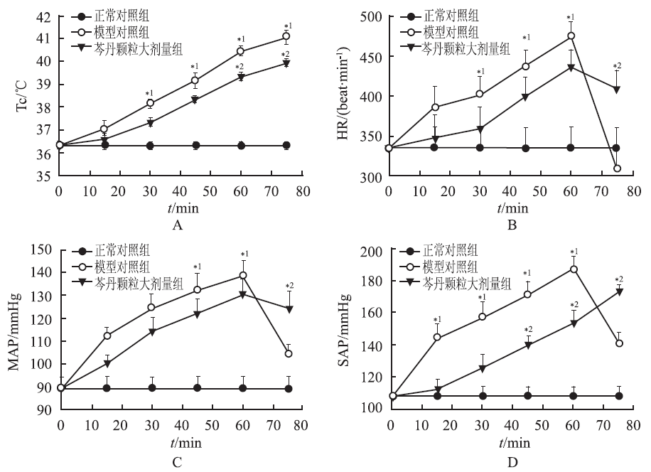
与正常对照组比较,模型对照组大鼠入仓15 min后Tc、MAP、HR、SAP开始升高,30~60 min明显升高(P<0.05),60 min后除Tc外,其他各指标开始降低,75 min 明显降低(P<0.05);与模型对照组比较,芩丹颗粒大剂量组大鼠入仓后Tc、MAP、HR、SAP升高速度和幅度均降低,60和75 min Tc明显降低(P<0.05),75 min时MAP、HR降低,但降低幅度明显低于正常对照组。见图1。
表1
Tab.1
表1
表1
5组大鼠SOD、MDA、TNOS、iNOS的含量比较
Tab.1
Comparison of the content of SOD,MDA,TNOS and iNOS among five groups of rats x¯±s,n=15
组别
MDA(μmol·L-1)
SOD
TNOS
iNOS
(U·mL-1)
正常对照组
7.59±1.02
387.06±57.51
26.59±1.43
13.27±1.28
模型对照组
11.66±2.25*1
291.22±51.17*1
35.33±1.17*1
23.66±2.05*1
芩丹颗粒小剂量组
11.10±2.16
313.62±42.59
33.62±2.59
22.10±2.35
芩丹颗粒中剂量组
9.28±1.35
333.29±52.37
32.81±1.78
20.24±1.85
芩丹颗粒大剂量组
7.90±1.57*2
373.51±38.78*2
31.59±1.63
17.20±1.57*2
Compared with normal control group,*1P<0.05;compared with model control group,*2P<0.05
与正常对照组比较,*1P<0.05;与模型对照组比较,*2P<0.05
表1
5组大鼠SOD、MDA、TNOS、iNOS的含量比较
Tab.1
Comparison of the content of SOD,MDA,TNOS and iNOS among five groups of rats x¯±s,n=15
表2
Tab.2
表2
表2
5组大鼠中暑时间和中暑后存活时间比较
Tab.2
Comparison of the heat stroke starting time and survival time among five groups of rats min,x¯±s,n=15
组别
MAP达到 峰值时间
MAP由峰值下降 25 mmHg时间
存活时间
正常对照组
≥480
≥480
≥480
模型对照组
60.33±2.36*1
75.66±3.26*1
85.17±3.85*1
芩丹颗粒小剂量组
61.58±2.27
77.27±2.45
86.23±3.85
芩丹颗粒中剂量组
65.61±1.97
80.33±3.07
87.26±1.97
芩丹颗粒大剂量组
75.52±3.01*2
85.33±2.62*2
95.25±3.16*2
Compared with normal control group,*1P<0.05;compared with model control group,*2P<0.05
与正常对照组比较,*1P<0.05;与模型对照组比较,*2P<0.05
表2
5组大鼠中暑时间和中暑后存活时间比较
Tab.2
Comparison of the heat stroke starting time and survival time among five groups of rats min,x¯±s,n=15
Fig.1
Comparison of Tc(A)、HR(B)、MAP(C) and SAP(D) among three groups of rats(x¯±s,n=15) Compared with normal control group,*1P<0.05;compared with model control group,*2P<0.05
Fig.2
Histopathological changes of livers in three groups of rats(HE staining,×400) A. normal control group; B. model control group; C.high-dose Qindan group
Focuses on heat stroke, which is a life-threatening illness characterized by an elevated core body temperature that rises above 40 070705C. Dysfunction of the central nervous system that results in delirium, convulsions, or coma; How heat stroke is often fatal, despite adequate lowering of the body temperature and aggressive treatment; Responses; Pathophysiology; Clinical and metabolic manifestations; Treatment.
LEON LR, BOUCHAMAA.Heat stroke[J]. Compr Physiol,2015,5(2):611-647.
Focuses on heat stroke, which is a life-threatening illness characterized by an elevated core body temperature that rises above 40 070705C. Dysfunction of the central nervous system that results in delirium, convulsions, or coma; How heat stroke is often fatal, despite adequate lowering of the body temperature and aggressive treatment; Responses; Pathophysiology; Clinical and metabolic manifestations; Treatment.
ZHAO YQ, GAO JT, LIU SH, et al.Geranylgeranylacetone preconditioning may attenuate heat-induced inflammation and multiorgan dysfunction in rats[J]. J Pharm Pharmacol,2010,62(1):99-105.
ABSTRACT Geranylgeranylacetone, an acyclic isoprenoid, is a non-toxic inducer of heat shock protein (HSP)70. HSP70 overproduction is associated with heat tolerance in rats. This study aimed to investigate whether geranylgeranylacetone preconditioning of rats reduced heat-induced inflammation and multiple organ dysfunction. Anaesthetised rats were given vehicle or geranylgeranylacetone (800 mg/kg) orally. After 48 h they were exposed to ambient temperature of 43 degrees C for 70 min to induce heatstroke. Another group of rats kept at room temperature were used as normothermic controls. Vehicle-treated rats all succumbed to heat stress; their survival time was 25 +/- 4 min. Pretreatment with geranylgeranylacetone significantly increased survival time to 92 +/- 15 min. Compared with normothermic controls, all vehicle-treated heatstroke rats displayed hepatic and renal dysfunction (e.g. increased plasma levels of serum urea nitrogen, creatinine, aspartate aminotransferase, alanine aminotransferase and alkaline phosphatase) and active inflammation (e.g. increased plasma and brain levels of interleukin-1 beta, tumour necrosis factor-alpha and interleukin-6). These heat-stress response indicators were all significantly suppressed by geranylgeranylacetone pretreatment. In addition, the plasma and brain levels of interleukin-10 (an anti-inflammatory cytokine) and brain levels of HSP70 were significantly increased after geranylgeranylacetone preconditioning during heatstroke. Geranylgeranylacetone preconditioning attenuates heat-induced inflammation and multiorgan dysfunction in rats.
ALZEER AH, AL-ARIFIA, WARSY AS, et al.Nitric oxide production is enhanced in patients with heat stroke[J]. Intens Care Med, 1999, 25(1):58-62.
Objective: To determine whether nitric oxide (NO) production is increased in heat stroke (HS) patients. Design: A prospective analysis of nitrite and nitrate (NO · 2 /NO 3 ) levels in ten HS patients was performed at the HS center in Makkah, Saudi Arabia. Methods: Plasma (NO · 2 /NO 3 ) levels were determined spectrophotometrically before cooling (0 time), and at 6, 12, and 24 h post-cooling. Results: The mean level of NO in the ten HS victims before cooling was significantly higher than in eight control patients (35.6 ± 37.0 vs 3.0 ± 4.2 μmol/l; p < 0.01). The levels were higher in non-survivors than in survivors. NO also correlated positively with the Acute Physiology and Chronic Health Evaluation II score ( r = 0.72, p < 0.018). There was no correlation between the NO level before cooling and blood pressure, rectal temperature, or cooling time. Conclusion: HS is associated with excessive NO production, the magnitude of which is proportional to the severity of illness. NO may be an important mediator and integral part of the pathophysiological processes resulting in HS and may be a central factor linking the neurological and cardiovascular abnormalities observed in HS.
SHARMA HS, WESTMANJ, ALMP, et al.Involvement of nitric oxide in the pathophysiology of acute heat stress in the rat - influence of a new antioxidant compound H-290/51[J]. Thermoregulation,1997, 813(1):581-590.
Abstract The possibility that nitric oxide (NO) is involved in the pathophysiology of brain injury caused by heat stress (HS) was examined using immunohistochemistry of a constitutive isoform of neuronal nitric oxide synthase (c-NOS) in a rat model. In addition, to discover the role of oxidative stress in inducing c-NOS activity in HS, the effect of a new antioxidant H-290/51 on HS-induced expression of c-NOS immunoreactivity was examined. Subjection of conscious young animals to a 4-h HS in a biological oxygen demand (BOD) incubator at 38 degrees C resulted in marked upregulation of c-NOS in the cerebral cortex and hippocampus of stressed rats compared to normal rats kept at room temperature (21 +/- 1 degrees C). The c-NOS immunoreactivity was found in distorted neurons located in the edematous regions not normally showing c-NOS activity. Pretreatment with H-290/51 significantly attenuated the upregulation of c-NOS in animals subjected to HS, and the signs of neuronal distortion and edema were less pronounced. These results suggest that HS has the capacity to induce upregulation of c-NOS, and these effects can be reduced by prior treatment with H-290/51, indicating a possible neuroprotective effect of antioxidants in thermal brain injury.
CHANG CP, LEE CC, CHEN SH, et al.Aminoguanidine protects against intracranial hypertension and cerebral ischemic injury in experimental heatstroke[J]. J Pharmacol Sci, 2004,95(1):56-64.
Abstract The aim of the present study was to ascertain whether aminoguanidine attenuated intracranial hypertension and cerebral ischemic injury in experimental heatstroke. Urethane-anesthetized rats were exposed to heat stress (ambient temperature of 43 degrees C) to induce heatstroke. Control rats were exposed to 24 degrees C. Mean arterial pressure, cerebral perfusion pressure, and cerebral blood flow after the onset of heatstroke were all significantly lower than in control rats. However, colonic temperature, intracranial pressure, heart rate, cerebral inducible nitric oxide synthase (iNOS)-dependent NO, and neuronal damage score were greater after the onset of heatstroke. Aminoguanidine (30 micromol/kg, i.v.; 30 min before the start of heat exposure) pretreatment significantly attenuated the heatstroke-induced hyperthermia, arterial hypotension, intracranial hypertension, cerebral ischemia and neuronal damage, and increased iNOS-dependent NO formation in the brain. The extracellular concentrations of ischemic (e.g., glutamate and lactate/pyruvate ratio) and damage (e.g., glycerol) markers in the hypothalamus were also increased after the onset of heatstroke. Aminoguanidine pretreatment significantly attenuated the increase in hypothalamic ischemia and damage markers associated with heatstroke. Delaying onset of aminoguanidine administration (i.e., 0 or 30 min after the start of heat exposure) reduced the preventive efficiency on heatstroke-induced hyperthermia, arterial hypotension, intracranial hypertension, cerebral ischemia, and increased iNOS-dependent NO formation in brain. These results suggest that aminoguanidine protects against heatstroke-induced intracranial hypertension and cerebral ischemic injury by inhibition of cerebral iNOS-dependent NO production.
CHENG BC, CHANG CP, LIN MT, et al.Inhibition of neuronal nitric oxide synthase causes attenuation of cerebrovascular dysfunction in experimental heatstroke[J]. Neuropharmacology,2007, 52(2):297-305.
Abstract The present study was performed to assess the prophylactic effect of 7-nitroindazole (7-NI), an inhibitor of neuronal nitric oxide synthase (nNOS), in an animal model of heatstroke. Anesthetized rats, immediately before the start of heat stress, were divided into two major groups and given the following: vehicle solution (1 mL per kg body weight) or 7-NI (5-20mg/mL per kg body weight) intraperitoneally. They were exposed to ambient temperature of 43 degrees C to induce heatstroke. Another group of rats were exposed to room temperature (24 degrees C) and used as normothermic controls. Their physiologic and biochemical parameters were continuously monitored. When the vehicle-pretreated rats underwent heat stress, their survival time values were found to be 21-25 min. Pretreatment with intraperitoneal doses of 7-NI significantly improved survival during heatstroke (55-164 min). As compared to those of normothermic controls, all vehicle-pretreated heatstroke animals displayed higher levels of core temperature, intracranial pressure, nitric oxide metabolite (NO(2)(-)), glutamate, glycerol, lactate/pyruvate ratio, neuronal damage score and nNOS expression in the hypothalamus, and tumor necrosis factor-alpha (TNF-alpha) in the serum. In contrast, all vehicle-pretreated heatstroke animals had lower levels of mean arterial pressure, cerebral perfusion pressure, cerebral blood flow, and brain PO(2). Administration of 7-NI before the start of heat exposure significantly reduced the hyperthermia, intracranial hypertension, nNOS-dependent NO(2)(-), glutamate, glycerol, lactate/pyruvate ratio, and neuronal damage score in the hypothalamus, as well as overproduction of TNF-alpha in the serum that occurred during heatstroke. The data show that reduction of nNOS-dependent NO(2)(-) with 7-NI causes attenuation of cerebrovascular dysfunction, hyperthermia, and TNF-alpha overproduction during heatstroke in the rat.
CHEN SH,LIN MT,CHANG CP,et al.Ischemic and oxidative damage to the hypothalamus may be responsible for heat stroke[J].Curr Neuropharmacol, 2013,11(2): 129-140.
The hypothalamus may be involved in regulating homeostasis, motivation, and emotional behavior by controlling autonomic and endocrine activity. The hypothalamus communicates input from the thalamus to the pituitary gland, reticular activating substance, limbic system, and neocortex. This allows the output of pituitary hormones to respond to changes in autonomic nervous system activity. Environmental heat stress increases cutaneous blood flow and metabolism, and progressively decreases splanchnic blood flow. Severe heat exposure also decreases mean arterial pressure (MAP), increases intracranial pressure (ICP), and decreases cerebral perfusion pressure (CPP = MAP - ICP), all of which lead to cerebral ischemia and hypoxia. Compared with normothermic controls, rodents with heatstroke have higher hypothalamic values of cellular ischemia (e.g., glutamate and lactate-to-pyruvate ratio) and damage (e.g., glycerol) markers, pro-oxidant enzymes (e.g., lipid peroxidation and glutathione oxidation), proinflammatory cytokines (e.g., interleukin-1 and tumor necrosis factor-), inducible nitric oxide synthase-dependent nitric oxide, and an indicator for the accumulation of polymorphonuclear leukocytes (e.g., myeloperoxidase activity), as well as neuronal damage (e.g., apoptosis, necrosis, and autophagy) after heatstroke. Hypothalamic values of antioxidant defenses (e.g., glutathione peroxidase and glutathione reductase), however, are lower. The ischemic, hypoxic, and oxidative damage to the hypothalamus during heatstroke may cause multiple organ dysfunction or failure through hypothalamic-pituitary-adrenal axis mechanisms. Finding the link between the signaling and heatstroke-induced hypothalamic oxidative and ischemic damage might allow us to clinically attenuate heatstroke. In particular, free radical scavengers, heat shock protein-70 inducers, hypervolemic hemodilution, inducible nitric oxide synthase inhibitors, progenitor stem cells, flutamide, estrogen, interleukin-1 receptor antagonists, glucocorticoid, activated protein C, and baicalin mitigate preclinical heatstroke levels.
LEON LR,DINEENS,BLAHA MD, et al.Attenuated thermoregulatory, metabolic, and liver acute phase protein response to heat stroke in TNF receptor knockout mice[J]. Am J Physiol Regul Integr Comp Physiol, 2013,305(12):1421-1432.
Tumor necrosis factor (TNF) is considered an adverse mediator of heat stroke (HS) based on clinical studies showing high serum levels. However, soluble TNF receptors (sTNFR; TNF antagonists) were higher in survivors than nonsurvivors and TNFR KO mice showed a trend towards increased mortality suggesting TNF has protective actions for recovery. We delineated TNF actions in HS by comparing thermoregulatory, metabolic and inflammatory responses between B6129F2 (WT) and TNFR KO mice. Prior to heat exposure, TNFR KO mice showed ~0.4C lower core temperature (Tc; radiotelemetry), ~10% lower metabolic rate (Mr; indirect calorimetry) and reduced plasma IL-1 and sIL-1RI than WT mice. KO mice selected warmer temperatures than WT mice in a gradient, but remained hypothermic. In the calorimeter, both genotypes showed a similar heating rate, but TNFR KO maintained lower Tc and Mr than WT mice for a given heat exposure duration and required ~30 min longer to reach maximum Tc (42.4C). Plasma IL-6 increased at ~3h of recovery in both genotypes, but KO mice showed a more robust sIL-6R response. Higher sIL-6R in the KO mice was associated with delayed liver p-STAT3 protein expression and attenuated serum amyloid A3 (SAA3) gene expression suggesting the acute phase response (APR) was attenuated in these mice. Our data suggest that the absence of TNF signaling induced a regulated hypothermic state in the KO mice, TNF-IL-1 interactions may modulate Tc and Mr during homeostatic conditions, and TNF modulates the APR during HS recovery through interactions with the liver IL-6-STAT3 pathway of SAA3 regulation.
SHEN KH, LIN CH, CHANG HK, et al.Premarin can act via estrogen receptors to rescue mice from heatstroke-induced lethality[J].Shock, 2008, 30(6):668-674.
The present study was conducted to assess whether Premarin, a water-soluble estrogen sulfate, can act via estrogen receptors (ERs) to rescue mice from heat-induced lethality. Unanesthetized, unrestrained mice were exposed to ambient temperature of 42.4 degrees C to induce heatstroke (HS). Another group of mice was exposed to room temperature (24 degrees C) and used as normothermic controls. They were given isotonic sodium chloride solution, Premarin (0.1 - 1.0 mg/kg of body weight, i.p.), or Premarin (1 mg/kg of body weight, i.p.) plus the nonselective ER antagonist ICI 182, 780 (0.25 mg/kg of body weight, i.p.) 1 h after the termination of heat stress. Their physiologic and biochemical parameters were continuously monitored. Mice that survived on day 4 of heat treatment were considered survivors. When the vehicle-treated mice underwent heat, the fraction survival and core temperature at +4 h of body heating were found to be 0 of 12 and 34.4 degrees C +/- 3 degrees C, respectively. Administration of Premarin (1 mg/kg) 1 h after the cessation of heat stress rescued the mice from heat-induced death (fraction survival, 12/12) and reduced the hypothermia (core temperature, 37.3 degrees C). The beneficial effects of Premarin in ameliorating lethality and hypothermia can be abolished by simultaneous administration of ICI 182, 780. Both IL-10 (an anti-inflammatory cytokine) and estradiol in the serum were increased significantly in heat-stressed mice administered Premarin compared with vehicle-treated HS group. Heat-induced apoptosis, as indicated by terminal deoxynucleotidyl-transferase-mediated alpha UDP-biotin nick end-labeling staining, in the spleen, liver, and kidney were significantly reduced by Premarin. The increased levels of cellular ischemia (e.g., glutamate, lactate-to-pyruvate ratio, and nitrite) and damage (e.g., glycerol) markers and iNOS expression in the hypothalamus during HS were decreased significantly by Premarin therapy. The levels of proinflammatory cytokines (e.g., IL-1 beta and TNF-alpha) and renal and hepatic dysfunction markers in plasma that are up-regulated in heat stressed mice were significantly lower in Premarin-administered mice. The data indicate that Premarin may act via ERs to rescue mice form HS-induced lethality.
LIN MT, LIU HH, YANG YL.Involvement of interleukin-1 receptor mechanisms in development of arterial hypotension in rat heatstroke[J]. Am J Physiol,1997, 273(4 Pt 2): H2072-H2077.
Rats, under urethan anesthesia, were exposed to a high ambient temperature (42 degrees C) to induce heatstroke and to assess the hemodynamic changes associated with heatstroke. Compared with normothermic controls, rats with heatstroke showed higher values of colonic temperature, heart rate, and plasma levels of interleukin (IL)-1 but lower values of R wave amplitude, P-R and Q-T intervals, systolic wave amplitude, diastolic and dicrotic wave duration, mean arterial pressure, stroke volume, and cardiac output. Animals injected intravenously with an IL-1-receptor antagonist at the time of heatstroke induction were protected from some of the cardiovascular effects of heatstroke, such as depressed ventricular depolarization, decreased stroke volume, decreased cardiac output, and arterial hypotension. The hemodynamic changes associated with heatstroke could be mimicked by IL-1beta administration. Other cardiovascular parameters such as total peripheral vascular resistance were unaffected by heatstroke induction or IL-1beta treatment. The results indicate that a selective decline in stroke volume or ventricular depolarization resulting from increased plasma levels of IL-1 may be an important mechanism signaling arterial hypotension or circulatory failure in rat heatstroke.
HELWIG BG, LEON LR.Tissue and circulating expression of IL-1 family members following heat stroke[J]. Physiol Genomics, 2011,43(19):1096-1104.
Interleukin-1 (IL-1) is thought to have a significant role in the pathophysiology of heat (HS), although little is known regarding the actions or expression patterns of the IL-1 family. This study tested the hypotheses that following HS IL-1 family is dynamic, while loss of IL-1 enhances recovery. IL-1 family expression was determined in plasma, spleen, and liver from C57BL/6J (n=24 control, n=20 HS) at maximum core temperature (Tc,Max), hypothermia, and 24 h post-HS (24 h). Soluble IL-1 receptor subtype I (sIL-1RI) expression peaked at 24 h (14,659.01±2,016.28 pg/ml, P<0.05), while sIL-1RII peaked at hypothermia (19,099.30±1,177.07 pg/ml). IL-1α in the spleen (ninefold) and liver (fourfold) along with IL-1RI (threefold spleen and fivefold liver) were maximal at hypothermia. Spleen IL-1β peaked at Tc,Max (fourfold) but at hypothermia (fourfold) in liver. of the IL-1 family member peaked (2.5-fold) at Tc,Max but was similar at all other time points. Subsequent studies revealed that despite accruing a greater heating area (298±16 vs. 247±13°C·min, P<0.05), IL-1RI knockout (KO) (n=14) showed an attenuated hypothermia depth (28.5±0.2 vs. 27.3±0.5°C, P<0.05) and duration (675±82 vs. 1,283±390 min, P<0.05) with a higher 24 h Tc (36.9 vs. 34.1°C, P<0.05) compared with C57BL/6J (n=8). The current results demonstrate that following HS IL-1 family is altered and IL-1RI KO display Tc responses consistent with a more rapid recovery.
LIN XJ, MEI GP, LIUJ, et al.Therapeutic effects of melatonin on heatstroke-induced multiple organ dysfunction syndrome in rats[J]. J Pineal Res,2011,50(4):436-444.
Abstract:68 Melatonin reportedly exerts beneficial effects to attenuate multiple organ dysfunction syndrome (MODS) in septic shock. Heatstroke resembles septic shock in many aspects. Thus, this study was performed on the anesthetized rats by using heat exposure to induce heatstroke-associated MODS. We evaluated the effect of melatonin, a versatile molecule synthesized in the pineal gland and in many organs, in heatstroke rats and showed that melatonin (0.2–5.065mg/kg of body weight, i.v., immediately after the start of heat stress) significantly (i) attenuated hyperthermia, hypotension and hypothalamic ischemia and hypoxia, (ii) reduced plasma index of the toxic oxidizing radicals like nitric oxide metabolites and hydroxyl radicals, (iii) diminished plasma index of hepatic and renal dysfunction like creatinine, blood urea nitrogen, alanine aminotransferase, aspartate aminotransferase, alkaline phosphatase, and lactate dehydrogenase, (iv) attenuated plasma systemic inflammation response molecules like soluble intercellular and lesion molecule-1, E-selectin, tumor necrosis factor-alpha, interleukin (IL)-1β, and IL-6, (v) promoted plasma levels of an anti-inflammatory cytokine IL-10, (vi) reduced an index of infiltration of polymorphonuclear neutrophils in the lung like myeloperoxidase activity, and (vii) promoted the survival time to fourfold compared with the heatstroke alone group. Thus, melatonin could be a novel agent for the treatment of heatstroke animals or patients in the early stage.
RODRIGUEZ-FERNANDEZM, GROSMANB, DOYLE FJ, et al.Modeling the intra- and extracellular cytokine signaling pathway under heat stroke in the liver[J]. PLoS One,2013,8(9):e73393.
Heat stroke (HS) is a life-threatening illness induced by prolonged exposure to a hot environment that causes central nervous system abnormalities and severe hyperthermia. Current data suggest that the pathophysiological responses to heat stroke may not only be due to the immediate effects of heat exposure per se but also the result of a systemic inflammatory response syndrome (SIRS). The observation that pro- (e.g., IL-1) and anti-inflammatory (e.g., IL-10) cytokines are elevated concomitantly during recovery suggests a complex network of interactions involved in the manifestation of heat-induced SIRS. In this study, we measured a set of circulating cytokine/soluble cytokine receptor proteins and liver cytokine and receptor mRNA accumulation in wild-type and tumor necrosis factor (TNF) receptor knockout mice to assess the effect of neutralization of TNF signaling on the SIRS following HS. Using a systems approach, we developed a computational model describing dynamic changes (intra- and extracellular events) in the cytokine signaling pathways in response to HS that was fitted to novel genomic (liver mRNA accumulation) and proteomic (circulating cytokines and receptors) data using global optimization. The model allows integration of relevant biological knowledge and formulation of new hypotheses regarding the molecular mechanisms behind the complex etiology of HS that may serve as future therapeutic targets. Moreover, using our unique modeling framework, we explored cytokine signaling pathways with three in silico experiments (e.g. by simulating different heat insult scenarios and responses in cytokine knockout strains in silico).

 , 万朋
, 万朋
