Objective To observe the effect of qixiantang decoction on asthma model mice and to explore its mechanism of phosphatase gene (PTEN)-up-regulation. Methods A total of 28 healthy female BALB/c mice were divided into 4 groups according to the random number table(n=7): normal control group, model control group, qixiantang decoction group, and dexamethasone group. The mice were sensitized with ovalbumin (OVA) for asthma model. Qixiantang decoction group was treated with drug after OVA sensitization. Hematoxylin-eosin (H-E) staining was applied to observe the pulmonary inflammation in mice, and periodic acid Schiff (PAS) staining was used to examine airway mucus secretion. ELISA was used to detect the concentration of serum IgE. Real-time quantitative PCR was used to examine IL-13 and IL-5 gene expression changes in lung tissues of mice. Western blotting was used to detect the expression of PTEN and SIRT1 protein in lung tissues.Results The lung tissue inflammatory infiltration and mucus secretion in model control group were higher than normal control group (P<0.01), and that in the qixiantang decoction group. The level of serum IgE in model control group [(6.67±2.59) pg·mL-1)] was significantly higher than normal control group [(0.27±0.05) pg·mL-1, P<0.01] ,and that in the qixiantang decoction group [(3.52±1.44) pg·mL-1,P<0.05]. The expression of PTEN and SIRT1 in lung tissue of model control group were significantly lower than normal control group, and that of qixiantang decoction group. The expression of IL-5 and IL-13 mRNA of qixiantang decoction group was significantly lower (P<0.05). ConclusionQixiantang decoction could significantly ameliorate inflammation in asthmatic mice by regulate IgE、IL-5、IL-13 expression,and might up-regulate PTEN expression via SIRT1 signal.
Key words:
decoction
;
Asthma
;
Phosphatase gene
哮喘已成为影响全球3亿多人的公共卫生问题,其发病率越来越高,迫切需要开发有效的治疗药物[1]。治疗哮喘的传统方法为使用糖皮质激素和支气管扩张药,然而患者的复发情况依然没有得到理想的控制,生活质量下降。本课题组通过大量的中医临床治疗观察发现,中药方剂在治疗哮喘方面确实有可靠的疗效,因此探讨中药方剂的作用机制对寻找新的哮喘疗法具有重要意义。本课题组选择在哮喘临床治疗中已取得肯定疗效的自拟芪仙汤(主要由补肾要药巴戟天、淫羊藿和益气要药黄芪等组成)进行动物实验以探究其治疗哮喘的作用机制。张力蛋白同源10号染色体丢失的磷酸酶基因(phosphatase gene ,PTEN)是近年来新发现的一个与哮喘发病相关的基因[2]。PTEN基因和沉默信息调节因子1(silent information regulator 1,SIRT1)基因在哮喘的发病机制中具有重要作用,为此本实验拟通过支气管哮喘小鼠模型,进一步观察芪仙汤对哮喘小鼠的作用,并初步探讨PTEN基因表达上调的机制。
图1
4组小鼠肺组织病理特征 (×200) A. 正常对照组;B. 模型对照组;C. 芪仙汤组;D. 地塞米松组
Fig.1
Pathological features of lung tissues in four groups of mice ( ×200) A. normal control group; B. model control group; C. qixiantang decoction group; D. dexamethasone group
图2
4组小鼠肺组织中IL-5和IL-13基因的表达 (x¯±s,n=7) A. 正常对照组;B. 模型对照组;C. 芪仙汤组;D. 地塞米松组;与正常对照组比较,*1P<0.05;与模型对照组比较, *2P<0.05
Fig.2
Gene expression of IL-13 and IL-5 in lung tissues of four groups of mice (x¯±s,n=7) A. normal control group; B. model control group; C. qixiantang decoction group; D. dexamethasone group;Compared with normal control group, *1P<0.05;compared with model control group, *2P<0.05
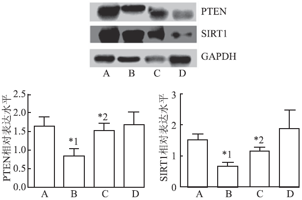
图3
4组小鼠肺组织中PTEN和SIRT1的表达 (x¯±s,n=7) A. 正常对照组;B. 模型对照组;C. 芪仙汤组;D. 地塞米松组; 与正常对照组比较,*1P<0.05;与模型对照组比较, *2P<0.05
Fig.3
Expression of PTEN and SIRT1 in lung tissue of four groups of mice (x¯±s,n=7) A. normal control group; B. model control group; C. qixiantang decoction group; D. dexamethasone group; Compared with normal control group, *1P<0.05;compared with model control group, *2P<0.05
MATHIAS CB.Natural killer cells in the development of asthma[J]. Current Allergy Asthma Reports, 2015, 15(2): 1-8.
ABSTRACT Asthma is an immune-mediated disease of the airways characterized by reversible airway obstruction, bronchial eosinophilic inflammation, and airway hyperresponsiveness (AHR). The immune dysregulation in asthma has been attributed to the involvement of diverse immune cells that contribute to the immunopathology of the disease. Natural killer (NK) cells play critical roles in host defense against viruses and various cancers. Accumulating evidence demonstrates additional important roles for these cells in T cell priming, dendritic cell maturation, and the development of inflammation, all of which have the potential to enhance or dampen allergic responses. The ability of NK cells to produce Th2-type cytokines and their pivotal role in combating respiratory infections which cause airway dysfunction in asthmatics further suggest that they may directly contribute to the immunopathogenesis of allergic airway disease. In this review, we examine emerging evidence and discuss the putative roles of NK cells in the sensitization, progression, and resolution of asthma.
IKENOUET, INOKIK, ZHAOB, et al.PTEN acetylation modulates its interaction with PDZ domain[J]. Cancer Res, 2008, 68(17): 6908-6912.
The suppressor gene is frequently inactivated in . As a major suppressor, function must be tightly regulated. Both and association have been reported to regulate activity. In addition, the COOH terminus of has a typical motif that interacts with several . In this report, we show that is acetylated on (402), which is in the COOH-terminal motif. We show that plays a major role in acetylation, whereas the deacetylase is mainly responsible for deacetylation. Interestingly, (402) acetylation modulates interaction with , indicating a potential role of acetylation in regulating function.
BARNES PJ.Th2 cytokines and asthma: an introduction[J]. Respir Res, 2001, 2(2): 64-65.
In this issue of Respiratory Research we focus on Th2 cytokines and their potential role in allergic diseases, such as asthma. John Steinke and Larry Borish [4] discuss the role of IL-4 in the pathogenesis of asthma and make the point that this is an upstream cytokine that regulates allergic inflammation by promoting Th2 cell differentiation and IgE synthesis. Early studies with an IL-4 antagonist, soluble recombinant IL-4 receptor (altrakincept), show therapeutic benefit as a steroid-replacing agent in moderately severe asthma [5] and longer term clinical trials are now underway. IL-5 is discussed by Scott Greenfeder and colleagues [6]. IL-5 is a cytokine that is highly specific for eosinophilic inflammation and antibodies that block IL-5 actions are effective in reducing eosinophilic inflammation and airway hyperresponsiveness (AHR) in various species. Recently, studies of a humanised anti-IL-5 monoclonal antibody (mepomizulab) in asthmatic patients have confirmed its extraordinary efficacy in reducing eosinophils in the circulation and the airways, but surprisingly no reduction in response to allergen or in AHR [7]. This result has been confirmed in a preliminary clinical trial of asthmatic patients whose symptoms were not controlled with inhaled corticosteroids and who showed no clinical improvement with anti-IL-5 antibody, despite a marked suppression of circulating eosinophils [8]. These studies confirm the importance of IL-5 in eosinophilic inflammation in man, but question the role of eosinophils in asthma. IL-13 has many actions similar to those of IL-4 and also regulates IgE production but, unlike IL-4, it does not regulate T cell differentiation to Th2 cells and T lymphocytes do not respond to IL-13. The role of IL-13 in asthma was recently reviewed in this journal by Marsha Wills-Carp [9]. IL-9 has been less intensively investigated than the other Th2 cytokines, but appears to amplify Th2-cell-mediated responses, as reviewed by Roy Levitt and colleagues
The article discusses the drug Bosatria (mepolizumab) manufactured by London-based GlaxoSmithKline (GSK), which targets the pro-inflammatory cytokine interleukin-5 (IL-5) to curb severe asthma. Other drugs discussed include Teva Pharmaceutical's Cinquil (reslizumab), MedImmune's Benralizumab for eosinophilic inflammation, Regeneron and Sanofi's Dupilumab that inhibits IL-4 and IL-13.
MENZELLAF, LUSUARDIM, GALEONEC, et al.Tailored therapy for severe asthma[J]. Multidiscip Respira Med , 2015, 10(1): 1-8.
Abstract Patients with severe asthma or COPD have often a suboptimal symptom control due to inadequate treatment. A better understanding of pathogenetic mechanisms, phenotypes, endotypes and the new technologies available in the fields of molecular biology and immunogenetics have made it possible to synthesize specific monoclonal antibodies virtually able to interact with any target antigen, or to open a way for new therapeutic target options. At the moment, the only biologic drug available in clinical practice is omalizumab. To overcome the limits of omalizumab, the research has focused on new monoclonal antibodies presenting higher avidity for IgE (e.g. ligelizumab and lumiximab) and ability to interact also with low affinity IgE receptor (Fc系RII). At present, many new biological drugs with different mechanisms of action and targets are matter of research. It is very important to identify the asthmatic phenotype in order to select the most appropriate drug for the individual patient. The most promising agents are targeted against cytokines of Th2 pattern and related receptors, such as IL-2 (daclizumab) and IL-13 (lebrikizumab) or IL-5 in patients with hypereosinophilia (mepolizumab, reslizumab and benralizumab). Other interesting drugs have as a target TNF-伪 or its soluble receptor (infliximab, golimumab and etanercept) or IL-1 (canakinumab), a cytokine with an important systemic proinflammatory action. Finally, the discovery of increased levels of C5a in the airways of asthmatic patients has led to the synthesis of a specific monoclonal antibody (eculizumab). Further help should come from the identification of biomarkers that can guide in choosing the best treatment for the individual patient, such as IgE for omalizumab or periostin for lebrikizumab.
DU WJ,DONG JC,CAIC.Effects of icariin on Bcl-2 and Bax protein expressions and eosinophils apoptosis in bronchial asthmatic mice[J]. Chin J Integ Tradi West Med, 2011,31(9):1248-1253.
Objective To study the effects of icarrin on Bcl-2 and Bax protein expressions and eosinophils apoptosis in bronchial asthmatic mice.Methods 48 female Balb/c mice were randomly divided into 6 groups,i.e.,the normal control group,the model group,the Dexamethasone group,the low dose icariin group,the middle dose icariin group,and the high dose icariin group,8 mice in each group.Bronchial asthma in mice were induced by intraperitioneal sensitization and challenged with nebulized ovalbumin(OVA).The mice of each treatment group were administrated with different doses of icariin by peritoneal injection from the first asthma sensitization(the 3rd week after the modeling) to the day before killing once every other day,while mice in the normal control group were administrated with physiological saline.The mice were killed after 6 weeks of treatment.The apoptosis of eosinophils and the Bcl-2 and Bax protein expressions of the lung tissues were detected by TUNEL and immunohistochemical assay respectively.Results As compared with the model group,the apoptosis ratio of eosinophils were higher in the rest four treatment groups(P0.05).The Bcl-2 protein positive areas in the lung tissues and the airway wall were significantly lowered(P0.05).The Bax protein positive area significantly increased(P0.05).Conclusion In bronchial asthmatic mice,icariin could enhance the apoptosis of eosinophils and lessen their infiltration by decreasing the expression of Bcl-2 protein and increasing the expression of Bax protein in lung.
LIB, DUANX, XUC, et al.Icariin attenuates glucocorticoid insensitivity mediated by repeated psychosocial stress on an ovalbumin-induced murine model of asthma[J]. Int Immunopharm , 2014, 19(2): 381-390.
Evidence shows that psychosocial stress exacerbates asthma, but there is little intervention to alleviate negative effects of psychosocial stress on asthma. We investigated the role of icariin in anti-inflammation and anti-anxiety potential in a murine model combined psychosocial stress with allergic exposure. The results indicated that icariin administered remarkable increased activity in the center of the open field, reversed airway hyperresponsivenesss, reduced inflammatory cytokine infiltration to the lung and whole body and also in part recovered glucocorticoid responsiveness. Furthermore, our data also showed that icariin significantly inhibited increases of corticosterone and markedly increased glucocorticoid receptor mRNA and protein expression in the lungs of mice exposed to both stress and allergen. Collectively, we speculate that inducing glucocorticoid receptor modulation might be the potential mechanisms of icariin to facilitate corticosteroid responsiveness of cytokine production.
WEIY, LIUB, SUNJ, et al.Regulation of Th17/Treg function contributes to the attenuation of chronic airway inflammation by icariin in ovalbumin-induced murine asthma model[J]. Immunobiology, 2015, 220(6): 789-797.
Icariin which is a flavonoid glucoside isolated from Epimedium brevicornu Maxim, has been reported to have anti-osteoporotic, anti-inflammatory and anti-depressant-like activities. In this study, we observed the effect of icariin on airway inflammation of ovalbumin (OVA)-induced murine asthma model and the associated regulatory mode on T-helper (Th)17 and regulatory T (Treg) cell function. Our data revealed that chronic OVA inhalation induced a dramatic increase in airway resistance ( R L ) and decrease in the lung dynamic compliance (Cdyn), and icariin and DEX treatment caused significant attenuation of such airway hyperresponsiveness (AHR). BALF cell counts demonstrated that icariin and DEX led to a prominent reduction in total leukocyte as well as lymphocyte, eosinophil, neutrophil, basophil and monocyte counts. Histological analysis results indicated that icariin and DEX alleviated the inflammatory cells infiltrating into the peribronchial tissues and goblet cells hyperplasia and mucus hyper-production. Flow cytometry test demonstrated that icariin or DEX administration resulted in a significant percentage reduction in CD4+ROR纬t+ T cells and elevation of CD4+Foxp3+ T cells in BALF. Furthermore, icariin or DEX caused a significant reduction in IL-6, IL-17 and TGF-尾 level in BALF. Unfortunately, icariin had no effect on IL-10 level in BALF. Western blot assay found that icariin or DEX suppressed ROR纬t and promoted Foxp3 expression in the lung tissue. qPCR analysis revealed that icariin and DEX resulted in a notable decrease in ROR纬t and increase in Foxp3 mRNA expression in isolated spleen CD4+ T cell. In conclusion, our results suggested that icariin was effective in the attenuation of AHR and chronic airway inflammatory changes in OVA-induced murine asthma model, and this effect was associated with regulation of Th17/Treg responses, which indicated that icariin may be used as a potential therapeutic method to treat asthma with Th17/Treg imbalance phenotype.
PARK YN, LEE YJ, CHOI JH, et al.Alleviation of OVA-induced airway inflammation by flowers of Inula japonica in a murine model of asthma[J].Biosci Biotech Biochem , 2011, 75(5): 871-876.
The flowers of Inula japonica (Inulae Flos) have long been used in traditional medicine for treating inflammatory diseases. The effects on OVA-induced asthmatic mice of an Inulae Flos extract (IFE) were evaluated in this study. The anti-asthmatic effects of IFE were determined by observing eosinophil recruitment, airway hyper-responsiveness (AHR), Th2 cytokine and IgE levels, and lung histopathology. The IFE treatment effectively reduced the percentage of eosinophils and Th2 cytokines in the bronchoalveolar lavage fluid (BALF) when compared to the levels in OVA-induced mice. IFE also suppressed AHR induced by aerosolized methacholine in OVA-induced mice. The results of the histopathological studies indicate that inflammatory cell infiltration and mucus hypersecretion were both inhibited by the IFE administration when compared to the effect on OVA-induced mice. The IFE treatment also suppressed the serum IgE levels and decreased Th2 cytokines in the supernatant of cultured splenocytes. These results suggest that IFE may have therapeutic potential against asthma.
NAM KW, OH GT, SEO EK, et al.Nuclear factor kappaB-mediated down-regulation of adhesion molecules: possible mechanism for inhibitory activity of bigelovin against inflammatory monocytes adhesion to endothelial cells[J]. J Ethnopharm , 2009, 123(2): 250-256.
The flowers of Inula britannica L. var. chinensis (Rupr.) Reg. (Compositae) are used in traditional medicine to treat asthma, chronic bronchitis, and acute pleurisy in China and Korea. However, the pharmacological actions of Inula britannica L. var. chinensis on endothelial cells and inflammatory monocytes are not clear. In this study, we investigated whether bigelovin, a sesquiterpene lactone isolated from the flowers of Inula britannica L. var. chinensis, inhibits monocyte adhesion and adhesion molecule expression in brain endothelial cells. We measured tumor necrosis factor-alpha (TNF-alpha)-enhanced Raw264.7 monocyte binding to brain endothelial cells and the levels of cell adhesion molecules, including vascular adhesion molecule-1 (VCAM-1), intracellular adhesion molecule-1 (ICAM-1), and endothelial-selectin (E-selectin) on the surface of brain endothelial cells. Bigelovin significantly inhibited these in a dose-dependent manner without affecting cell viability. Furthermore, bigelovin suppressed the nuclear factor kappaB (NF-kappaB) promoter-driven luciferase activity, NF-kappaB activation, and degradation of NF-kappaB inhibitor protein alpha (IkappaBalpha). These results indicate that bigelovin inhibits inflammatory monocyte adhesion to endothelial cells and the expression of VCAM-1, ICAM-1, and E-selectin by blocking IkappaBalpha degradation and NF-kappaB activation.
SHAO CR, CHEN FM, TANG YX.Clinical and experimental study on Ligusticum wallichii mixture in preventing and treating bronchial asthma[J].中国中西医结合杂志, 1994, 14(8): 465-468.
HU JZ, HUANG JH, XIAO ZM, et al.Tetramethy-lpyrazine accelerates the function recovery of traumatic spinal cord in rat model by attenuating inflammation[J]. J Neurolog Sci , 2013, 324(1): 94-99.
In the present study, we explored the effects of tetramethylpyrazine (TMP), an alkaloid extracted from the Chinese herbal medicine Ligusticum wallichii Franchat (chuanxiong), on a rat model of contusion spinal cord injury (SCI). The contusion SCI model was induced in rats by a modified Allen's weight-drop method with a severity of 502g02×025002mm impacting on the T10 segment. In the TMP treatment group, rats were injected intraperitoneally (i.p.) with TMP (20002mg/kg), every 2402h for 502days, starting half an hour after contusion SCI. The control group was treated with saline. Compared with the control group, the TMP group significantly ameliorated the recovery of hindlimb function of rats. TMP treatment significantly reduced the expression of macrophage migration inhibitory factor, nuclear factor κappa B, pro-inflammatory cytokine interleukin-18 and neutrophil infiltration. On the other hand, TMP enhanced the expression of inhibitor κappa B and anti-inflammation cytokine interleukin-10. In conclusion, our results demonstrate that TMP inhibits the development of inflammation and tissue injury associated with spinal cord contusion in rats which may improve the rats' hindlimb function.
MAC, WANGY, DONGL, et al.Anti-inflammatory effect of resveratrol through the suppression of NF-κB and JAK/STAT signaling pathways[J]. Acta Biochim Biophysica Sinica, 2015, 47(3): 207-213.
Resveratrol, the most important ingredient extracted from Polygonum cuspidatum, exerts cytoprotective effects via anti-inflammatory actions in vitro. In this study, we investigated this effect of resveratrol on the lipopolysaccharide (LPS)-induced inflammatory response and its underlying molecular mechanism of action in RAW264.7 murine macrophages. Results showed that resveratrol down-regulated the expression of inducible nitric oxide synthase (iNOS) and interleukin-6 (IL-6), therefore, suppressed the production of nitric oxide and the secretion of IL-6 in LPS-stimulated RAW264.7 cells in a dose-dependent manner. Resveratrol also inhibited the translocation of high-mobility group box 1 (HMGB1) from the nucleus to the cytoplasm and of nuclear transcription factor kappa-B (NF-魏B) p65 from the cytoplasm to the nucleus; it suppressed the phosphorylation of I魏B伪. Furthermore, these actions were mediated by suppressing the phosphorylation of signal transducer and activator of transcription (STAT)-1 and -3. In conclusion, these data indicate that resveratrol exerts anti-inflammatory effects, at least in part by reducing the release of HMGB1 and modulating the NF-魏B and Janus kinase/STAT signaling pathways. Resveratrol could potentially be developed as a useful agent for the chemoprevention of inflammatory diseases. 漏 The Author 2015. Published by ABBS Editorial Office in association with Oxford University Press on behalf of the Institute of Biochemistry and Cell Biology, Shanghai Institutes for Biological Sciences, Chinese Academy of Sciences.
ZHANGL, LIY, GUZ, et al.Resveratrol inhibits enterovirus 71 replication and pro-inflammatory cytokine secretion in rhabdosarcoma cells through blocking IKKs/NF-κB signaling pathway[J]. PLoS One,2015, 10(2):1-13.
Polydatin and resveratrol, as major active components in Polygonum cuspidatum , have anti-inflammatory, antioxidant and antitumor functions. However, the effect and mechanism of polydatin and resveratrol on enterovirus 71 (EV71) have not been reported. In this study, resveratrol revealed strong antiviral activity on EV71, while polydatin had weak effect. Neither polydatin nor resveratrol exhibited influence on viral attachment. Resveratrol could effectively inhibit the synthesis of EV71/VP1 and the phosphorylation of IKKα, IKKβ, IKKγ, IKBα, NF-κB p50 and NF-κB p65, respectively. Meanwhile, the remarkably increased secretion of IL-6 and TNF-α in EV71-infected rhabdosarcoma (RD) cells could be blocked by resveratrol. These results demonstrated that resveratrol inhibited EV71 replication and cytokine secretion in EV71-infected RD cells through blocking IKKs/NF-κB signaling pathway. Thus, resveratrol may have potent antiviral effect on EV71 infection.
CHENG, TANG JH, NIZ, et al.Antiasthmatic effects of resveratrol in ovalbumin-induced asthma model mice involved in the upregulation of PTEN[J]. Biol Pharm Bull, 2015,38(4):507-513.
Resveratrol, a natural polyphenolic compound known for its antioxidative and antiinflammatory effects, exerts antiasthmatic effects, although the mechanism underlying these effects remains elusive. The phosphatase and tensin homology deleted on chromosome ten gene (PTEN) is involved in the pathogenesis of asthma, and PTEN overexpression in asthmatic mice improved asthma symptoms. To investigate whether the antiasthmatic mechanisms of resveratrol correlated with the upregulation of PTEN expression, an ovalbumin (OVA)-induced murine asthma model was used to determine the effectiveness of resveratrol treatment. PTEN mRNA and protein expression was assessed with real-time polymerase chain reaction (PCR) and immunochemistry. To determine whether airway remodeling occurred, the inner airway wall, mucous layer, and smooth muscle areas were each determined using an image analysis system. The lung epithelial cell line 16HBE was used to study the regulation of PTEN expression levels by resveratrol in vitro. Our data demonstrated that resveratrol inhibited OVA-induced airway inflammation and airway remodeling in asthmatic mice. PTEN expression was decreased in the murine asthma model, although the expression of PTEN was restored following treatment with resveratrol. Correlation efficiency analysis showed that PTEN expression was associated with the degree of airway remodeling. Further in vitro studies demonstrated that the inhibition of Sirtuin 1 (SIRT1) activity by a SIRT1 inhibitor and RNA interference decreased PTEN protein expression, while resveratrol attenuated the decreases in PTEN expression induced by the SIRT1 inhibitor. These data suggest the mechanism of the antiasthmatic effects of resveratrol in an OVA-induced murine asthma model, which resulted in the upregulation of PTEN via SIRT1 activation.
CHOIJ, LEE KT, CHOI MY, et al.Antinociceptive anti-inflammatory effect of monotropein isolated from the root of morinda officinalis[J]. Biol Pharm Bull, 2005, 28(10): 1915-1918.
Abstract The root of Morinda officinalis (Rubiaceae) is used to treat rheumatoid arthritis and impotence in the traditional Oriental medicine. To identify the antinociceptive anti-inflammatory components of this crude drug, we adopted an activity-directed fractionation approach. The active fraction of the BuOH extract of M. officinalis root was subjected to silica gel and ODS column chromatography to yield two diterpenes, compounds 1 and 2 and these were identified as monotropein and deacetylasperulosidic acid, respectively. The iridoid glycoside, monotropein, was tested for its anti-inflammatory antinociceptive effects using hot plate- and writhing antinociceptive assays and by using carrageenan-induced anti-inflammatory assays in mice and rats. Pretreatment with monotropein (at 20, 30 mg/kg/d, p.o.) significantly reduced stretching episodes and prolonged action time in mice. It also significantly reduced acute paw edema by carrageenan in rats. These results indicate that monotropein contributes to the antinociceptive and anti-inflammatory action of Morinda officinalis root.
SHIN JS, YUN KJ, CHUNG KS, et al.Monotropein isolated from the roots of Morinda officinalis ameliorates proinflammatory mediators in RAW 264.7 macrophages and dextran sulfate sodium (DSS)-induced colitis via NF-κB inactivation[J]. Food Chem Toxicol, 2013, 53(2): 263-271.
[本文引用:0]
[23]
WANGF, WUL, LIL, et al.Monotropein exerts protective effects against IL-1-induced apoptosis and catabolic responses on osteoarthritis chondrocytes[J]. Int Immunopharm, 2014, 23(2): 575-580.
Osteoarthritis, characterized by a loss of articular cartilage accompanied with inflammation, is the most common age-associated degenerative disease. Monotropein, an iridoids glycoside isolated from the roots of Morinda officinalis How, has been demonstrated to exhibit anti-inflammatory activity. In the present study, monotropein was firstly to exhibit cartilage protective activity by down regulating the pro-inflammatory cytokines in the knee synovial fluid in vivo. The anti-apoptotic and anti-catabolic effects of monotropein on rat OA chondrocytes treated by IL-1尾 were investigated in vitro. In cultured chondrocytes, monotropein attenuated apoptosis in a dose-dependent manner in response to IL-1尾 stimulation. Moreover, treatment with monotropein, the expressions of MMP-3 and MMP-13 were significantly decreased, the expression of COL2A1 was increased. Taken together, these findings suggested that monotropein exerted anti-apoptosis and anti-catabolic activity in chondrocytes, which might support its possible therapeutic role in OA.
CAIC, ZHANGH, LE JJ, et al.Inflammatory airway features and hypothalamic-pituitary-adrenal axis function in asthmatic rats combined with chronic obstructive pulmonary disease[J]. Chin Med J, 2010, 123(13): 1720-1726.
Abstract Bronchial asthma (BA) and chronic obstructive pulmonary disease (COPD) are both inflammatory airway diseases with different characteristics. However, there are many patients who suffer from both BA and COPD. This study was to evaluate changes of inflammatory airway features and hypothalamic-pituitary-adrenal (HPA) axis function in asthmatic rats combined with COPD. Brown Norway (BN) rats were used to model the inflammatory airway diseases of BA, COPD and COPD + BA. These three models were compared and evaluated with respect to clinical symptoms, pulmonary histopathology, airway hyperresponsiveness (AHR), inflammatory cytokines and HPA axis function. The inflammatory airway features and HPA axis function in rats in the COPD + BA model group were greatly influenced. Rats in this model group showed features of the inflammatory diseases BA and COPD. The expression of inflammatory cytokines in this model group might be up or downregulated when both disease processes are present. The levels of corticotrophin releasing hormone mRNA and corticosterone in this model group were both significantly decreased than those in the control group (P < 0.05). BN rat can be used as an animal model of COPD + BA. By evaluating this animal model we found that the features of inflammation in rats in this model group seem to be exaggerated. The HPA axis functions in rats in this model group have been disturbed or impaired, which is prominent at the hypothalamic level.
Sirtuin1 () is a NAD-dependent class III deacetylase (HDAC), and regulates pulmonary immune/inflammatory system and the process mainly through . could become a potential target for treatment of due to participating in the development of a variety of . In this paper, physiological characteristics, biological activities, modification regulations and its relationship with chronic obstructive , and are reviewed.
YAOH, RAHMANI.Perspectives on translational and therapeutic aspects of SIRT1 in inflammaging and senescence[J]. Biochem Pharmacol , 2012, 84(10): 1332-1339.
Sirtuin1 (SIRT1), a type III protein deacetylase, is considered as a novel anti-aging protein involved in regulation of cellular senescence/aging and inflammation. SIRT1 level and activity are decreased during lung inflammaging caused by oxidative stress. The mechanism of SIRT1-mediated protection against inflammaging is associated with the regulation of inflammation, premature senescence, telomere attrition, senescence associated secretory phenotype, and DNA damage response. A variety of dietary polyphenols and pharmacological activators are shown to regulate SIRT1 so as to intervene the progression of type 2 diabetes, cancer, cardiovascular diseases, and chronic obstructive pulmonary disease associated with inflammaging. However, recent studies have shown the non-specific regulation of SIRT1 by the aforementioned pharmacological activators and polyphenols. In this perspective, we have briefly discussed the role of SIRT1 in regulation of cellular senescence and its associated secretory phenotype, DNA damage response, particularly in lung inflammaging and during the development of chronic obstructive pulmonary diseases. We have also discussed the potential directions for future translational therapeutic avenues for SIRT1 in modulating lung inflammaging associated with senescence in chronic lung diseases associated with increased oxidative stress.
WANGY, LID, MAG, et al.Increases in peripheral SIRT1: a new biological characteristic of asthma[J]. Respirology, 2015,doi:10.1111/resp.12558.
Abstract BACKGROUND AND OBJECTIVE: Silent information regulator 1 (SIRT1) is a class III histone deacetylase that exerts both anti-inflammatory and anti-aging effects. However, no data are available regarding SIRT1 expression in patients with asthma. Here, we studied SIRT1 levels in the serum of patients with asthma and analysed the distribution of SIRT1 in both the serum and the lungs in an asthmatic mouse model to determine its clinical significance. METHODS: Serum SIRT1 levels, total immunoglobulin E (IgE) levels and peripheral blood eosinophil percentages as well as pulmonary function were quantified in 97 patients with asthma and 118 healthy volunteers. BALB/c mice were sensitized and challenged using ovalbumin (OVA) to produce the asthmatic model, and SIRT1 levels in both the serum and the lung tissues were subsequently measured. RESULTS: The serum SIRT1 levels were significantly elevated in the patients with asthma compared with the controls. Serum SIRT1 levels positively correlated with total IgE levels and negatively correlated with pulmonary function. In the OVA-sensitized and challenged mice, an increased serum SIRT1 level was confirmed, whereas decreased SIRT1 expression was observed in the lung tissues. CONCLUSIONS: These data indicate that lung SIRT1 expression decreased while serum SIRT1 increased in the setting of asthma. Serum SIRT1 levels correlate positively with both IgE levels and negatively with pulmonary function, suggesting that increased peripheral SIRT1 levels represent a new biological characteristic of asthma. Increased serum SIRT1 may be an auxiliary index for the diagnosis of asthma and elevating lung SIRT1 levels may be a new strategy for asthma therapy. 漏 2015 Asian Pacific Society of Respirology.
IKENOUET,INOKIK,ZHAOB, et al.PTEN acetylation modulates its interaction with PDZ domain[J]. Cancer Res,2008,68(17):6908-6912.
The suppressor gene is frequently inactivated in . As a major suppressor, function must be tightly regulated. Both and association have been reported to regulate activity. In addition, the COOH terminus of has a typical motif that interacts with several . In this report, we show that is acetylated on (402), which is in the COOH-terminal motif. We show that plays a major role in acetylation, whereas the deacetylase is mainly responsible for deacetylation. Interestingly, (402) acetylation modulates interaction with , indicating a potential role of acetylation in regulating function.
SHRESTHAS,YANGK,GUYC, et al.Treg cells require the phosphatase PTEN to restrain TH1 and TFH cell responses[J]. Nat Immunol,2015,16(2):178-187.
The interplay between effector T cells and regulatory T cells (T reg cells) is crucial for adaptive immunity, but how T reg cells control diverse effector responses is elusive. We found that the phosphatase PTEN links T reg cell stability to repression of type 1 helper T cell (T H 1 cell) and follicular helper T cell (T FH cell) responses. Depletion of PTEN in T reg cells resulted in excessive T FH cell and germinal center responses and spontaneous inflammatory disease. These defects were considerably blocked by deletion of interferon- 纬 , indicating coordinated control of T H 1 and T FH responses. Mechanistically, PTEN maintained T reg cell stability and metabolic balance between glycolysis and mitochondrial fitness. Moreover, PTEN deficiency upregulates activity of the metabolic checkpoint kinase complex mTORC2 and the serine-threonine kinase Akt, and loss of this activity restores functioning of PTEN-deficient T reg cells. Our studies establish a PTEN-mTORC2 axis that maintains T reg cell stability and coordinates T reg cell鈥搈ediated control of effector responses.
Silent mating type information regulation 2 homolog 1 (SIRT1) plays a critical role in reactive oxygen species-triggered apoptosis in mouse embryonic stem (mES) cells. Here, we investigated a possible role for the PTEN/Akt/JNK pathway in the SIRT1-mediated apoptosis pathway in mES cells. Akt was activated by removal of anti-oxidant 2-mercaptoethanol in SIRT1(-/-) mES cells. Since PTEN is a negative regulator of Akt and its activity can be modulated by acetylation, we investigated if SIRT1 deacetylated PTEN to downregulate Akt to trigger apoptosis in anti-oxidant-free culture conditions. PTEN was hyperacetylated and excluded from the nucleus in SIRT1(-/-) mES cells, consistent with enhanced Akt activity. SIRT1 deficiency enhanced the acetylation/phosphorylation level of FOXO1 and subsequently inhibited the nuclear localization of FOXO1. Cellular acetylation levels were enhanced by DNA-damaging agent, not by removal of anti-oxidant. c-Jun NH2-terminal kinase (JNK) was activated by removal of anti-oxidant in SIRT1-dependent manner. Although p53 acetylation was stronger in SIRT1(-/-) mES cells, DNA-damaging stress activated phosphorylation and enhanced cellular levels of p53 irrespective of SIRT1, whereas removal of anti-oxidant slightly activated p53 only with SIRT1. Expression levels of Bim and Puma were increased in anti-oxidant-free culture conditions in an SIRT1-dependent manner and treatment with JNK inhibitor blocked induction of Bim expression. DNA-damaging agent activated caspase3 regardless of SIRT1. Our data support an important role for SIRT1 in preparing the PTEN/JNK/FOXO1 pathway to respond to cellular reactive oxygen species.
QUY, ZHANGJ, WUS, et al.SIRT1 promotes proliferation and inhibits apoptosis of human malignant glioma cell lines[J]. Neurosci Lett , 2012, 525(2): 168-172.
In mammalian cells, SIRT1 decreases PTEN acetylation and inactivates the AKT pathway in a SIRT1 deacetylase-dependent manner. However, the function of SIRT1 in glioma was unknown. SIRT1 reexpression or knockdown was induced in human glioma cell lines. The cell synchronization, BrdU labeling and mitotic index were detected. Subsequently, cell cycle, cell viability, apoptosis, cell growth and proliferation were analyzed. Our work identified that SIRT1-knockdown significantly delayed mitotic entry of glioma cells, inhibited its growth and proliferation, and promoted its apoptosis. The apoptosis was related to PTEN/PI3K/AKT signaling pathway. The results showed that SIRT1 might be a promoter factor on tumorigenesis of glioma through PTEN/PI3K/AKT signaling pathway.
Nuclear factor kappaB-mediated down-regulation of adhesion molecules: possible mechanism for inhibitory activity of bigelovin against inflammatory monocytes adhesion to endothelial cells
Monotropein isolated from the roots of Morinda officinalis ameliorates proinflammatory mediators in RAW 264.7 macrophages and dextran sulfate sodium (DSS)-induced colitis via NF-κB inactivation
2013
Monotropein exerts protective effects against IL-1-induced apoptosis and catabolic responses on osteoarthritis chondrocytes

 , 陈清阁, 倪振华, 赖一鸣, 王雄彪

