Objective To investigate the effects of S-allylcysteine (SAC), on nitric oxide (NO) production and antioxidant enzyme activities in hyperlipidemic rats. Methods Male Wistar rats were randomly divided into seven groups. Five groups including normal control group (normal diet), model control group (high-fat diet, HFD) and SAC low,medium,high treated group (high-fat diet+25,50,100 mg·kg-1SAC) were sacrificed after 4 weeks dosing,while the other two groups including L-arginine group (normal diet+ 20 mg·kg-1L-arginine) and SAC+L-arginine group (50 mg·kg-1 SAC+20 mg·kg-1L-arginine) were sacrificed at 4 h after dosing. The serum, livers and kidneys were collected. The levels of NO, the activities of nitric oxide synthase (NOS), antioxidant enzymes in vivo and L-arginine contents in serum were determined.Results Comparing with model control group, the activities of total NOS in serum and liver were significantly reduced in SAC-treated groups (P<0.05). The level of L-arginine in SAC-treated groups was (8.25±1.15), (7.76±1.24) and (7.22±1.64) μg·mL-1, respectively. Compared with model control group, the level of L-arginine were significantly reduced in SAC-treated groups (P<0.05). Comparing with L-arginine group, the activities of total NOS (T-NOS) and iNOS were reduced in SAC+L-arginine group. SAC treatment (100 mg·kg-1) significantly increased the activities of superoxide dismutase (SOD) (P<0.01) and the level of glutathione (GSH) (P<0.01), and decreased the level of malondialdehyde (MDA) in serum and liver of hyperlipidemic rats. Conclusion These data suggest that SAC inhibits the NO production by reducing iNOS activity, arginine concentration and exhibited antioxidant activity, which may play a pharmacologically important role in protection from oxidative injury and pathogenesis of atherosclerosis.
Fig.1
Comparison of NO level and NOS activity of the serum, liver and kidney among five groups of rats (x¯±s,n=6) Compared with normal control group, *1P<0.01,*4P<0.05; compared with model control group, *2P<0.05, *3P<0.01
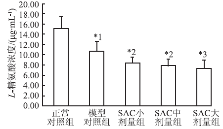
Fig.2
Comparison of the serum concentration of L-arginine among five groups of rats (x¯±s,n=6) Compared with normalcontrol group, *1P<0.05;compared with model control group, *2P<0.05, *3P<0.01
Fig.3
Comparison of NO level and NOS activity of the serum, liver and kidney among three groups of rats (x¯±s,n=6) Compared with normal control group, *1P<0.01,*4P<0.05; compared with L-arginine group, *2P<0.01, *3P<0.05
表1
5组大鼠血清中TC、TG、Vc等浓度与 SOD、CAT、GSH-Px活性比较
Tab.1
Comparison of the serum concentration of TC,TG and Vc as well as the activity of SOD, CAT and GSH-Px among five groups of rats x¯±s,n=6
组别
TC
TG
Vc/ (μg·mL-1)
MDA/ (μmol·L-1)
GSH/ (mg·L-1)
SOD
CAT
GSH-Px/ (U·mL-1·min-1)
mmol·L-1
(U·mL-1)
正常对照组
1.84±0.19
0.88±0.10
3.70±0.34
5.82±0.32
407.03±31.41
263.47±14.49
10.19±1.58
410.48±31.30
模型对照组
4.31±0.84*1
1.17±0.17*1
3.30±0.48
8.01±0.70*1
328.73±31.15*1
213.38±27.29*1
6.06±1.09*2
709.01±45.64*1
SAC
小剂量组
2.79±0.48*2
1.05±0.17
4.16±0.11*2
6.47±0.61*2
379.55±20.18*2
239.44±8.74
7.77±2.38
664.75±40.09
中剂量组
2.48±0.28*2
0.85±0.14*2
4.37±0.21*2
5.81±0.51*2
400.51±22.33*2
250.20±9.08*3
8.46±1.86*3
632.18±30.75*2
大剂量组
2.33±0.22*2
0.88±0.16*3
4.52±0.32*2
5.22±0.46*2
417.70±16.70*2
263.04±11.06*2
8.50±0.92*3
663.55±33.94*2
Compared with normal control group, *1P<0.01;compared with model control group, *2P<0.01, *3P<0.05
与正常对照组比较,*1P<0.01;与模型对照组比较, *2P<0.01, *3P<0.05
表1
5组大鼠血清中TC、TG、Vc等浓度与 SOD、CAT、GSH-Px活性比较
Tab.1
Comparison of the serum concentration of TC,TG and Vc as well as the activity of SOD, CAT and GSH-Px among five groups of rats x¯±s,n=6
表2
Tab.2
表2
表2
5组大鼠肝脏中MDA、GSH水平与 SOD、CAT、GSH-Px活性比较
Tab.2
Comparison of the level of MDA, GSH and the activity of SOD,CAT and GSH-Px in liver among five groups of rats x¯±s,n=6
组别
MDA/(μmol·g-1)
GSH/(mg·g-1)
SOD
GSH-Px
CAT
(U·mg-1)
正常对照组
2.06±0.33
29.94±1.81
273.14±26.16
34.47±4.10
29.84±4.57
模型对照组
3.97±0.55*1
20.59±0.46*1
185.74±16.50*1
25.86±3.33*1
21.03±2.63*1
SAC
小剂量组
3.39±0.99
21.06±1.63
192.81±12.67
27.05±2.10
22.27±2.40
中剂量组
2.68±0.26*2
25.92±1.39*2
208.15±4.92*3
28.54±2.84
24.18±1.67
大剂量组
2.28±0.33*2
27.82±1.28*2
218.76±13.33*3
31.79±3.66*3
24.37±3.12
Compared with normal control group, *1P<0.01;compared with model control group, *2P<0.01, *3P<0.05
与正常对照组比较,*1P<0.01;与模型对照组比较, *2P<0.01, *3 P<0.05
表2
5组大鼠肝脏中MDA、GSH水平与 SOD、CAT、GSH-Px活性比较
Tab.2
Comparison of the level of MDA, GSH and the activity of SOD,CAT and GSH-Px in liver among five groups of rats x¯±s,n=6
表3
Tab.3
表3
表3
5组大鼠肾脏中MDA、GSH水平与 SOD、CAT、GSH-Px活性比较
Tab.3
Comparison of the level of MDA, GSH and the activity of SOD,CAT and GSH-Px in kidneys among five groups of rats x¯±s,n=6
组别
MDA/ (μmol·g-1)
GSH/ (mg·g-1)
SOD
GSH-Px
CAT
(U·mg-1)
正常对照组
1.58±0.22
35.70±4.55
222.88±20.64
27.99±5.73
25.89±4.19
模型对照组
2.03±0.11*1
27.91±2.11*1
164.33±24.09*1
22.30±1.53*2
18.22±4.74*2
SAC
小剂量组
1.61±0.37*3
24.00±4.14
176.38±31.37
22.54±3.15
21.03±1.68
中剂量组
1.55±0.28*4
27.09±4.22
182.68±37.51
22.34±3.68
22.12±4.40
大剂量组
1.46±0.11*4
32.93±2.52*4
208.46±11.73*4
25.86±4.76
23.71±4.83
Compared with normal control group,*1P<0.01, *2P<0.05;compared with model control group, *3P<0.05, *4P<0.01
GUTIERREZ-ESCOBAR AJ, ARENAS AF, VIILLORIAL-GUERREROY, et al. Toxoplasma gondii: molecular cloning and characterization of a nitric oxide synthase-like protein[J]. Exp Parasitol, 2008, 119(3): 358-363.
Toxoplasma gondii has a nitrite production and a putative nitric oxide synthase (NOS) motif genomic sequence. In order to demonstrate that this sequence is functional and could be involved in the metabolism of l -arginine derivatives, we constructed a baculovirus carrying the previously identified Toxoplasma NOS-like DNA sequence . The recombinant protein was expressed into insect Sf9 cells and his activity was tested in serial microplate colorimetric assays. The protein produced 21nmol/min/ml nitrites per microgram of protein and followed Michaelis–Menten kinetics, with a K m for l -arginine of 2.3mM. Furthermore, the optimal pH, temperature and incubation time for the recombinant Toxoplasma NOS-like protein were established. Toxoplasma NOS runs as a band of 11.6kDa on tricine–sodium dodecyl sulfate–polyacrylamide gel electrophoresis. Our results indicate that the recombinant protein derived from the putative genomic sequence, at the chromosome 1b of T. gondii, is able to produce nitrites from l -arginine as substrate.
HUANGQ, HUANGC, ZHAOY, et al.LPS-stimulated RAW264.7 macrophage CAT-2-mediated L-arginine uptake and nitric oxide biosynthesis is inhibited by omega fatty acid lipid emulsion[J]. J Surg Res, 2013, 179(1): e211-e217.
ABSTRACT Omega-3 fatty acid (ω-3 FA) lipid emulsion has been reported to inhibit nitric oxide (NO) production and alter inducible nitric oxide synthase (iNOS) protein expression in lipopolysaccharide (LPS)-stimulated murine macrophages. However, the role of cellular uptake of l-arginine and iNOS transcription in ω-3 FA emulsion-induced inhibition of NO has not been explored. In addition, cationic amino acid transporter-2 (CAT-2) can regulate iNOS activity. The effect of ω-3 FA emulsion on CAT-2 expression is unknown. In the present study, we hypothesized that ω-3 FA emulsion pretreatment would decrease the production of NO in LPS-stimulated macrophages and that this effect would occur through alterations in the cellular uptake of l-arginine and CAT-2 expression, in addition to iNOS expression.
LIJ, BILLIAR TR.Nitric oxide IV determinants of nitric oxide protection and toxicity in liver[J]. Am J Physiol, 1999, 276(5): G1069-G1073.
Whereas nitric (NO) produced by constitutive endothelial NO synthase is protective to the liver, NO produced by the () can be either toxic or protective depending on the conditions. The availability of selective inhibitors and lacking various isoforms made it possible to begin to elucidate the precise roles of NO in the liver. Under conditions of redox stress, induced NO contributes to hepatic damage. However, in acute inflammatory conditions associated with cytokine exposure, NO acts as a potent inhibitor of in the liver. Our current understanding of the mechanisms by which NO exerts both hepatoprotective and hepatotoxic actions is discussed in this themes article.
RODRIGUEZ-RAMOST, CARPIOY, BOLIVARJ, et al.An inducible nitric oxide synthase (NOS) is expressed in hemocytes of the spiny lobster Panulirus argus: cloning, characterization and expression analysis[J]. Fish Shellfish Immunol, 2010, 29(3): 469-479.
Nitric oxide (NO) is a free radical gas involved in a variety of physiological processes in invertebrates, such as neuromodulation, muscle contraction and host defense. Surprisingly, little is known about the involvement of NO synthase (NOS) in the immune system of crustaceans. This work is focused on the study of the NOS gene of the spiny lobster Panulirus argus, a crustacean with commercial interest, and its relationship with the immune response to a microbial elicitor. A NOS full-length DNA was isolated from hemocytes by reverse transcription-polymerase chain reaction (RT-PCR) using degenerated primers. The open reading frame (ORF) encodes a protein of 1200 amino acids, with an estimated molecular mass of 135.9 kDa, which contains the conserved domains and binding motifs of NOS found in a variety of organisms. NOS gene expression in lobster gills, heart, stomach, digestive gland, abdominal muscle, gut and hemocytes was studied by Real Time quantitative PCR (Real Time qPCR). The expression was higher in hemocytes, heart and gills. In addition, when lobster hemocytes were exposed in vitro to Escherichia coli O55:B5 lipopolysaccharide (LPS), an increase in the NOS activity and also in the NOS gene expression evaluated by Real Time qPCR was observed, thus demonstrating the presence of an inducible crustacean NOS by a microbial elicitor of the immune response. The information is relevant in providing basic knowledge for further studies of crustacean defense mechanisms.
BULTH.Nitricoxide and atherosclerosis: possible implications for therapy[J]. Mol Med Today, 1996, 2(12): 510-518.
[本文引用:1]
[6]
PERROTTAI, BRUNELLIE, SCIANGULAA, et al.Inducible and endothelial nitric oxide synthase expression in human atherogenesis: an immunohistochemical and ultrastructural study[J]. Cardiovasc Pathol, 2009, 18(6): 361-368.
Our data confirm and extend previous findings of a direct relationship between dysregulation of nitric oxide pathway and atherosclerosis, suggesting another possible mechanism by which nitric oxide synthase system abnormalities may promote vascular dysfunction during human atherogenesis. Changes in nitric oxide production might be the primary step in the development of atheroma.
BUTTERY LD, SPRINGALL DR, CHESTER AH, et al.Inducible nitric oxide synthase is present within human atherosclerotic lesions and promotes the formation and activity of peroxynitrite[J]. Lab Invest, 1996, 75(1): 77-85.
KIM JW, KANG KW, OH GT, et al.Induction of hepatic inducible nitric oxide synthase by cholesterol in vivo and in vitro[J]. Exp Mol Med, 2002, 34(2): 137-144.
Cholesterol-rich diet impairs endothelial NO synthase (eNOS) and enhances inducible NOS (iNOS) expression. In this study, we investigated effects of cholesterol on iNOS expression in high-fat-fed rat models, HepG2 and RAW264.7 cells. The high-fat diet increased the plasma total cholesterol level 6-7 fold and low-density lipoprotein cholesterol level (LDL-C) approximately 70 fold and slightly increased the level of lipid peroxidation as determined by thiobarbituric acid-reactive substance assay. The high-fat diet also increased plasma nitric oxide (NO) concentrations up to 5 fold, and induced iNOS mRNA expression in liver. The contractile responses of the endothelium-denuded thoracic aortic rings to phenylephrine were significantly damaged in high-fat-fed rats when assessed by organ chamber study. Treatment with estrogen for 4 days failed to reduce iNOS expressions as well as aortic contractility, although it improved lipid profiles. In cultured HepG2 or murine macrophage RAW264.7 cells, 3 days treatment with either 25-hydroxycholesterol or 7-ketocholesterol induced iNOS mRNA expression, as determined by RT-PCR. Our data suggested that the chronic exposure of hepatocytes and macrophage cells to high concentration of cholesterol or oxysterols may induce iNOS expression and subsequent synthesis of NO, which may be important in the pathogenesis of atherosclerosis.
VAN DO VIJVER L P, KARDINAAL AF, GROBBEE DE, et al. Lipoprotein oxidation, antioxidants and cardiovascular risk: epidemiologic evidence[J]. Prostaglandins Leukot Essent Fatty Acids, 1997, 57(4/5): 479-487.
Abstract This review summarizes the scientific evidence for a possible role of antioxidants in the prevention of coronary heart disease (CHD). Dietary antioxidants include vitamin E, vitamin C and beta-carotene, whereas selenium is an integral part of the antioxidant enzyme glutathione peroxidase. Experimental studies suggest that the oxidation of low-density lipoproteins (LDL) in the vessel wall plays an important role in the development of atherosclerotic lesions. The resistance of LDL to oxidation is increased by antioxidant supplementation, at least in vitro. Epidemiological studies have not demonstrated unequivocally that a high intake of antioxidants leads to a decreased risk of CHD. Studies on dietary intake and serum levels of antioxidants do point in the direction of a preventive effect of antioxidants, whereas the results of intervention studies are less conclusive. Beta-carotene supplementation is not associated with any decrease in CHD; high doses of vitamin E may be beneficial, but results from large trials are to be awaited. General preventive measures based on antioxidant supplementation are not yet justifiable.
SARAVANANG, PONMURUGANP.Attenuation of streptozotocin-induced alterations in acetylcholinesterase and antioxidant system by S-allylcysteine in rats[J]. Food Bioscience, 2013, 4(1): 31-37.
The present study was designed to investigate the effect of the administration of S-allylcysteine (SAC) (150聽mg/kg body weight for 45 days) on acetylcholinesterase (AChE) activity and antioxidant levels in the brain tissues of streptozotocin-induced diabetic rats. The levels of glucose, TBARS, hydroperoxide and acetylcholinesterase were increased significantly whereas the levels of plasma insulin, reduced glutathione, superoxide dismutase and catalase were decreased in experimental diabetic rats. Administration of SAC to diabetic rats reverted all these parameters. The effect of SAC was compared with glyclazide, a well-known antioxidant and antihyperglycemic drug. In conclusion, the present findings showed that treatment with SAC prevents the increase in AChE activity, lipid peroxidation, and consequently improves the antioxidant system in diabetic rats, indicating that this compound can be considered as possible therapeutics to be investigated in brain disorders associated with the diabetes.
KIM KM, CHUN SB, KOO MS, et al.Differential regulation of NO availability from macrophages and endothelial cells by the garlic component S-allylcysteine[J]. Free Radic Biol Med, 2001, 30(7): 747-756.
Garlic has been used as a traditional medicine for prevention and treatment of cardiovascular diseases. However, the molecular mechanism of garlic鈥檚 pharmacological action has not been clearly elucidated. We examined here the effect of garlic extract and its major component, S-allyl cysteine (SAC), on nitric oxide (NO) production by macrophages and endothelial cells. The present study demonstrates that these reagents inhibited NO production through the suppression of iNOS mRNA and protein expression in the murine macrophage cell line RAW264.7, which had been stimulated with LPS and IFN纬. The garlic extract also inhibited NO production in peritoneal macrophages, rat hepatocytes, and rat aortic smooth muscle cells stimulated with LPS plus cytokines, but it did not inhibit NO production in iNOS-transfected AKN-1 cells or iNOS enzyme activity. These reagents suppressed NF-魏B activation and murine iNOS promoter activity in LPS and IFN纬-stimulated RAW264.7 cells. In contrast, these reagents significantly increased cGMP production by eNOS in HUVEC without changes in activity, protein levels, and cellular distribution of eNOS. Finally, garlic extract and SAC both suppressed the production of hydroxyl radical, confirming their antioxidant activity. These data demonstrate that garlic extract and SAC, due to their antioxidant activity, differentially regulate NO production by inhibiting iNOS expression in macrophages while increasing NO in endothelial cells. Thus, this selective regulation may contribute to the anti-inflammatory effect and prevention of atherosclerosis by these reagents.
CALAPAIG, CRUPIA, FIRENZUOLIF, et al.Neuroprotective effects of Ginkgo biloba extract in brain ischemia are mediated by inhibition of nitric oxide synthesis[J]. Life Sci , 2000, 67(22): 2673-2683.
We studied the effects of pre-treatment (15 days) with oral administration of Ginkgo biloba extract (Ph-Gb 37.5-150 mg/kg) on brain malonildialdehyde (MDA), brain edema, brain nitrite and nitrate and delayed neuronal death following transient cerebral ischemia in the Mongolian gerbil. Survival was not modified, however, pre-treatment with Ginkgo biloba significantly and in a dose-dependent way reduced post-ischemic brain MDA levels and post-ischemic brain edema. Delayed neuronal death in the CA1 of the hippocampus was attenuated by the highest dose of the extract. Increase of nitrite and nitrate was observed after cerebral ischemia in the hippocampus and it was dose-dependently reduced in animals pretreated with Ph-Gb, thus suggesting that neuroprotective effects of Ginkgo biloba may be due to an inhibitory action on nitric oxide formation.
YAGIK.Lipid peroxides and related radicals in clinical medicine[J]. Adv Exp Med Biol, 1994, 366(1): 1-15.
In 1980, when the author organized an international symposium on “Lipid Peroxides in Biology and Medicine” 1 , he was convinced that the research on the significance of lipid peroxides and their related free radicals in medicine had gotten off to a good start. Thereafter, many valuable results on this topic have been accumulated, and the problem has become of practical importance in clinical medicine. Thus, the present symposium seems to be timely organized.
IQBALZ, MIDGLEY JM, WATSON DG, et al.Effect of oral administration of vitamin C on human aqueous humor ascorbate concentration[J]. Acta Pharmacol Sin, 1999, 20(10): 879-883.
AIM: To study oral administration of vitamin C on human aqueous humour ascorbate concentration. METHODS: High performance liquid chromatography (HPLC) coupled with electrochemical detector (ECD) was used. The effect of oral administration of various doses of ascorbic acid, 0 (control), 1.0, 1.5, 2.0, 3.0, and 5.0 g, on its concentration in aqueous humour, obtained from volunteer cataract patients was studied. RESULTS: The concentration of ascorbic acid in aqueous humour of control group (without administration of vitamin-C tablet or drug containing ascorbic acid was (254 +/- 119) mg.L-1. This study revealed that the administration of 2.0 g of ascorbic acid saturate the aqueous humour and further increase in the dose (3.0 g and 5.0 g) did not increase its concentration in aqueous humour, although its concentration was increased in plasma. CONCLUSION: Oral administration of 2.0 g of Vc is sufficient to saturate the aqueous humour where it may be helpful in controlling the intra-ocular pressure.
PIJ, KUMAGAIY, SUNG, et al.Improved method for simultaneous determination of L-arginine and its mono- and dimethylated metabolites in biological samples by high-performance liquid chromatography[J]. J Chromatogr B Biomed Sci Appl, 2000,742(1):199-203.
ABSTRACT An improved method has been developed for the determination of L-arginine and its methylated metabolites, N(G)-monomethyl-L-arginine (L-NMMA), N(G),N(G)-dimethyl-L-arginine (asymmetric DMA, ADMA) and N(G),N(G)'-dimethyl-L-arginine (symmetric DMA, SDMA) in biological samples. Extraction of these compounds with a strong cation-exchange resin AG50W-X8 with L-homoarginine (2-amino-6-guanidinohexanoic acid) as an internal standard gave a recovery of more than 70% except for SDMA from plasma samples. After extracted samples were converted to fluorescent derivatives with o-phthalaldehyde (OPA) in an alkaline medium, the following high-performance liquid chromatographic separation with a ODS column (wide-pore size, 300 A) was successfully performed with an isocratic mobile phase system. The method permits quantitative determination of L-arginine and its methylated metabolites at concentrations as low as 4 microM and 0.18 microM, respectively. Using this method, the levels of L-arginine, L-NMMA, ADMA and SDMA in human plasma, urine and rat tissue were determined.
SHARMAS, SINGHM, SHARMA PL.Mechanism of attenuation of diabetes mellitus and hypercholesterolemia induced vascular endothelial dysfunction by protein tyrosine phosphatase inhibition[J]. Vascul Pharmacol, 2011, 54(3/6): 80-87.
FERSONO, DESSYC, MONIOTTES, et al.Hyper-cholesterolemia decreases nitric oxide production by promoting the interaction of caveolin and endothelial nitric oxide synthase[J]. J Clin Invest, 1999, 103(6): 897-905.
An inducible nitric oxide synthase (NOS) is expressed in hemocytes of the spiny lobster Panulirus argus: cloning, characterization and expression analysis
Improved method for simultaneous determination of L-arginine and its mono- and dimethylated metabolites in biological samples by high-performance liquid chromatography
Mechanism of attenuation of diabetes mellitus and hypercholesterolemia induced vascular endothelial dysfunction by protein tyrosine phosphatase inhibition

 , 张容
, 张容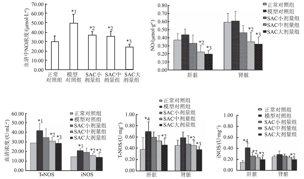

