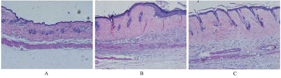
Objective To evaluate the inhibitory effect of asiaticoside on bleomycin-induced skin cicatrization. Methods Thirty male C57BL/6 mice were randomly divided into three groups: negative control group,model control group,and asiaticoside group,ten in each group.In model control group and asiaticoside group,1 mg·mL-1 bleomycin was subcutaneously injected into the dorsal skin of mice every day;4 h later,1 mL 0.9% sodium chloride solution 1 mL asiaticoside (20 mg·mL-1) was injected into the lesion skin in the model control group and the asiaticoside group,respectively.In the negative control group,the same volume of 0.9% sodium chloride solution was subcutaneously injected into the dorsal skin of the mice at the two time points every day.After 21 days,skin specimens were harvested to observe the histomorphology and detect myofibroblast proliferation and expression of inflammatory factors. Results The skin scar was significantly attenuated in the asiaticoside group as compared with the model control group,and the dermal thickness measured exhibited a gradual decrease in asiaticoside group.The expression of α-antismooth muscle antisbidy and infiltration of inflammatory cells were significantly lower in the asiaticoside group than in the model control group. Conclusion Asiaticoside inhibits the development of skin scar of mice by regulating proliferation and differentiation of myofibroblasts and down-regulating inflammatory cells.
Fig.2
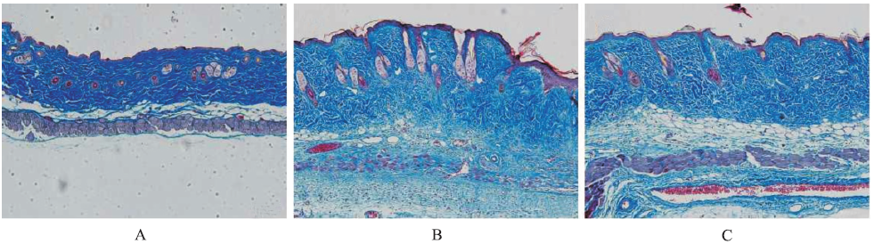
Cutaneous histomorphology of three groups of mice(Masson’s Trichome staining,×100) A.negative control group;B.model control group;C.asiaticoside group
Fig.3
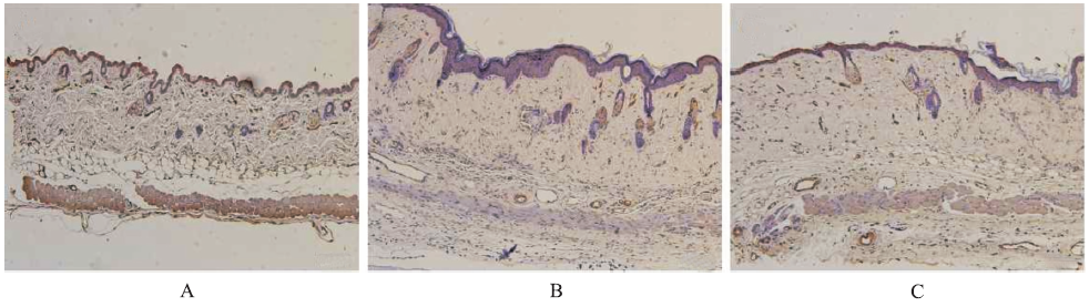
Detection on α-SMA protein expression in skin tissue of three groups of mice(immunohistochemical staining,×100) A.negative control group;B. model control group;C.asiaticoside groups
Fig.4
Detection on inflammatory cytokines in skin tissue of three groups of mice Compared with negative control group,*1P<0.01;compared with model control group,*2P<0.01
YUN KJ,KIM JY,KIM JB,et al.Inhibition of LPS indu-ced NO and PGE2 production by asiatic acid via NF-kappa B inactivation in RAW 264.7 macrophages: possible involvement of the IKK and MAPK pathways[J].Int Immunopharmacol,2008, 8(3): 431-441.
In the present study, we investigated the effect of asiatic acid (the aglycon of asiaticoside) and asiaticoside isolated from the leaves of Centella asiatica (Umbelliferae) on LPS-induced NO and PGE 2 production in RAW 264.7 macrophage cells. Asiatic acid more potently inhibited LPS-induced NO and PGE 2 production than asiaticoside. Consistent with these observations, the protein and mRNA expression levels of inducible iNOS and COX-2 enzymes were inhibited by asiatic acid in a concentration-dependent manner. In addition, asiatic acid dose-dependently reduced the production of IL-6, IL-1β and TNF-α in LPS-stimulated RAW 264.7 macrophage cells. Furthermore, asiatic acid inhibited the NF-κB activation induced by LPS, and this was associated with the abrogation of IκB-α degradation and with subsequent decreases in nuclear p65 and p50 protein levels. Moreover, the phosphorylations of IKK, p38, ERK1/2, and JNK in LPS-stimulated RAW 264.7 cells were suppressed by asiatic acid in a dose-dependent manner. These results suggest that the anti-inflammatory properties of asiatic acid might be the results from the inhibition of iNOS, COX-2, IL-6, IL-1β, and TNF-α expressions through the down-regulation of NF-κB activation via suppression of IKK and MAP kinase (p38, ERK1/2, and JNK) phosphorylation in RAW 264.7 cells.
HUANG YH,ZHANG SH,ZHEN RX, et al.Asiaticoside inducing apoptosis of tumor cells and enhancing anti-tumor activity of vincristine[J].Cancer,2004,23(12):1599-1604.
BACKGROUND OBJECTIVE: Asiaticoside (ATS), a triterpene extracted from Centella asiatica (L.) Urban, a traditional Chinese herb,possesses good wound healing activities because of its stimulative effect on collagen synthesis. Recently, the anti tumor effect of asiaticiside has been reported. This study was to examine the induction of apoptosis in cancer cells, and the enhancement of vincristine (VCR) cytotoxicity by asiaticoside. METHODS: MTT assay was used to evaluate inhibitory effect of asiaticoside combined with vincristine on proliferation of several cancer cell lines, including KB, KBv200, MCF 7, and MCF 7/ADM. Cell cycle, and apoptosis of KB cells were analyzed by flow cytometry; apoptosis induction was also proved by electrophoresis,and morphologic assessment; the expression of apoptosis , and cell cycle related proteins were determined by Western blot. RESULTS: The IC50 values of asiaticoside for KB, KBv200, MCF 7, and MCF 7/ADM cells detected by MTT assay were (1.11卤0.13) mg/ml, (1.82卤0.08) mg/ml, (1.58卤0.15) mg/ml, and (3.25卤0.46) mg/ml, respectively. Multidrug resistant KBv200, and MCF 7/ADM cancer cells displayed similar sensitivity to asiaticoside as their parental counterparts (KB, and MCF 7 cells). Moreover, asiaticoside induced apoptosis in KB cells. At sub cytotoxicity concentration, asiaticoside showed synergistic effect with vincristine in these 4 cell lines. The apoptosis rates were much higher in asiaticoside plus vincristine groups than in vincristine or asiaticoside groups. Bcl 2 phosphorylation levels were higher in the combination groups than in vincristine or asiaticoside alone groups. The activated caspase 3 protein was only presented in the combination groups. Asiaticoside plus vincristine enhanced S G2/M arrest, up regulated Cyclin B1 protein expression, and down regulated P34cdc2 protein expression in KB cells. CONCLUSION: Asiaticoside,as a biochemical modulator,may induce apoptosis,and enhance anti tumor activity of vincristine in cancer cells, might be useful in cancer chemotherapy.
YAMAMOTOT,KURODAM,NISHIOKAK.Animal model of sclerotic skin Ⅲ: histopathological comparison of bleomycin-induced scleroderma in various mice strains[J].Arch Dermatol Res,2000,292(11):535-541.
Abstract We have recently established a mouse model for scleroderma by repeated local bleomycin treatment. In this study, we compared the susceptibility to bleomycin in the development of dermal sclerosis among Balb/c, C3H/He, C57BL/6J, A/J, DBA/2, B10.BR, B10.A, and B10.D2 mouse strains. After either bleomycin or PBS treatment, skin from the injection site was histologically examined. Dermal sclerosis was induced by bleomycin treatment for 4 weeks in all of the strains examined. In particular, C3H/He, DBA/2, B10.D2 and B10.A mice developed intense dermal sclerosis characterized by deposition of homogeneous material in the dermis and thickened collagen bundles. Dermal thickness showed a more than twofold increase following bleomycin treatment, as compared with PBS treatment, except in C57BL/6J and DBA/2 mice. In A/J, C3H/He, B10.A, and B10.D2 mice, dermal thickness showed a more than 2.5-fold increase. Mast cell numbers in sclerotic skin were significantly greater than in PBS-treated skin in Balb/c and B10.A mice after 4 weeks of treatment. We also examined whether bleomycin treatment for 3 weeks could induce dermal sclerosis in C3H mice. Histological examination revealed that epidermal thickness as well as dermal sclerosis was increased in C3H mice following bleomycin treatment for 3 weeks. Increased hydroxyproline content as well as mRNA expression of alpha1(I) collagen, as determined by Northern blot analysis, were observed following bleomycin treatment. Taken together, we conclude that C3H/He and B10.A mouse strains are bleomycin-'susceptible', and these strains are considered to be a suitable experimental model of bleomycin-induced scleroderma.
SHIHB,BAYATA.Genetics of keloid scarring[J].Arch Dermatol Res,2010,302(5): 319-339.
Keloid scarring, also known as keloid disease (KD), is a common, abnormally raised fibroproliferative cutaneous lesion that can occur following even minor skin trauma. The aetiopathogenesis of KD has remained an enigma todate compounded by an ill-defined clinical management. There is strong evidence suggesting a genetic susceptibility in individuals affected by KD, including familial heritability, common occurrence in twins and high prevalence in certain ethnic populations. This review aims to address the genetic aspects of KD that have been described in present literature that include inheritance patterns, linkage studies, case-揷ontrol association studies, whole genome gene expression microarray studies and gene pathways that were significant in KD. In addition to our clinical and scientific background in KD, we used search engines, Scopus, Scirus and PubMed, which searched for key terms covering various genetic aspects of KD. Additionally, genes reported in seven whole genome gene expression microarray studies were separately compared in detail. Our findings indicate a varied inheritance pattern in KD (predominantly autosomal dominant), linkage loci (chromosomes 2q23 and 7p11), several human leukocyte antigen (HLA) alleles (HLA-DRB1*15, HLA-DQA1*0104, DQ-B1*0501 and DQB1*0503), negative candidate gene case-揷ontrol association studies and at least 25 dysregulated genes reported in multiple microarray studies. The major pathways reportedly proposed to be involved in KD include apoptosis, mitogen-activated protein kinase, transforming growth factor-尾, interleukin-6 and plasminogen activator inhibitor-1. In summary, involvement of more than one gene is likely to be responsible for susceptibility to KD. A better understanding of the genes involved in KD may potentially lead to the development of more effective diagnostic, therapeutic and prognostic measures.
JACKSON WM,NESTI LJ,TUAN RS.Mesenchymal stem cell therapy for attenuation of scar formation during wound healing[J].Stem Cell Res Ther,2012,3(3):20-22.
Abstract Scars are a consequence of cutaneous wound healing that can be both unsightly and detrimental to the function of the tissue. Scar tissue is generated by excessive deposition of extracellular matrix tissue by wound healing fibroblasts and myofibroblasts, and although it is inferior to the uninjured skin, it is able to restore integrity to the boundary between the body and its environment. Scarring is not a necessary process to repair the dermal tissues. Rather, scar tissue forms due to specific mechanisms that occur during the adult wound healing process and are modulated primarily by the inflammatory response at the site of injury. Adult tissue-derived mesenchymal stem cells, which participate in normal wound healing, are trophic mediators of tissue repair. These cells participate in attenuating inflammation in the wound and reprogramming the resident immune and wound healing cells to favor tissue regeneration and inhibit fibrotic tissue formation. As a result, these cells have been considered and tested as a likely candidate for a cellular therapy to promote scar-less wound healing. This review identifies specific mechanisms by which mesenchymal stem cells can limit tissue fibrosis and summarizes recent in vivo studies where these cells have been used successfully to limit scar formation.
WANGQ,DONGY,GENGS,et al.Photodynamic therapy inhibits the formation of hypertrophic scars in rabbit ears by regulating metalloproteinases and tissue inhibitor of metalloproteinase-1[J].Clin Exp Dermatol,2014,39(2):196-201.
BACKGROUND: scarring (HS) is a chronic skin condition, and inhibition of normal fibroblast plays an important role in its . Photodynamic therapy () is known to inhibit of proliferation in blood vessels and fibroblasts in scar tissue, with no significant adverse reactions reported. AIM: To investigate the effect of in the ear model of HS, and the specific mechanism of action of .: We assessed the clinical and histopathological appearance of ears with HS with and without . In addition, mRNA levels of matrix metalloproteinase (MMP)-2, , and tissue inhibitor of metalloproteinase ()-1, and concentration of-were all measured to confirm senescence.: Our data indicate that can accelerate fibroblast by increasing the ratio of MMPs to , in addition to promoting of and , thereby inhibiting HS . These effects lasted for up to 60 days, and induced no significant adverse local or systemic reactions. The efficacy of the treatment can be maximized by applying an appropriately high concentration of aminolaevulinic acid. CONCLUSIONS: can induce senescence in fibroblasts, and may constitute a useful treatment for pathological scarring.
ALVES CC,TORRINHAS RS,GIORGIR,et al.TGF-β1 expression in wound healing is acutely affected by experimental malnutrition and early enteral feeding[J].Int Wound J,2014,11(5):533-539.
Inhibition of LPS indu-ced NO and PGE production by asiatic acid via NF-kappa B inactivation in RAW 264.7 macrophages: possible involvement of the IKK and MAPK pathways
Photodynamic therapy inhibits the formation of hypertrophic scars in rabbit ears by regulating metalloproteinases and tissue inhibitor of metalloproteinase-1

 , 杨琳
, 杨琳
