中国科技论文统计源期刊 中文核心期刊
美国《化学文摘》《国际药学文摘》
《乌利希期刊指南》
WHO《西太平洋地区医学索引》来源期刊
日本科学技术振兴机构数据库(JST)
第七届湖北十大名刊提名奖
美国《化学文摘》《国际药学文摘》
《乌利希期刊指南》
WHO《西太平洋地区医学索引》来源期刊
日本科学技术振兴机构数据库(JST)
第七届湖北十大名刊提名奖

 , 程晓华, 徐文炜, CHENG Xiaohua, XU Wenwei
目的 观察齐墩果酸体外肠吸收特征及P-糖蛋白介导跨膜转运的机制。方法 建立齐墩果酸细胞摄取量以及转运量高效液相色谱-质谱联用仪的定量检测方法,噻唑蓝法确定齐墩果酸对Caco-2细胞的安全浓度范围,考察不同药物浓度、孵育时间、介质pH值、体系温度对Caco-2细胞摄取齐墩果酸的影响;通过构建Caco-2细胞单层模型,评价P-糖蛋白抑制药维拉帕米对齐墩果酸跨膜转运的影响,计算其表观渗透系数(
Objective To explore the absorption characteristics and mechanism of
Caco-2 细胞模型重现性好,与药物体内吸收具有良好的相关性,近年来被国内外学者广泛用于研究药物体外肠道吸收过程与机制,也是目前研究体外肠吸收的经典模型[6]。笔者在本研究采用Caco-2细胞模型研究小肠上皮细胞对OA的摄取、跨膜转运及外排作用,评价药物浓度、孵育时间、介质pH值、体系温度对细胞摄取的影响,以及P-糖蛋白(P-gp)抑制药对细胞跨膜转运吸收的影响,以期阐明OA的吸收机制,为合理设计其剂型和深入研究其药动学提供实验依据。
Caco-2细胞株购于美国典型菌种保藏中心(American Type Culture Collection,ATCC),实验中使用第30~40代细胞。
达尔伯克改良伊格尔培养基(Dulbecco's Modified Eagle Medium,DMEM)(GIBCO公司,批号:31800-022),胎牛血清(Hyclone公司,批号:NXJ0709),0.1%胰蛋白酶(GIBCO公司,批号:27250018),谷氨酰胺(GIBCO公司,批号:25030),非必需氨基酸(GIBCO公司,批号:1232254),维拉帕米(Sigma公司,批号:20120816),罗丹明123 (Sigma公司,批号:R06154),OA(中国食品药品检定研究院,含量:98%,批号:201206),格列喹酮对照品(中国食品药品检定研究院,批号:130824,含量:99.2%),乙腈(TEDIA公司,色谱纯,批号:14025017),胰蛋白酶-EDTA(Sigma公司,批号:S10054),醋酸铵(天津福晨化学试剂厂,批号:20140415),水为超纯水,其他试剂均为分析纯。
MCO20-AIC型二氧化碳(CO2)培养箱(日本三洋公司),CKX-41型倒置光学显微镜(日本奥林巴斯公司),EVOM细胞电位仪(美国WPI公司),GS-15R高速冷冻离心机(德国Sigma公司),LCMS-2010EV型高效液相色谱-质谱联用仪(HPLC-MS,日本岛津公司);LCMSsolution色谱工作站(日本岛津公司)。
将Caco-2细胞培养于高糖DMEM培养基中(含10%胎牛血清、非必需氨基酸、1%
色谱柱:Shim-pack VP-ODS C18(150 mm×2.1 mm,5 μm);流动相:乙腈-0.1%醋酸铵=68∶32,流速:0.2 mL·min-1,柱温:35 ℃;采用选择性负离子检测及电喷雾离子化(electrospray ionization,ESI);检测对象:OA,
取对数生长期Caco-2细胞,调整细胞密度至5×10⁴个·mL-1接种于96孔培养板,24 h后换液,实验组分别加入不同浓度OA培养液,调零空白组加入等体积培养液,每组设4个复孔,继续培养12和24 h后每孔加入噻唑蓝(MTT),每孔加二甲亚砜(DMSO)溶液150 μL,空气恒温振荡器振荡10 min,待结晶物充分溶解,在酶标仪上选择波长570 nm,空白孔调零,测定各孔吸光度值。细胞存活率(%)=(实验组吸光度值/对照组吸光度值)×100%。
取对数生长期Caco-2细胞,调整细胞密度为1×10⁴个·mL-1,接种培养于24孔培养板,实验前2 h缓慢吸弃旧培养液,加入预热Hank'平衡盐溶液(hank's balanced salt solution,HBSS)1.0 mL荡洗细胞3次,最后置于37 ℃培养箱温孵30 min,缓慢吸弃HBSS溶液,洗去细胞单分子层表面杂质,考察药物浓度、孵育时间、体系温度以及介质pH值对药物摄取的影响。不同因素各组均设4个复孔,加入药液1 mL,培养24 h后弃去药液,加入4℃HBSS缓冲液终止摄取。每孔加入超纯水1 mL,细胞刮取器刮下细胞,超声粉碎细胞,取上清液200 μL,加入乙腈,离心,取上清液20 μL用于HPLC-MS分析,另取200 μL进行细胞蛋白质含量测定,OA摄取量以μg·mg-1蛋白质为单位。
取符合转运实验要求的Caco-2细胞模型,试验前用预热HBSS清洗生长有Caco-2细胞的Transwell板3次,置于37 ℃培养箱温孵30 min,考察药物浓度、反应时间、转运方向及P-gp抑制药维拉帕米对细胞转运OA的影响。当转运方向为AP→BL时,AP中加入OA供试溶液0.4 mL作为供给液,BL中加入空白HBSS 1.0 mL作为接收液。当转运方向为BL→AP时,AP中加入空白HBSS0.4 mL作为接收液,BP中加入供试药液1.0 mL。将Transwell板置于37 ℃、转速50 r·min-1双层空气恒温振荡器中孵育,分别于10,20,40,60,90,120 min吸取接收液,同时补加空白HBSS 200 μL,每组设4个平行孔。取转运样品200 μL,加入乙腈,离心10 min,取上清液进行HPLC-MS分析,计算其表观渗透系数(
细胞的摄取量=
成功建立了测定OA含量专属性强、灵敏度高的HPLC-MS分析方法,OA保留时间约2.3 min,转运液及细胞悬液均无杂质干扰样品的测定。结果见
MTT实验结果表明,随着OA浓度增加,细胞存活率逐渐下降,呈剂量依赖性,培养时间对细胞存活率基本无影响。在5~40 μg·mL-1范围内细胞的存活率>80%,OA的浓度>40 μg·mL-1,部分细胞出现细胞毒作用并伴随细胞形态学改变,结果见
Caco-2细胞对OA的摄取量随培养时间的延长而变化,0~40 min细胞对其摄取呈增加趋势,40 min后逐渐趋于饱和,细胞摄取量无显著增加(
表1 OA不同浓度和作用时间对Caco-2细胞存活率的影响
Tab.1
Effects of different concentration and incubation time of OA on the survival rate of Caco-2 cells
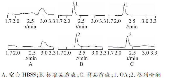
图1
样品检测质谱图
A.blank HBSS;B.standard solution;C.sample solution;1.OA;2.gliquidone
Fig.1 HPLC-MS chromatograms of the sample
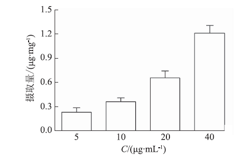
将Caco-2细胞分别与5,10,20,40 μg·mL-1的OA供试液培养40 min,考察浓度对细胞摄取OA的影响,结果见图3。摄取量随浓度的增加呈线性上升,细胞对OA的摄取可能主要以被动扩散的方式进行。
将40 μg·mL-1OA供试液在pH值为5,6,7.4,8的条件下培养40 min,测定加药40 min后细胞对OA的吸收量,考察pH值对细胞吸收OA的影响。结果在pH值为5,6,7.4,8条件下,细胞对OA摄取量分别为(1.27±0.25),(1.31±0.35),(1.36±0.33),(1.41±0.28)μg·mL-1,组间比较差异无统计学意义(
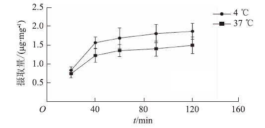
分别考察37 ℃与4 ℃时,作用不同时间Caco-2细胞对40 μg·mL-1OA的摄取量,结果见
取OA供试液,分别加入Caco-2细胞单层膜的AP侧和BL侧,不同浓度OA在不同时间点跨膜转运(AP→BL)过程中的转运量变化情况见
为证实是否存在P-gp外排作用对OA转运的影响,转运液中加入P-gp抑制药维拉帕米 (Ver,100 μmol·L-1),孵育30 min后加入40 μg·mL-1OA进行转运实验。结果见
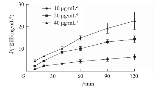
图5
OA浓度与作用时间对AP→BL转运量的影响(
Fig.5
Effects of time and concentration of OA on AP→BL transport volume (
表2 OA浓度对Papp和PDR的影响
Tab.2
Effects of concentration of OA on Papp and PDR
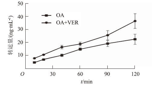
图6
Ver对AP→BL转运量的影响(
Fig.6
Effects of Ver on AP→BL transportvolumn of OA(
表3
Ver对
Tab.3
Effects of P-gp on
Caco-2细胞来源于人结肠癌细胞,其结构和生化特点类似于人类小肠上皮细胞,体外培养一定时间后可以分化成具有多种药物载体和酶的小肠微绒毛结构,能够在细胞水平提供关于药物分子通过小肠黏膜的吸收、代谢信息,因此被广泛用于研究药物吸收机制[7]。
本研究结果显示,在0~24 h、OA浓度为5~40 μg·mL-1范围内,Caco-2细胞存活率均>80%,因此选取40 μg·mL-1为最大药物浓度应用于本实验,以避免浓度过高导致细胞死亡,影响对药物吸收的判断。通过建立灵敏度高、特异性强的细胞摄取液和转运液中OA浓度的HPLC-MS测定方法,评价不同药物浓度、反应时间、介质pH值等因素对OA摄取以及P-gp抑制药对OA跨膜转运的影响。研究结果表明,OA摄取存在时间和浓度依赖性,随着OA浓度上升细胞摄取量呈线性增加,表明OA主要以被动扩散方式被摄取。快速摄取在开始时出现,40 min后细胞内外液浓度达到相对平衡,摄取量趋于平缓并达到相对饱和,不同介质pH值对OA的摄取量无显著影响;细胞摄取量随着温度的升高而降低,呈现一定的温度依赖性,4 ℃时OA细胞摄取量明显大于37 ℃,预示OA细胞摄取可能受外排蛋白质影响,当温度4 ℃时,外排蛋白质活力下降,外排作用减弱,故药物摄取量增加。
The authors have declared that no competing interests exist.

