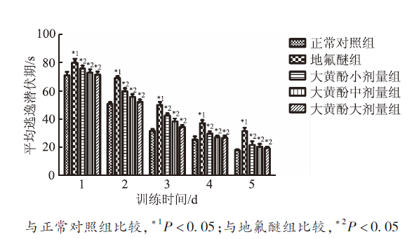
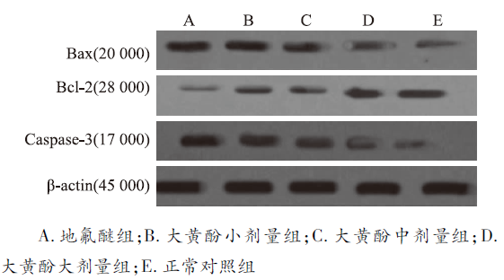
Objective To investigate the influence of inhalation anesthestic desfluran (Des) on the learning and memory abilities of rats and the protective role of chrysophanol. Methods Totally, 50 male and 50 female rats, aging 24 months and weighing (500±10) g, were randomly divided into five groups: normal control group, Des group, low-, medium- and high-dose Chr group (0.1, 1.0 and 10.0 mg·kg-1), with 20 rats in each group. After anesthetization for 24 h, the Morris water maze was used to investigate the abilities of learning and memory of rats. The amount of Aβ1-42 was determined by ELISA assay, and the apoptosis of rat hippocampal neurons in five group was observed by TUNEL assay. Furthermore, the expression levels of Bcl-2, Bax and Caspase-3 were examined by Western blotting. The activity of acetylcholinesterase in each rats hippocampus was determined using iron trichloride chromogenic spectrophotometer colorimetric analysis method. Results Compared with the normal control group, the mean escape latency of the rats in Des group was significantly prolonged; the spatial exploring time (29.85±4.51) s was reduced; the apoptotic rate of neurons (0.742±0.052)%, the amount of Aβ1-42 peptide (9 618.72±1 076.43) pmol·g-1 , the expression levels of Caspase-3 (1.132±0.217), and Bax (1.298±0.209) were increased; the expression of Bcl-2 (0.318±0.038) were reduced; the activity of acetylcholinesterase (96.38±7.62) U·mL-1 was increased. Compared with the Des group, the rats in all Chr groups obtained shorter escape latency and longer spatial exploring time; the amount of Aβ1-42 peptide and the expression levels of Caspase-3 and Bax were down-regulated; the activity of acetylcholinesterase was reduced. In addition, chrysophanol improved the abilities of learning and memory of anesthetic rats in a dose-dependent manner. Conclusion Chrysophanol could improve the abilities of the learning and memory of rats after desflurane anesthesia, along with inhibition of Aβ deposition.
图2
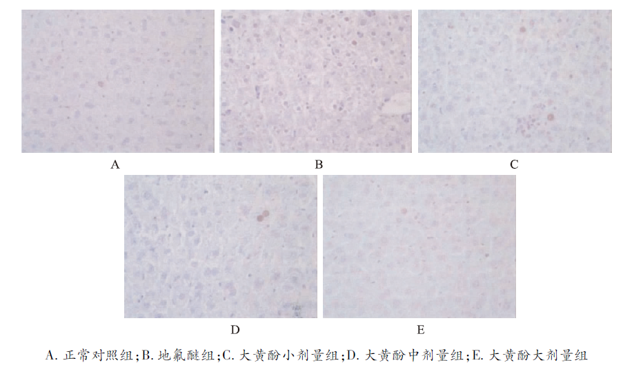
5组老年大鼠海马组织细胞凋亡比较(×200) A.normal control group;B.desfluran group;C.low-dose ahrysophanol group ;D.medium-dose hrysophanol group;E.high-dose hrysophanol group
Fig.2
Comparison of cell apoptosis in hippocampus among five groups of aged rats(×200)
LINF,ZHANGC,CHENX,et al.Chrysophanol affords neuroprotection against microglial activation and free radical-mediated oxidative damage in BV2 murine microglia[J].Int J Clin Exp Med,2015,8(3):3447-3455.
Abstract In this study, chrysophanol, isolated from a marine fungus, was examined for its protective effects against inflammatory responses and oxidative stress in BV2 microglia. Chrysophanol was studied to assess its capabilities of protecting against lipopolysaccharide (LPS)-induced inflammatory responses in BV2 cells. It was found that chrysophanol reduced the level of nitric oxide (NO) and prostaglandin-E2 (PGE2) production by diminishing reducing the expression of inducible NO synthase (iNOS) and cyclooxygenase-2 (COX-2). Assessment of the inhibitory activities of chrysophanol on the generation of pro-inflammatory cytokines was also performed. Furthermore, Chrysophanol treatment significantly reduced intracellular reactive oxygen species (ROS)-mediated cell damage and inhibited DNA oxidation in BV2 cells. Moreover, antioxidative mechanisms by of chrysophanol were evaluated investigated by measuring the expression levels of antioxidative enzymes such superoxide dismutase (SOD) and glutathione (GSH). Therefore, results suggested that chrysophanol has potential antioxidant and anti-inflammatory activities in microglia and further might be a useful therapeutic agent for the treatment of neurodegenerative diseases.
SHANJ,SUNL,WANGD,et al.Comparison of the neuroprotective effects and recovery profiles of isoflurane,sevoflurane and desflurane as neurosurgical pre-conditioning on ischemia/reperfusion cerebral injury[J].Int J Clin Exp Pathol,2015,8(2):2001-2009.
Abstract There are a few reports regarding the comparison of these anesthetic agents, but previous studies mainly focus on the veterinary anesthesiology. Less attention has been focused comparing the effectiveness of these inhalational anesthetic agents in neurosurgery. This lack of interest is regretful particularly considering the fact that anesthetics during neurosurgery are an issue of extreme sensitivity and subtlety, where the cerebral oxygenation process plays a significant role in the neuroprotective mechanisms. The purpose of this retrospective study is to contribute to the existing knowledge of the comparative studies of the volatile anesthetic agents such as isoflurane, sevoflurane and desflurane by evaluating the maintenance and emergence characteristics after volatile anesthetics-induced preconditioning with isoflurane, sevoflurane or desflurane for inpatient ischemia/reperfusion cerebral injury during cerebral or neural surgeries. The aim was to investigate their neuroprotective mechanisms and effects by analyzing and comparing the superiority of each agent in a Chinese patient population, in terms of faster emergence, and early and intermediate recovery. The intraoperative haemodynamic profiles and postoperative adverse effects of these three agents were also systematically analyzed. We found that sevoflurane, when compared with isoflurane and desflurane, provided anesthesia with similar hemodynamic stability but allowed for a smoother, more rapid emergence and better quality of induction and recovery to surgical patients under clinical conditions, particularly to those who were experiencing substantial cerebral vasodilation. Sevoflurane offers several advantages, including a relative lack of airway irritation, a more rapid onset and recovery, and greater hemodynamic stability than other potent inhaled agents. These properties would appear to afford sevoflurane significant clinical potential.
HEJ,ZHANGY,XUER,et al.Effect of desflurane versus sevoflurane in pediatric anesthesia: a meta-analysis[J].J Pharm Pharm Sci,2015,18(2):199-206.
ABSTRACT To compare the effect of desflurane versus sevoflurane in pediatric anesthesia by conducting meta-analysis. Studies were searched from PubMed, Medline, Springer, Elsevier Science Direct, Cochrane Library and Google Scholar up to July 2014. Weighted mean difference (WMD) or risk ratio (RR) and 95% confidence intervals (CIs) were considered as effect sizes. Heterogeneity across studies was assessed by Cochran Q test and I2 statistic. The random effects model was performed in the meta-analysis when heterogeneity was observed, or the fixed effect model was used. Review Manager 5.1 software was applied for the meta-analysis. A total of 11 studies (13 comparisons) involving 1,273 objects were included in this meta-analysis. No heterogeneity was observed between studies for any comparison but for postoperative extubation time. The results showed significant differences between desflurane and sevoflurane groups for postoperative extubation time (WMD = -3.87, 95%CI = -6.14 to -1.60, P 0.05) were detected for discharge from the recovery room, oculocardiac reflex, nausea and vomiting and severe pain. Desflurane may have less adverse effects than sevoflurane when used in pediatric anesthesia with significantly shorter postoperative extubation time, eye opening time and awakening time as well as slighter agitation. This article is open to POST-PUBLICATION REVIEW. Registered readers (see "For Readers") may comment by clicking on ABSTRACT on the issue's contents page.
JAKOBSSONJ.Desflurane: a clinical update of a third-generation inhaled anaesthetic[J].Acta Anaesthesiol Scand,2012,56(4):420-432.
Available volatile anaesthetics are safe and efficacious; however, their varying pharmacology provides small but potentially clinically important differences. Desflurane is one of the third eneration inhaled anaesthetics. It is the halogenated inhaled anaesthetic with the lowest blood and tissue solubilities, which promotes its rapid equilibration and its rapid elimination following cessation of administration at the end of anaesthesia. The low fat solubility of desflurane provides pharmacological benefits, especially in overweight patients and in longer procedures by reducing slow compartment accumulation. A decade of clinical use has provided evidence for desflurane's safe and efficacious use as a general anaesthetic. Its benefits include rapid and predictable emergence, and early recovery. In addition, the use of desflurane promotes early and predictable extubation, and the ability to rapidly transfer patients from the operating theatre to the recovery area, which has a positive impact on patient turnover. Desflurane also increases the likelihood of patients, including obese patients, recovering their protective airway reflexes and awakening to a degree sufficient to minimise the stay in the high dependency recovery area. The potential impact of the rapid early recovery from desflurane anaesthesia on intermediate and late recovery and resumption of activities of daily living requires further study.
BUTTERFIELD DA,SWOMLEY AM,SULTANAR.Amyloid beta-peptide (1-42)-induced oxidative stress in Alzheimer disease: importance in disease pathogenesis and progression[J].Antioxid Redox Signal,2013,19(8):823-835.
ABSTRACT Alzheimer disease (AD) is an age-related neurodegenerative disease characterized by cognitive impairment that affects the life and activities of the affected individuals and their families. One of the main histopathological hallmarks of AD is the presence of senile plaques (SP). The main component of SP is amyloid beta-peptide (A) that is derived from the proteolytic cleavage of amyloid precursor protein (APP). Studies conducted so far are consistent with the notion that methionine present at 35 position of A is critical to A-induced oxidative stress, and neurotoxicity. This review focuses on importance of methionine in the Abeta-induced oxidative stress. Further, we also discussed about signatures of oxidatively modified proteins, identified using redox proteomics approach, during the progression of AD. However, the exact relationship of the specifically oxidatively modified proteins in AD pathogenesis is poorly understood. Further studies are needed to address if the therapies directed towards oxidative stress and the associated target proteins might help to delay or prevent the progression and pathogenesis of AD.
JIANGJ,JIANGH.Effect of the inhaled anesthetics isoflurane,sevoflurane and desflurane on the neuropathogenesis of Alzheimer's disease (review)[J].Mol Med Rep,2015,12(1):3-12.
The incidence of Alzheimer's disease (AD) in individuals >65 years of age is 13% and ~66million individuals in this age group undergo surgery annually under anesthesia. It is therefore important to determine whether commonly used inhaled anesthetics induce cytotoxicity, which may lead to neurodegeneration. Findings from several studies suggest that the anesthetics, isoflurane, sevoflurane and desflurane, may activate caspases, increase the synthesis and accumulation of amyloid (A) protein, and induce hyperphosphorylation of tau proteins, all of which are cellular responses consistent with the neuropathogenesis of AD. Other studies have arrived at different and occasionally contradictory conclusions. The present review attempts to resolve this discrepancy by reviewing previous studies, which have investigated the effects of commonly used inhaled anesthetics on the synthesis and accumulation of A, tau pathology and cognitive function. The possible underlying mechanism was also reviewed. However, several aspects of this phenomenon remain to be elucidated. Further studies are required to fully examine anesthesiainduced neurotoxicity and elucidate the effect of inhaled anesthetics on the onset and progression of AD.
CALLAWAY JK,JONES NC,ROYSE AG,et al.Memory impairment in rats after desflurane anesthesia is age and dose dependent[J].J Alzheimers Dis,2015,44(3):995-1005.
Post-operative (POCD) predominantly affects the elderly who suffer and concentration deficits after anesthesia and surgery. Animal studies have demonstrated alone may contribute to POCD but results are variable and little is known about common other than . The present study investigated dose-dependence of anesthesia in young adult and aged . We hypothesize higher concentrations of will result in impairment in the water maze and that impairment will be worse in aged . Effects of anesthesia (1 or 1.5 , 4 h) , or sham exposure on were investigated in young adult (3 months) and aged (20-24 months) at 1, 4, and 12 weeks post-exposure. The Morris water maze was used to assess acquisition and retention of spatial reference . Latency to find the hidden platform and swimming speed were compared between treatments. Aged showed significant impairment in task acquisition after exposure to 1.5 , but not 1.0 when tested 1 week following exposure. Latency to find the platform and distance travelled were significantly longer in aged given 1.5 (latency: F(1,108) = 19.71, p < 0.0001; distance: F(1,108) = 5.79, p = 0.018). Deficits were not long-lasting and were no longer present at 4 or 12 weeks. In contrast, young adult performed equally as well as sham-exposed control irrespective of dose. This study showed the effects of on and in the water maze are age and dose dependent and are brief in duration.
ENGELHARDK,WERNERC,REEKERW,et al.Desflurane and isoflurane improve neurological outcome after incomplete cerebral ischaemia in rats[J].Br J Anaesth,1999,83(3):415-421.
ABSTRACT We have investigated the effects of isoflurane and desflurane on neurological outcome in a rat model of incomplete cerebral ischaemia. We studied 40 non-fasted male Sprague-Dawley rats, anaesthetized, intubated and ventilated mechanically with isoflurane and nitrous oxide in oxygen (FlO2 0.3). Arterial and venous catheters were inserted for measurement of arterial pressure, drug administration and blood sampling. A biparietal electroencephalogram (EEG) was recorded continuously using subdermal platinum electrodes. At completion of surgery, administration of isoflurane was discontinued (with the exception of those animals receiving isoflurane as treatment) and rats were allowed an equilibration period of 30 min according to the following procedure: group 1 (n = 10), 66% nitrous oxide in oxygen and fentanyl (bolus 10 micrograms kg-1 i.v. followed by infusion at a rate of 25 micrograms kg-1 h-1); group 2 (n = 10), 1.0 MAC of isoflurane in oxygen (FlO2 0.3) and air; groups 3 and 4 (n = 10 per group), 1.0 MAC or 1.5 MAC of desflurane in oxygen (FlO2 0.3) and air, respectively. Ischaemia was produced by combined unilateral common carotid artery ligation and haemorrhagic hypotension to 35 mm Hg for 30 min. Functional neurological deficit was evaluated for 3 days after cerebral ischaemia. At baseline, brain electrical activity was higher with fentanyl-nitrous oxide, 1.0 MAC of isoflurane and 1.0 MAC of desflurane (groups 1-3) compared with 1.5 MAC of desflurane (group 4). Neurological outcome was improved in isoflurane and desflurane anaesthetized animals (groups 2-4), regardless of the concentration used compared with fentanyl-nitrous oxide anaesthesia (group 1). The increase in plasma epinephrine and norepinephrine concentrations during ischaemia was significantly higher in fentanyl-nitrous oxide anaesthetized animals (group 1) compared with animals who received volatile anaesthetics (groups 2-4). These data suggest that cerebral protection produced by isoflurane and desflurane appears to be related to reduction in sympathetic activity rather than suppression of cerebral metabolic rate.
LAURENJ,GIMBEL DA,NYGAARD HB,et al.Cellular prion protein mediates impairment of synaptic plasticity by amyloid-beta oligomers[J].Nature,2009,457(7233):1128-1132.
[本文引用:1]
[10]
YATABET,YAMASHITAK,YOKOYAMAM.Influence of desflurane on postoperative oral intake compared with propofol[J].Asia Pac J Clin Nutr,2014,23(3):408-412.
Abstract Postoperative oral intake is an important predictor of early postoperative recovery, and anesthesia is known to influence this intake. We compared the influences of desflurane anesthesia and propofol anesthesia on early postoperative oral intake retrospectively. The subjects included a consecutive series of patients who received general anesthesia with propofol or desflurane between June and December 2013. The total amount of calories and proteins taken orally and the incidence of postoperative nausea and vomiting (PONV) on postoperative days (POD) 0, 1, and 2 were collected. A total of 147 patients were analyzed. The desflurane (Des) and the propofol (Pro) groups included 52 and 95 patients, respectively. The incidence of PONV on POD 0, 1, and 2 did not show significant intergroup differences. Total calorie intake on POD 1 and 2 was not significantly different between the 2 groups (1117±508 vs. 1036±549 kcal/day, p=0.39 and 1504±368 vs. 1437±433 kcal/day, p=0.35, respectively). Total amount of protein via oral intake on POD 1 and 2 were not significantly different between the two groups (45.9±21.1 vs. 43.8±22.8 g/day, p=0.60 and 61.3±15.0 vs. 58.9±18.0 g/day, p=0.42, respectively). These findings suggest that desflurane and propofol affect postoperative oral intake in a similar fashion. These results should be confirmed in a future prospective study.
CHEN DL,ZHANGP,LINL,et al.Protective effect of Bajijiasu against beta-amyloid-induced neurotoxicity in PC12 cells[J].Cell Mol Neurobiol,2013,33(6): 837-850.
Beta-amyloid peptide (Aβ), a major protein component of senile plaques associated with Alzheimer’s disease (AD), is also directly neurotoxic. Mitigation of Aβ-induced neurotoxicity is thus a possible therapeutic approach to delay or prevent onset and progression of AD. This study evaluated the protective effect of Bajijiasu (β- d -fructofuranosyl (2–2) β- d -fructofuranosyl), a dimeric fructose isolated from the Chinese herb Radix Morinda officinalis , on Aβ-induced neurotoxicity in pheochromocytoma (PC12) cells. Bajijiasu alone had no endogenous neurotoxicity up to 20002μM. Brief pretreatment with 10–4002μM Bajijiasu (202h) significantly reversed the reduction in cell viability induced by subsequent 2402h exposure to Aβ 25–35 (2102μM) as measured by MTT and LDH assays, and reduced Aβ 25–35 -induced apoptosis as indicated by reduced annexin V-EGFP staining. Bajijiasu also decreased the accumulation of intracellular reactive oxygen species and the lipid peroxidation product malondialdehyde in PC12 cells, upregulated expression of glutathione reductase and superoxide dismutase, prevented depolarization of the mitochondrial membrane potential (Ψm), and blocked Aβ 25–35 -induced increases in [Ca 2+ ] i . Furthermore, Bajijiasu reversed Aβ 25–35 -induced changes in the expression levels of p21, CDK4, E2F1, Bax, NF-κB p65, and caspase-3. Bajijiasu is neuroprotective against Aβ 25–35 -induced neurotoxicity in PC12 cells, likely by protecting against oxidative stress and ensuing apoptosis.
VILA-REALH,COELHOH,ROCHAJ,et al.Peptidomimetic beta-secretase inhibitors comprising a sequence of amyloid-beta peptide for Alzheimer's disease[J].J Med Chem,2015,58(14):5408-5418.
Alzheimer's disease is a grave social problem in an aging population. A major problem is the passage of drugs through the blood-brain barrier. This work tests the hypothesis that the conjugation of peptidomimetic β-secretase inhibitors with a fragment of amyloid-β peptide facilitates entrance into the central nervous system. HVR-3 (compound 4), one of the conjugation products, was found to be as potent as OM00-3, a known peptidomimetic inhibitor, 4-fold more selective toward β-secretase 1 in relation to β-secretase 2 and 3-fold more resistant to in vitro metabolization in human serum. Its intravenous administration to mice and Wistar rats generated an active metabolite recovered from the rodent's brains.
HARCHA PA,VARGASA,YIC,et al.Hemichannels are required for amyloid beta-peptide-induced degranulation and are activated in brain mast cells of APPswe/PS1dE9 mice[J].J Neurosci,2015,35(25):9526-9538.
Mast cells (MCs) store an array of proinflammatory mediators in secretory granules that are rapidly released upon activation by diverse conditions including amyloid beta (A) peptides. In the present work, we found a rapid degranulation of cultured MCs through a pannexin1 hemichannel (Panx1 HC)-dependent mechanism induced by A 25-35 peptide. Accordingly, A 25-35 peptide also increased membrane current and permeability, as well as intracellular Ca(2+) signal, mainly via Panx1 HCs because all of these responses were drastically inhibited by Panx1 HC blockers and absent in the MCs of Panx1(-/-) mice. Moreover, in acute coronal brain slices of control mice, A尾25-35 peptide promoted both connexin 43 (Cx43)- and Panx1 HC-dependent MC dye uptake and histamine release, responses that were only Cx43 HC dependent in Panx1(-/-) mice. Because MCs have been found close to amyloid plaques of patients with Alzheimer's disease (AD), their distribution in brain slices of APPswe/PS1dE9 mice, a murine model of AD, was also investigated. The number of MCs in hippocampal and cortical areas increased drastically even before amyloid plaque deposits became evident. Therefore, MCs might act as early sensors of amyloid peptide and recruit other cells to the neuroinflammatory response, thus playing a critical role in the onset and progression of AD.
YUF,GONGP,HUZ,et al.Cu(Ⅱ) enhances the effect of Alzheimer's amyloid-beta peptide on microglial activation[J].J Neuroinflammation,2015,12(1):122-125.
ABSTRACT Aggregated forms of amyloid-β (Aβ) peptides are important triggers for microglial activation, which is an important pathological component in the brains of Alzheimer's patients. Cu(II) ions are reported to be coordinated to monomeric Aβ, drive Aβ aggregation, and potentiate Aβ neurotoxicity. Here we investigated whether Cu(II) binding modulates the effect of Aβ on microglial activation and the subsequent neurotoxicity. Aβ peptides were incubated with Cu(II) at an equimolar ratio to obtain the Cu(II)-Aβ complex. Primary and BV-2 microglial cells were treated with Cu(II)-Aβ, Aβ, or Cu(II). The tumor necrosis factor-α (TNF-α) and nitric oxide levels in the media were determined. Extracellular hydrogen peroxide was quantified by a fluorometric assay with Amplex Red. Mitochondrial superoxide was detected by MitoSOX oxidation. Incubation of Cu(II) with Aβ confers different chemical properties on the resulting complex. At the subneurotoxic concentrations, Cu(II)-Aβ (but not Aβ or Cu(II) alone) treatment induced an activating morphological phenotype of microglia and induced the microglial release of TNF-α and nitric oxide as well as microglia-mediated neuronal damage. Cu(II)-Aβ-triggered microglial activation was blocked by nuclear factor (NF)-κB inhibitors and was accompanied with NF-κB activation. Moreover, Cu(II)-Aβ induced hydrogen peroxide release, which was not affected by NADPH oxidase inhibitors. Mitochondrial superoxide production was increased after Cu(II)-Aβ stimulation. N-acetyl-cysteine, a scavenger of reactive oxygen species (ROS), inhibited Cu(II)-Aβ-elicited microglial release of TNF-α and nitric oxide as well as the microglia-mediated neurotoxic effect. Our observations suggest that Cu(II) enhances the effect of Aβ on microglial activation and the subsequent neurotoxicity. The Cu(II)-Aβ-triggered microglial activation involves NF-κB activation and mitochondrial ROS production.
PARK SE,KIM ND,YOO YH.Acetylcholinesterase plays a pivotal role in apoptosome formation[J].Cancer Res,2004,64(8): 2652-2655.
[本文引用:1]
[16]
TURKANH,AYDINA,SAVALA,et al.Oxidative and antioxidative effects of desflurane and sevoflurane on rat tissue in vivo[J].Arh Hig Rada Toksikol,2011,62(2): 113-119.
General anaesthetics are often used in patients who are under oxidative stress due to a critical illness or surgical trauma. Some anaesthetics may worsen oxidative stress and some may act as antioxidants. The aim of this study was to evaluate liver, brain, kidney, and lung tissue oxidative stress in rats exposed to desflurane and sevoflurane and in unexposed rats. The animals were divided in three groups: control (received only air); sevoflurane (8 %), and desflurane (4 %). After four hours of exposure, we evaluated the levels of malondialdehyde (MDA), superoxide dismutase (SOD), glutathione peroxidase (GSH-Px), Cu, and Zn. Exposure to either of the anaesthetics significantly increased lung MDA levels compared to control (Mann-Whitney U test; P<0.05), probably because it is the tissue directly exposed to anaesthetic gases. Oxidative stress and antioxidant activity in other tissues varied between the desflurane and sevoflurane groups. Our results suggest that anaesthesiologist should not only be aware of the oxidative or antioxidative potential of anaesthetics they use, but should also base their choices on organs which are the most affected by their oxidative action
Comparison of the neuroprotective effects and recovery profiles of isoflurane,sevoflurane and desflurane as neurosurgical pre-conditioning on ischemia/reperfusion cerebral injury


