中图分类号:
R979.1
文献标识码:
A
文章编号:
1004-0781(2017)07-0727-04
摘要:
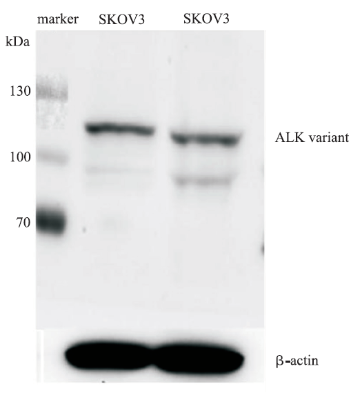
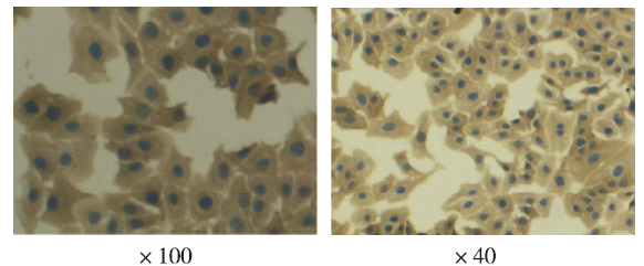
目的 探索人卵巢浆液性腺癌细胞系SKOV3中间变性淋巴瘤激酶(ALK)的表达及其对克唑替尼的反应。方法 采用蛋白免疫印迹法(Western blotting)及免疫细胞化学法检测ALK蛋白在人卵巢癌细胞系SKOV3中的表达;细胞增殖实验检测ALK抑制药克唑替尼对SKOV3细胞增殖的影响。结果 Western blotting结果显示,SKOV3细胞中有ALK蛋白表达;免疫细胞化学结果显示,ALK在SKOV3细胞质中有表达。细胞增殖实验显示,克唑替尼≤106 nmol·L-1时SKOV3细胞活力与对照组比值均接近1,达到107 nmol·L-1时SKOV3细胞活力值接近64%。结论 人卵巢癌细胞系SKOV3细胞中有ALK蛋白表达,但其对克唑替尼治疗不敏感。
关键词:
克唑替尼
;
癌
;
卵巢
;
SKOV3细胞
;
间变性淋巴瘤激酶
Abstract:
Objective To explore the expression of anaplastic lymphoma kinase(ALK) in human ovarian serous adenocarcinoma cell line SKOV3 and to further investigate the effect of crizotinib on SKOV3 cells. Methods The expression of ALK in SKOV3 cells were examined by Western blotting and immunocytochemical methods,and the effect of ALK inhibitor crizotinib on proliferation of SKOV3 cells were evaluated by cell proliferation assay. Results Both Western blotting and immunocytochemical methods showed the expression of ALK in SKOV3 cells.The results of cell proliferation assay suggested that the cell viability of SKOV3 cells was close to 1 when the drug concentration was less than 106 nmol·L-1,and close to 64% when the drug concentration was up to 107nmol·L-1. Conclusion ALK is overexpressed in human ovarian serous adenocarcinoma cell line SKOV3,but SKOV3 cells were insensitive to the therapy of ALK inhibitor crizotinib.
Key words:
Crizotinib
;
Cancer
;
ovarian
;
Cell line SKOV3
;
Aanaplastic lymphoma kinase
卵巢癌是女性生殖系统常见恶性肿瘤之一。据估计,2015年我国有约5.2万例卵巢癌新发病例,近2.3万例死于该病[1]。卵巢癌严重危害女性健康,具有起病隐匿、早期不易发现、易转移、预后差等特点。尽管卵巢癌对手术和化学治疗(化疗)的初始有效率很高,但容易复发并出现化疗耐药[2]。了解卵巢癌发生的分子遗传学机制,寻找治疗的分子靶点有可能克服化疗耐药问题。分子靶向治疗选择性高,不良反应轻,已经成为恶性肿瘤治疗中越来越重要的组成部分,可能成为改善卵巢癌预后的有效方法之一。间变性淋巴瘤激酶(anaplastic lymphoma kinase,ALK)是受体酪氨酸激酶家族成员,于1994年在间变性大细胞淋巴瘤(anaplastic large cell lymphoma,ALCL)的一个亚型中被发现[3]。随后,在多种肿瘤中发现ALK基因重排,证明ALK是强力致癌驱动基因[4-5]。克唑替尼是一种小分子酪氨酸激酶抑制药,是第一个针对ALK的靶向治疗药物。目前克唑替尼已被国内外批准用于棘皮动物微管相关蛋白4/ALK(Echinoderm microtubule-associated protein-like 4/ALK,EML4-ALK)融合基因阳性的非小细胞肺癌的治疗。近年有研究报道在卵巢癌患者组织标本中检测到ALK融合基因表达[6-7]。浆液性腺癌是卵巢癌最常见的病理类型,目前笔者尚未见文献报道人卵巢浆液性腺癌细胞系SKOV3中是否存在ALK表达以及是否对ALK抑制药治疗敏感。笔者在本实验中采用免疫细胞化学法及蛋白质免疫印迹法探索ALK 蛋白在SKOV3细胞中的表达,并进一步验证该细胞是否对ALK抑制药克唑替尼治疗敏感。
CHENW,ZHENGR,BAADE PD,et al.Cancer statistics in China,2015[J].CA Cancer J Clin,2016,66(2):115-132.
<p>With increasing incidence and mortality, cancer is the leading cause of death in China and is a major public health problem. Because of China's massive population (1.37 billion), previous national incidence and mortality estimates have been limited to small samples of the population using data from the 1990s or based on a specific year. With high-quality data from an additional number of population-based registries now available through the National Central Cancer Registry of China, the authors analyzed data from 72 local, population-based cancer registries (2009-2011), representing 6.5% of the population, to estimate the number of new cases and cancer deaths for 2015. Data from 22 registries were used for trend analyses (2000-2011). The results indicated that an estimated 4292,000 new cancer cases and 2814,000 cancer deaths would occur in China in 2015, with lung cancer being the most common incident cancer and the leading cause of cancer death. Stomach, esophageal, and liver cancers were also commonly diagnosed and were identified as leading causes of cancer death. Residents of rural areas had significantly higher age-standardized (Segi population) incidence and mortality rates for all cancers combined than urban residents (213.6 per 100,000 vs 191.5 per 100,000 for incidence; 149.0 per 100,000 vs 109.5 per 100,000 for mortality, respectively). For all cancers combined, the incidence rates were stable during 2000 through 2011 for males (+0.2% per year; P = .1), whereas they increased significantly (+2.2% per year; P CA Cancer J Clin 2016;66:115 132. 2016 American Cancer Society.</b></p>
SCHMID BC,OEHLER MK.New perspectives in ovarian cancer treatment[J].Maturitas,2014,77(2):128-136.
Ovarian cancer (OC) is increasingly understood as a heterogeneous disease comprising distinct subtypes of different origin that vary significantly with regard to molecular biology and clinical behaviour. Despite some limited progress in its treatment over the last decade, currently there are few therapeutic options and overall survival remains poor. Increasing knowledge about the molecular biology of ovarian cancer has led to the development of targeted therapies which promise to be more effective and to provide the basis for personalized treatment. The most successful strategies so far are employing anti-angiogenics (VEGF antibodies, tyrosine kinase inhibitors and angiopoietin antagonists) and polyadenosine diphosphate-ribose polymerase (PARP) inhibitors. Other approaches target aberrant OC signalling such as the PI3K/Akt/mTOR network, the epidermal growth factor receptor, the WEE1 tyrosine kinase and the folate receptor alpha. Immunotherapy is another promising new approach against ovarian cancer. In this area, immunotherapeutic modulation by administering autologous immune cells, such as dendritic cells (DCs), to stimulate antitumour host responses is of special interest. Finally, there is now growing evidence from clinical studies showing a survival advantage for intraperitoneal (IP) chemotherapy when compared to conventional intravenous treatment in the adjuvant setting. New strategies such as pressurized IP aerosol chemotherapy might further improve the efficacy of this approach.
MORRIS SW,KIRSTEIN MN,VALENTINE MB,et al.Fusion of a kinase gene,ALK,to a nucleolar protein gene,NPM,in non-Hodgkin's lymphoma[J].Science,1995,267(5196):316-317.
[本文引用:2]
[4]
LINE,LIL,GUANY,et al.Exon array profiling detects EML4-ALK fusion in breast,colorectal,and non-small cell lung cancers[J].Mol Cancer Res,2009,7(9):1466-1476.
The echinoderm microtubule-associated protein-like 4 naplastic lymphoma kinase (EML4-ALK) fusion gene has been identified as an oncogene in a subset of non mall cell lung cancers (NSCLC). We used profiling of cancer genomes on an exon array to develop a novel computational method for the global search of gene rearrangements. This approach led to the detection of EML4-ALK fusion in breast and colorectal carcinomas in addition to NSCLC. Screening of a large collection of patient tumor samples showed the presence of EML4-ALK fusion in 2.4% of breast (5 of 209), 2.4% of colorectal (2 of 83), and in 11.3% of NSCLC (12 of 106). Besides previously known EML4-ALK variants 1 (E13; A20) and 2 (E20; A20), a novel variant E21; A20 was found in colorectal carcinoma. The presence of an EML-ALK rearrangement was verified by identifying genomic fusion points in tumor samples representative of breast, colon, and NSCLC. EML4-ALK translocation was also confirmed by fluorescence in situ hybridization assay, which revealed its substantial heterogeneity in both primary tumors and tumor-derived cell lines. To elucidate the functional significance of EML4-ALK, we examined the growth of cell lines harboring the fusion following EML4 and WLK silencing by small interfering RNA. Significant growth inhibition was observed in some but not all cell lines, suggesting their variable dependence on ALK-mediated cell survival signaling. Collectively, these findings show the recurrence of EML4-ALK fusion in multiple solid tumors and further substantiate its role in tumorigenesis.
KELLEHER FC,MCDERMOTTR.The emerging pathogenic and therapeutic importance of the anaplastic lymphoma kinase gene[J].Eur J Cancer,2010,46(13):2357-2368.
The anaplastic lymphoma kinase gene (ALK) is a gene on chromosome 2p23 that has expression restricted to the brain, testis and small intestine but is not expressed in normal lymphoid tissue. It has similarity to the insulin receptor subfamily of kinases and is emerging as having increased pathologic and potential therapeutic importance in malignant disease. This gene was originally established as being implicated in the pathogenesis of rare diseases including inflammatory myofibroblastic tumour (IMT) and ALK-positive anaplastic large cell lymphoma, which is a subtype of non-Hodgkin's lymphoma. Recently the number of diseases in which ALK is implicated in their pathogenesis has increased. In 2007, an inversion of chromosome 2 involving ALK and a fusion partner gene in a subset of non-small cell lung cancer was discovered. In 2008, publications emerged implicating ALK in familial and sporadic cases of neuroblastoma, a childhood cancer of the sympatho-adrenal system. Chromosomal abnormalities involving ALK are translocations, amplifications or mutations. Chromosomal translocations are the longest recognised ALK genetic abnormality. When translocations occur a fusion gene is created between ALK and a gene partner. This has been described in ALK-positive anaplastic large cell lymphoma in which ALK is fused to NPM (nucleolar protein gene) and in non-small cell lung cancer where ALK is fused to EML4 (Echinoderm microtubule-associated protein 4). The most frequently described partner genes in inflammatory myofibroblastic tumour are tropomyosin 3/4 (TMP3/4), however in IMTs a diversity of ALK fusion partners have been found, with the ability to homodimerise a common characteristic. Point mutations and amplification of the ALK gene occur in the childhood cancer neuroblastoma. Therapeutic targeting of ALK fusion genes using tyrosine kinase inhibition, vaccination using an ALK specific antigen and treatment using viral vectors for RNAi are emerging potential therapeutic possibilities.
RENH,TAN ZP,ZHUX,et al.Identification of anaplastic lymphoma kinase as a potential therapeutic target in ovarian cancer[J].Cancer Res,2012,72(13):3312-3323.
ABSTRACT Ovarian cancer is the leading cause of death from gynecologic cancer. Improvement in the clinical outcome of patients is likely to be achieved by the identification of molecular events that underlie the oncogenesis of ovarian cancer. Here we show that the anaplastic lymphoma kinase (ALK) is aberrantly activated in ovarian cancer. Using an unbiased and global phosphoproteomic approach, we profiled 69 Chinese primary ovarian tumor tissues and found ALK to be aberrantly expressed and phosphorylated in 4 tumors. Genetic characterization of these ALK-positive tumors indicated that full-length ALK expression in two serous carcinoma patients is consistent with ALK gene copy number gain, whereas a stromal sarcoma patient carries a novel transmembrane ALK fusion gene: FN1-ALK. Biochemical and functional analysis showed that both full-length ALK and FN1-ALK are oncogenic, and tumors expressing ALK or FN1-ALK are sensitive to ALK kinase inhibitors. Furthermore, immunohistochemical analysis of ovarian tumor tissue microarray detected aberrant ALK expression in 2% to 4% serous carcinoma patients. Our findings provide new insights into the pathogenesis of ovarian cancer and identify ALK as a potential therapeutic target in a subset of serous ovarian carcinoma and stromal sarcoma patients.
There have been major advances in our understanding of the cellular and molecular biology of the human malignancies that are collectively referred to as ovarian cancer. At a recent Helene Harris Memorial Trust meeting, an international group of researchers considered actions that should be taken to improve the outcome for women with ovarian cancer. Nine major recommendations are outlined in this Opinion article.
WILLMOTT LJ,FRUEHAUF JP.Targeted therapy in ovarian cancer[J].J Oncol,2010,2010(4):1-9.
[本文引用:0]
[12]
SCHMID BC,OEHLER MK.New perspectives in ovarian cancer treatment[J].Maturitas,2014,77(2):128-136.
Ovarian cancer (OC) is increasingly understood as a heterogeneous disease comprising distinct subtypes of different origin that vary significantly with regard to molecular biology and clinical behaviour. Despite some limited progress in its treatment over the last decade, currently there are few therapeutic options and overall survival remains poor. Increasing knowledge about the molecular biology of ovarian cancer has led to the development of targeted therapies which promise to be more effective and to provide the basis for personalized treatment. The most successful strategies so far are employing anti-angiogenics (VEGF antibodies, tyrosine kinase inhibitors and angiopoietin antagonists) and polyadenosine diphosphate-ribose polymerase (PARP) inhibitors. Other approaches target aberrant OC signalling such as the PI3K/Akt/mTOR network, the epidermal growth factor receptor, the WEE1 tyrosine kinase and the folate receptor alpha. Immunotherapy is another promising new approach against ovarian cancer. In this area, immunotherapeutic modulation by administering autologous immune cells, such as dendritic cells (DCs), to stimulate antitumour host responses is of special interest. Finally, there is now growing evidence from clinical studies showing a survival advantage for intraperitoneal (IP) chemotherapy when compared to conventional intravenous treatment in the adjuvant setting. New strategies such as pressurized IP aerosol chemotherapy might further improve the efficacy of this approach.
MOURALIJ,BENARDA,LOURENCO FC,et al.Anaplastic lymphoma kinase is a dependence receptor whose proapoptotic functions are activated by caspase cleavage[J].Mol Cell Biol,2006,26(16):6209-6222.
Anaplastic lymphoma kinase (ALK) is a receptor tyrosine kinase, initially discovered as part of the NPM-ALK fusion protein, resulting from the t(2;5) translocation that is frequently associated with anaplastic large-cell lymphomas. The native ALK protein is normally expressed in the developing and, at a weaker level, adult nervous system. We recently demonstrated that the oncogenic, constitutively kinase-activated NPM-ALK protein was antiapoptotic when expressed in Jurkat lymphoblastic cells treated with cytotoxic drugs. In contrast, we now show that Jurkat cells overexpressing the wild-type ALK receptor are more sensitive to doxorubicin-induced apoptosis than parental cells. Moreover, the ALK protein is cleaved during apoptosis in a caspase-dependent manner. Mutation of aspartic residues to asparagine allowed us to map the caspase cleavage site in the juxtamembrane region of ALK. In order to assess the role of ALK in neural cell-derived tissue, we transiently expressed ALK in the 13.S.1.24 rat neuroblast immortalized cell line. ALK expression led to apoptotic cell death of the neuroblasts. ALK ligation by specific activating antibodies decreased ALK-facilitated apoptosis in both lymphoid and neuronal cell lines. Moreover, ALK transfection reduced the survival of primary cultures of cortical neurons. Thus, ALK has a proapoptotic activity in the absence of ligand, whereas it is antiapoptotic in the presence of its ligand and when the kinase is intrinsically activated. These properties place ALK in the growing family of dependence receptors.
PULFORDK,MORRIS SW,TURTURROF.Anaplastic lymphoma kinase proteins in growth control and cancer[J].J Cell Physiol,2004,199(3):330-358.
Abstract Top of page Abstract ANAPLASTIC LYMPHOMA KINASE AS A RECEPTOR TYROSINE KINASE IDENTIFICATION OF ALK FULL-LENGTH ALK ALK FUSION PROTEINS ONCOGENIC MECHANISMS EMPLOYED BY ALK FUSION PROTEINS RELEVANCE OF EXPRESSION OF ALK PROTEIN FOR PATIENTS POTENTIAL FUTURE THERAPEUTIC APPROACHES FOR ALK-POSITIVE MALIGNANCIES LITERATURE CITED The normal functions of full-length anaplastic lymphoma kinase (ALK) remain to be completely elucidated. Although considered to be important in neural development, recent studies in Drosophila also highlight a role for ALK in gut muscle differentiation. Indeed, the Drosophila model offers a future arena for the study of ALK, its ligands and signalling cascades. The discovery of activated fusion forms of the ALK tyrosine kinase in anaplastic large cell lymphoma (ALCL) has dramatically improved our understanding of the pathogenesis of these lymphomas and enhanced the pathological diagnosis of this subtype of non-Hodgkin's lymphoma (NHL). Likewise, the realisation that a high percentage of inflammatory myofibroblastic tumours express activated-ALK fusion proteins has clarified the causation of these mesenchymal neoplasms and provided for their easier discrimination from other mesenchymal-derived inflammatory myofibroblastic tumour (IMT) mimics. Recent reports of ALK expression in a range of carcinoma-derived cell lines together with its apparent role as a receptor for PTN and MK, both of which have been implicated in tumourigenesis, raise the possibility that ALK-mediated signalling could play a role in the development and/or progression of a number of common solid tumours. The therapeutic targeting of ALK may prove to have efficacy in the treatment of many of these neoplasms. 2004 Wiley-Liss, Inc.
LAWRENCEB,PEREZ-ATAYDEA,HIBBARD MK,et al.TPM3-ALK and TPM4-ALK oncogenes in inflammatory myofibroblastic tumors[J].Am J Pathol,2000,157(2):377-384.
Inflammatory myofibroblastic tumors (IMTs) are neoplastic mesenchymal proliferations featuring an inflammatory infiltrate composed primarily of lymphocytes and plasma cells. The myofibroblastic cells in some IMTs contain chromosomal rearrangements involving the ALK receptor tyrosine-kinase locus region (chromosome band 2p23). ALK-which is normally restricted in its expression to neural tissues-is expressed strikingly in the IMT cells with 2p23 rearrangements. We now report a recurrent oncogenic mechanism, in IMTs, in which tropomyosin (TPM) N-terminal coiled-coil domains are fused to the ALK C-terminal kinase domain. We have cloned two ALK fusion genes, TPM4-ALK and TPM3-ALK, which encode approximately 95-kd fusion oncoproteins characterized by constitutive kinase activity and tyrosylphosphorylation. Immunohistochemical and molecular correlations, in other IMTs, implicate non-TPM ALK oncoproteins that are predominantly cytoplasmic or pre- dominantly nuclear, presumably depending on the subcellular localization of the ALK fusion partner. Notably, a TPM3-ALK oncogene was reported recently in anaplastic lymphoma, and TPM3-ALK is thereby the first known fusion oncogene that transforms, in vivo, both mesenchymal and lymphoid human cell lineages.
SODAM,CHOI YL,ENOMOTOM,et al.Identification of the transforming EML4-ALK fusion gene in non-small-cell lung cancer[J].Nature,2007,448(7153):561-566.
Elsevier’s Scopus, the largest abstract and citation database of peer-reviewed literature. Search and access research from the science, technology, medicine, social sciences and arts and humanities fields.
GEORGE RE,SANDAT,HANNAM,et al.Activating mutations in ALK provide a therapeutic target in neuroblastoma[J].Nature,2008,455(7215):975-978.
Neuroblastoma, an embryonal tumour of the peripheral sympathetic nervous system, accounts for approximately 15% of all deaths due to childhood cancer. High-risk neuroblastomas are rapidly progressive; even with intensive myeloablative chemotherapy, relapse is common and almost uniformly fatal. Here we report the detection of previously unknown mutations in the ALK gene, which encodes a receptor tyrosine kinase, in 8% of primary neuroblastomas. Five non-synonymous sequence variations were identified in the kinase domain of ALK, of which three were somatic and two were germ line. The most frequent mutation, F1174L, was also identified in three different neuroblastoma cell lines. ALK complementary DNAs encoding the F1174L and R1275Q variants, but not the wild-type ALK cDNA, transformed interleukin-3-dependent murine haematopoietic Ba/F3 cells to cytokine-independent growth. Ba/F3 cells expressing these mutations were sensitive to the small-molecule inhibitor of ALK, TAE684 (ref. 4). Furthermore, two human neuroblastoma cell lines harbouring the F1174L mutation were also sensitive to the inhibitor. Cytotoxicity was associated with increased amounts of apoptosis as measured by TdT-mediated dUTP nick end labelling (TUNEL). Short hairpin RNA (shRNA)-mediated knockdown of ALK expression in neuroblastoma cell lines with the F1174L mutation also resulted in apoptosis and impaired cell proliferation. Thus, activating alleles of the ALK receptor tyrosine kinase are present in primary neuroblastoma tumours and in established neuroblastoma cell lines, and confer sensitivity to ALK inhibition with small molecules, providing a molecular rationale for targeted therapy of this disease.
JANOUEIX-LEROSEYI,LEQUIND,BRUGIERESL,et al.Somatic and germline activating mutations of the ALK kinase receptor in neuroblastoma[J].Nature,2008,455(7215):967-970.
Neuroblastoma, a tumour derived from the peripheral sympathetic nervous system, is one of the most frequent solid tumours in childhood. It usually occurs sporadically but familial cases are observed, with a subset of cases occurring in association with congenital malformations of the neural crest being linked to germline mutations of the PHOX2B gene. Here we conducted genome-wide comparative genomic hybridization analysis on a large series of neuroblastomas. Copy number increase at the locus encoding the anaplastic lymphoma kinase (ALK) tyrosine kinase receptor was observed recurrently. One particularly informative case presented a high-level gene amplification that was strictly limited to ALK, indicating that this gene may contribute on its own to neuroblastoma development. Through subsequent direct sequencing of cell lines and primary tumour DNAs we identified somatic mutations of the ALK kinase domain that mainly clustered in two hotspots. Germline mutations were observed in two neuroblastoma families, indicating that ALK is a neuroblastoma predisposition gene. Mutated ALK proteins were overexpressed, hyperphosphorylated and showed constitutive kinase activity. The knockdown of ALK expression in ALK-mutated cells, but also in cell lines overexpressing a wild-type ALK, led to a marked decrease of cell proliferation. Altogether, these data identify ALK as a critical player in neuroblastoma development that may hence represent a very attractive therapeutic target in this disease that is still frequently fatal with current treatments.
CHENY,TAKITAJ,CHOI YL,et al.Oncogenic mutations of ALK kinase in neuroblastoma[J].Nature,2008,455(7215):971-974.
Nature is the international weekly journal of science: a magazine style journal that publishes full-length research papers in all disciplines of science, as well as News and Views, reviews, news, features, commentaries, web focuses and more, covering all branches of science and how science impacts upon all aspects of society and life.
DEBELENKO LV,RAIMONDI SC,DAWN,et al.Renal cell carcinoma with novel VCL-ALK fusion:new representative of ALK-associated tumor spectrum[J].Mod Pathol,2011,24(3):430-442.
Renal cell carcinoma represents a model for contemporary classification of solid tumors; however, unusual and unclassifiable cases exist and are not rare in children and young adults. The anaplastic lymphoma kinase (ALK) gene has recently been implicated in subsets of pulmonary, esophageal, breast, and colon cancers. These findings strengthen the importance of molecular classification of carcinomas across different organ sites, especially considering the evolving targeted anticancer therapies with ALK inhibitors. In the current study of six pediatric renal cell carcinomas, two cases exhibited structural karyotypic abnormalities involving the ALK locus on chromosomal band 2p23. Fluorescence in situ hybridization (FISH) studies were positive for an ALK rearrangement in one case, and subsequent 5' rapid amplification of cDNA ends analysis of this tumor revealed that the 3' portion of the ALK transcript encoding for the kinase domain was fused in frame to the 5' portion of vinculin (VCL, NM_003373). The new fusion gene is predicted to have an open reading frame of 4122 bp encoding for a 1374-aa oncoprotein; its expression was shown by immunoblotting with anti-VCL and anti-ALK antibodies in tumor tissue lysates. Immunohistochemistry with the same antibodies demonstrated cytoplasmic and subplasmalemmal localization of the oncoprotein determined by its N-terminal VCL portion. FISH with a custom-designed VCL-ALK dual-fusion probe set confirmed the presence of the fusion in neoplastic cells and demonstrated the potential clinical utility of this approach for detecting VCL-ALK in routinely processed tissue. The five remaining pediatric renal cell carcinomas did not show ALK rearrangement by FISH or ALK expression by immunohistochemistry. The data identify the kidney as a new organ site for ALK-associated carcinomas and VCL as a novel ALK fusion partner. The results should prompt further studies to advance the molecular classification of renal cell carcinoma and help to select patients who would benefit from appropriate targeted therapies.
Abstract Renal Medullary Carcinoma (RMC) is an aggressive malignancy that affects young black individuals with sickle cell trait. No effective treatment is available, resulting in an ominous clinical course, with overall survival averaging less than four months. We report rearrangement of the ALK receptor tyrosine kinase in a pediatric case of RMC harboring a t(2;10)(p23;q22) translocation. Mass spectrometry-based proteomic evaluation identified a novel ALK oncoprotein in which the cytoskeletal protein vinculin (VCL) was fused to the ALK kinase domain. The resulting VCL-ALK fusion does not contain known self-association domains, but includes the talin binding domains of vinculin. We demonstrate coprecipitation of strongly tyrosine phosphorylated talins with the VCL-ALK oncoprotein, suggesting that ALK oncogenic crossphosphorylation is mediated by interactions between neighboring VCL-ALK proteins on a talin scaffold. This report widens the spectrum of ALK-related tumors and ALK fusion partners, and provides a rationale for treating RMC with targeted ALK inhibitors. 2010 Wiley-Liss, Inc.
CHIARLER,VOENAC,AMBROGIOC,et al.The anaplastic lymphomakinase in the pathogenesis of cancer[J].Nat Rev Cancer,2008,8(1):11-23.
Tyrosine kinases are involved in the pathogenesis of most cancers. However, few tyrosine kinases have been shown to have a well-defined pathogenetic role in lymphomas. The anaplastic lymphoma kinase (ALK) is the oncogene of most anaplastic large cell lymphomas (ALCL), driving transformation through many molecular mechanisms. In this Review, we will analyse how translocations or deregulated expression of ALK contribute to oncogenesis and how recent genetic or pharmacological tools, aimed at neutralizing its activity, can represent the basis for the design of powerful combination therapies.
PEJOVICT,PANDE NT,MORIM,et al.Expression profiling of the ovarian surface kinome reveals candidate genes for early neoplastic changes[J].Transl Oncol,2009,2(4):341-349.
OBJECTIVES: We tested the hypothesis that co-coordinated up-regulation or down-regulation of several ovarian cell surface kinases may provide clues for better understanding of the disease and help in rational design of therapeutic targets. STUDY DESIGN: We compared the expression signature of 69 surface kinases in normal ovarian surface epithelial cells (OSE), with OSE from patients at high risk and with ovarian cancer. RESULTS: Seven surface kinases, ALK, EPHA5, EPHB1, ERBB4, INSRR, PTK, and TGF尾R1 displayed a distinctive linear trend in expression from normal, highrisk, and malignant epithelium. We confirmed these results using semiquantitative reverse transcription-polymerase chain reaction and tissue array of 202 ovarian cancer samples. A strong correlate was shown between disease-free survival and the expression of ERBB4. DNA sequencing revealed two novel mutations in ERBB4 in two cancer samples. CONCLUSIONS: A distinct subset of the ovarian surface kinome is altered in the transition from high risk to invasive cancer and genetic mutation is not a dominant mechanism for these modifications. These results have significant implications for early detection and targeted therapeutic approaches for women at high risk of developing ovarian cancer.

 , 刘俊
, 刘俊
