Objective To explore the effect of 14,15-epoxyeicosatrienoic acids (14,15-EET) on the inflammatory response of BV2 cells under oxygen and glucose depriviation/reoxygenation (OGD/R) conditions. Methods BV2 cells were randomly divided into three groups,blank control group,vehicle control group,and 14,15-EET group.Under treatment of 14,15-EET,the concentration of inflammatory factor in BV2 cell culture media was detected by ELISA at different time points (reoxygenation for 0,3,6,12,24 h) after OGD1h.The viability of BV2 cells was detected by MTT assay at different time points.At the same conditions,using Transwell migration experiment,migration ability of BV2 cells was observed. Results The 14,15-EET group had the lower levels of inflammatory factor secretion,lower viability and weaker ability of migration than the vehicle control group.The above results were most statistically significant at OGD1h/R12h. Conclusion 14,15-EET can inhibit the inflammation of BV2 cells induced by the injury of OGD reperfusion.
Fig.2
Comparison of cell proliferation among three groups of BV2 cells at different time points after OGD/R(x¯±s,n=5) Compared with blank control group,*1P<0.01;compared with vehicle control group,*2P<0.05,*3P<0.01
Fig.3
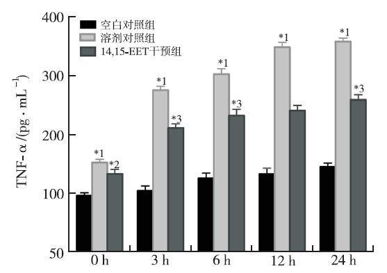
Comparison of TNF-α level in the supernatant among three groups of BV2 cells after OGD/R(x¯±s,n=5) Compared with blank control group,*1P<0.01;compared with vehicle control group,*2P<0.05,*3P<0.01
Fig.4
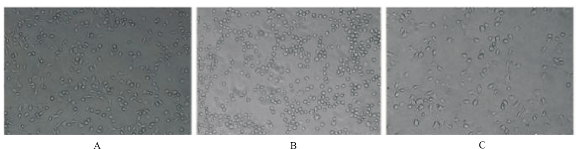
Crystal violet staining on cell migration of three groups of BV2 cells 12 h after OGD/R(×200) A.blank control group;B.vehicle control group;C.14,15-EET group
Fig.5
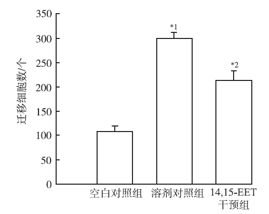
Comparison of the migratory number among three groups of BV2 cells 12 h after OGD/R(x¯±s,n=5) Compared with blank control group,*1P<0.05;compared with vehicle control group,*2P<0.05
FERRI CP,SCHOENBORNC,KALRAL,et al.Prevalence of stroke and related burden among older people living in Latin America,India and China[J].J Neurol Neurosurg Psychiatry,2011,82(10):1074-1082.
Despite the growing importance of stroke in developing countries, little is known of stroke burden in survivors. The authors investigated the prevalence of self-reported stroke, stroke-related disability, dependence and care-giver strain in Latin America (LA), China and India.Cross-sectional surveys were conducted on individuals aged 65+ (n=15鈥022) living in specified catchment areas. Self-reported stroke diagnosis, disability, care needs and care giver burden were assessed using a standardised protocol. For those reporting stroke, the correlates of disability, dependence and care-giver burden were estimated at each site using Poisson or linear regression, and combined meta-analytically.The prevalence of self-reported stroke ranged between 6% and 9% across most LA sites and urban China, but was much lower in urban India (1.9%), and in rural sites in India (1.1%), China (1.6%) and Peru (2.7%). The proportion of stroke survivors needing care varied between 20% and 39% in LA sites but was higher in rural China (44%), urban China (54%) and rural India (73%). Comorbid dementia and depression were the main correlates of disability and dependence.The prevalence of stroke in urban LA and Chinese sites is nearly as high as in industrialised countries. High levels of disability and dependence in the other mainly rural and less-developed sites suggest underascertainment of less severe cases as one likely explanation for the lower prevalence in those settings. As the health transition proceeds, a further increase in numbers of older stroke survivors is to be anticipated. In addition to prevention, stroke rehabilitation and long-term care needs should be addressed.
ZHANGB,PUS,ZHANGW,et al.Sex differences in risk factors,etiology,and short-term outcome of cerebral infarction in young patients[J].Atherosclerosis,2011,216(2):420-425.
Investigations to date have demonstrated that the underlying etiology, causes and burden of stroke may be different for women and men. However, data regarding sex differences among young cerebral ischemic stroke patients remains scarce. We conducted this study in 669 young Chinese adults with acute ischemic stroke as determined by the modified Rankin Scale at discharge. Stepwise multiple logistic regression analysis confirmed that NIHSS score (OR 1.277; 95% CI 1.179-1.383, p=0.000), diabetes mellitus (OR 0.121; 95% CI 0.0209-0.718, p=0.020) and serum glucose levels on admission (OR 1.135; 95% CI 0.997-1.293, p=0.046) independently predict short-term outcomes at discharge in young female patients with acute stroke, but the significant variables related to male patients appeared to be Apo A1 (OR 0.165; 95% CI 0.035-0.776, p=0.023) and NIHSS score on admission (OR 1.458; 95% CI 1.325-1.605, p=0.000). In our series, our data suggest that there are several sex differences for risk of cerebral infarction in young patients, which have important implications for the diagnosis, management and prognosis of stroke in young adults.
LIUY,WANY,FANGY,et al.Epoxyeicosanoid signaling provides multi-target protective effects on neurovascular unit in rats after focal ischemia[J].J Mol Neurosci,2016,58(2):254-265.
Multiple players are involved in the highly complex pathophysiologic responses after stroke. Therefore, therapeutic approaches that target multiple cellular elements of the neurovascular unit in the damage cascade hold considerable promise for the treatment of stroke. Cytochrome P450 (CYP) epoxygenases metabolize arachidonic acid to biologically active eicosanoids called epoxyeicosatrienoic acids (EETs), which are further converted by soluble epoxide hydrolase (sEH) to less bioactive diols. EETs have been shown to exert direct cytoprotective effects upon several individual components of the neurovascular unit under simulated ischemic conditions in vitro. However, the cellular mechanism underlying EET-mediated neuroprotective effects after ischemia remains to be clarified. In this study, we investigated the effects of 14,15-EET and 12-(3-adamantan-1-yl-ureido)dodecanoic acid (AUDA), a selective inhibitor of sEH, on multiple elements of neurovascular unit of the rat brain after middle cerebral artery occlusion-induced focal ischemia. The results showed that exogenous administration of 14,15-EET or AUDA could suppress astrogliosis and glial scar formation, inhibit microglia activation and inflammatory response, promote angiogenesis, attenuate neuronal apoptosis and infarct volume, and further promote the behavioral function recovery after focal ischemia. The results suggest that epoxyeicosanoid signaling is a promising multi-mechanism therapeutic target for the treatment of stroke.
SUNF,WANGX,MAOX,et al.Ablation of neurogenesis attenuates recovery of motor function after focal cerebral ischemia in middle-aged mice[J].PLoS One,2012,7(10):e46326.
by Fen Sun, Xiaomei Wang, XiaoOu Mao, Lin Xie, Kunlin Jin Depletion of neurogenesis worsens functional outcome in young-adult mice after focal cerebral ischemia, but whether a similar ef ...
GUOS,LO EH.Dysfunctional cell-cell signaling in the neurovascular unit as a paradigm for central nervous system disease[J].Stroke,2009,40(3 Suppl):4-7.
The fundamental premise of neuroprotection has historically focused on the prevention of neuronal death. However, despite tremendous advances in the molecular biology of intraneuronal mechanisms and pathways, a clinically effective neuroprotectant does not yet exist. This problem is especially clear for stroke, for which a large number of neuroprotection trials have failed. The concept of the neurovascular unit emphasizes that cell-cell signaling among the various neuronal, glial, and vascular compartments underlies the homeostasis of normal brain function. Conversely, dysfunctional signaling within the neurovascular unit should contribute to disease. This minireview surveys recent data that support this basic idea, with examples drawn from experimental models broadly relevant to stroke and neurodegeneration.
ZHANGL,ZHANG ZG,CHOPPM.The neurovascular unit and combination treatment strategies for stroke[J].Trends Pharmacol Sci,2012,33(8):415-422.
Tissue plasminogen activator (tPA) administered within 4.5h of symptom onset restores cerebral blood flow (CBF) and promotes neurological recovery of stroke patients. However, the narrow therapeutic time window and the risk of intracerebral hemorrhage after tPA treatment pose major hurdles to its clinical usage. In light of the failures of neuroprotective therapies in clinical trials, emerging concepts suggest that neuroprotection alone without restoration of tissue perfusion and vascular integrity may not be adequate for treatment of acute stroke. Here we review evidence of the use of adjuvant pharmacological agents to extend the therapeutic window for tPA via targeting the neurovascular unit and the underlying mechanisms of the combination therapy in experimental stroke.
INNAMORATO NG,LASTRES-BECKERI,CUADRADOA.Role of microglial redox balance in modulation of neuroinflammation[J].Curr Opin Neurol,2009,22(3):308-314.
This review discusses some of the emerging concepts on how modulation of redox homeostasis in microglia is crucial to restore its inactive state and modulate inflammation in neurologic diseases.Reactive oxygen species generated by microglia help to eliminate pathogens in the extracellular milieu but also act on microglia itself, altering the intracellular redox balance and functioning as second messengers in induction of proinflammatory genes. Recent findings indicate that restoration of redox balance may be determinant in driving microglia back to the resting state. Thus, deficiency of the transcription factor NF-E2-related factor-2 (Nrf2), guardian of redox homeostasis, results in exacerbated inflammatory response to neurotoxins whereas inducers of Nrf2 and its target heme oxygenase-1 downmodulate inflammation.New available information indicates that downregulation of microglia is a matter closely correlated with control of oxidative stress in this cell type and points to Nrf2 as a new therapeutic target for modulation of inflammation in neurodegenerative diseases.
YENARI MA,KAUPPINEN TM,SWANSON RA.Microg-lial activation in stroke:therapeutic targets[J].Neuroth-erapeutics,2010,7(4):378-391.
[本文引用:1]
[9]
STOLLG,JANDERS,SCHROETERM.Detrimental and bene-ficial effects of injury-induced inflammation and cyto-kine expression in the nervous system[J].Adv Exp Med Biol,2002,513:87-113.
Lesions in the nervous system induce rapid activation of glial cells and under certain conditions additional recruitment of granulocytes, T-cells and monocytes/macrophages from the blood stream triggered by upregulation of cell adhesion molecules, chemokines and cytokines. Hematogenous cell infiltration is not restricted to infectious or autoimmune disorders of the nervous system, but also occurs in response to cerebral ischemia and traumatic lesions. Neuroinflammation can cause neuronal damage, but also confers neuroprotection. Granulocytes occlude vessels during reperfusion after transient focal ischemia, while the functional role of T-cells and macrophages in stroke development awaits further clarification. After focal cerebral ischemia neurotoxic mediators released by microglia such as the inducible nitric oxide synthase (leading to NO synthesis) and the cytokines interleukin-1beta (IL-1beta) and tumor necrosis factor-alpha (TNF-alpha) are upregulated prior to cellular inflammation in the evolving lesion and functionally contribute to secondary infarct growth as revealed by numerous pharmacological experiments and by use of transgenic animals. On the other hand, cytokine induction remote from ischemic lesions involves NMDA-mediated signalling pathways and confers neuroprotection. After nerve injury T cells can rescue CNS neurons. In the peripheral nervous system neuroinflammation is a prerequisite for successful regeneration that is impeded in the CNS. In conclusion, there is increasing evidence that neuroinflammation represents a double edged sword. The opposing neurotoxic and neuroprotective properties of neuroinflammation during CNS injury provide arich and currently unexplored set of research problems.
ALOISIF.Immune function of microglia[J].Glia,2001,36:165-179.
[本文引用:1]
[11]
NAKAJIMAK,KOHSAKAS.Microglia:activation and their significance in the central nervous system[J].J Biochem,2001,130(2):169-175.
Microglia are resident monocyte-lineaged cells in the brain. Their characteristic feature is that they react to injury and diseases of the brain and become morphologically and functionally activated. Although some trigger molecules which activate microglia are predicted to be released from injured or affected cells, such molecules have not yet been identified. The main role of activated microglia is believed to be in brain defense, as scavengers of dead cells, and as immune or immunoeffector cells. Recent biochemical and neurobiological studies have further indicated that they significantly affect the pathological state and/or regulate the regenerative state and remodeling of the brain by producing a variety of biologically active molecules including cytotoxic and neurotrophic molecules.
MONSONEGOA,IMITOLAJ,ZOTAV,et al.Microglia-mediated nitric oxide cytotoxicity of T cells following amyloid beta-peptide presentation to Th1 cells[J].J Immunol,2003,171(5):2216-2224.
Alzheimer's disease is marked by progressive accumulation of amyloid beta-peptide (Abeta) which appears to trigger neurotoxic and inflammatory cascades. Substantial activation of microglia as part of a local innate immune response is prominent at sites of Abeta plaques in the CNS. However, the role of activated microglia as Abeta APCs and the induction of adaptive immune responses has not been investigated. We have used primary microglial cultures to characterize Abeta-Ag presentation and interaction with Abeta-specific T cells. We found that IFN-gamma-treated microglia serve as efficient Abeta APCs of both Abeta1-40 and Abeta1-42, mediating CD86-dependent proliferation of Abeta-reactive T cells. When cultured with Th1 and Th2 subsets of Abeta-reactive T cells, Th1, but not Th2, cells, underwent apoptosis after stimulation, which was accompanied by increased levels of IFN-gamma, NO, and caspase-3. T cell apoptosis was prevented in the presence of an inducible NO synthase type 2 inhibitor. Microglia-mediated proliferation of Abeta-reactive Th2 cells was associated with expression of the Th2 cytokines IL-4 and IL-10, which counterbalanced the toxic levels of NO induced by Abeta. Our results demonstrate NO-dependent apoptosis of T cells by Abeta-stimulated microglia which may enhance CNS innate immune responses and neurotoxicity in Alzheimer's disease. Secretion of NO by stimulated microglia may underlie a more general pathway of T cell death in the CNS seen in neurodegenerative diseases. Furthermore, Th2 type T cell responses may have a beneficial effect on this process by down-regulation of NO and the proinflammatory environment.
YANGL,ZHOUX,YANGJ,et al.Aspirin inhibits cytoto-xicity of prion peptide PrP106-126 to neuronal cells associated with microglia activation in vitro[J].J Neuroimmunol,2008,199(1/2):10-17.
[本文引用:0]
[14]
KAUSHALV,SCHLICHTER LC.Mechanisms of microglia-mediated neurotoxicity in a new model of the stroke penumbra[J].J Neurosci,2008,28(9):2221-2230.
Abstract After an ischemic stroke, neurons in the core are rapidly committed to die, whereas neuron death in the slowly developing penumbra is more amenable to therapeutic intervention. Microglia activation contributes to delayed inflammation, but because neurotoxic mechanisms in the penumbra are not well understood, we developed an in vitro model of microglia activation and propagated neuron killing. To recapitulate inflammatory triggers in the core, microglia were exposed to oxygen glucose-deprived neurons and astrocytes. To model the developing penumbra, the microglia were washed and allowed to interact with healthy naive neurons and astrocytes. We found that oxygen-glucose deprivation (OGD)-stressed neurons released glutamate, which activated microglia through their group II metabotropic glutamate receptors (mGluRs). Microglia activation involved nuclear factor kappaB (NF-kappaB), a transcription factor that promotes their proinflammatory functions. The activated microglia became neurotoxic, killing naive neurons through an apoptotic mechanism that was mediated by tumor necrosis factor-alpha (TNF-alpha), and involved activation of both caspase-8 and caspase-3. In contrast to some earlier models (e.g., microglia activation by lipopolysaccharide), neurotoxicity was not decreased by an inducible nitric oxide synthase (iNOS) inhibitor (S-methylisothiourea) or a peroxynitrite scavenger [5,10,15,20-tetrakis(N-methyl-4'-pyridyl)porphinato iron (III) chloride], and did not require p38 mitogen-activated protein kinase (MAPK) activation. The same microglia neurotoxic behavior was evoked without exposure to OGD-stressed neurons, by directly activating microglial group II mGluRs with (2S,2'R,3'R)-2-(2'3'-dicarboxycyclopropyl) glycine or glutamate, which stimulated production of TNF-alpha (not nitric oxide) and mediated TNF-alpha-dependent neurotoxicity through activation of NF-kappaB (not p38 MAPK). Together, these results support potential therapeutic strategies that target microglial group II mGluRs, TNFalpha overproduction, and NF-kappaB activation to reduce neuron death in the ischemic penumbra.
STREIT WJ.Microglia as neuroprotective,immunocompet-ent cells of the CNS[J].Glia,2002,40(2):133-139.
The role of glial cells is to support and sustain proper neuronal function and microglia are no exception to this. This viewpoint article emphasizes the fundamental interdependence of microglia and neurons and takes a look at the possibility of what could happen if microglial cells became dysfunctional as a result of aging, genetics, or epigenetics. Could microglial senescence be a factor in the pathogenesis of Alzheimer's and other neurodegenerative diseases? The cautious answer to that question is 'yes'. Future studies along these lines may provide novel insights into microglial involvement in neurodegenerative disease pathogenesis.
ILIFF JJ,JIAJ,NELSONJ,et al.Epoxyeicosanoid signal-ing in CNS function and disease[J].Prostaglandins,2010,91(3/4):68-84.
Epoxyeicosatrienoic acids (EETs) are arachidonic acid metabolites of cytochrome P450 epoxygenase enzymes recognized as key players in vascular function and disease, primarily attributed to their potent vasodilator, anti-inflammatory and pro-angiogenic effects. Although EETs’ actions in the central nervous system (CNS) appear to parallel those in peripheral tissue, accumulating evidence suggests that epoxyeicosanoid signaling plays different roles in neural tissue compared to peripheral tissue; roles that reflect distinct CNS functions, cellular makeup and intercellular relationships. This is exhibited at many levels including the expression of EETs-synthetic and -metabolic enzymes in central neurons and glial cells, EETs’ role in neuro-glio-vascular coupling during cortical functional activation, the capacity for interaction between epoxyeicosanoid and neuroactive endocannabinoid signaling pathways, and the regulation of neurohormone and neuropeptide release by endogenous EETs. The ability of several CNS cell types to produce and respond to EETs suggests that epoxyeicosanoid signaling is a key integrator of cell–cell communication in the CNS, coordinating cellular responses across different cell types. Under pathophysiological conditions, such as cerebral ischemia, EETs protect neurons, astroglia and vascular endothelium, thus preserving the integrity of cellular networks unique to and essential for proper CNS function. Recognition of EETs’ intimate involvement in CNS function in addition to their multi-cellular protective profile has inspired the development of therapeutic strategies against CNS diseases such as cerebral ischemia, tumors, and neural pain and inflammation that are based on targeting the cellular actions of EETs or their biosynthetic and metabolizing enzymes. Based upon the emerging importance of epoxyeicosanoids in cellular function and disease unique to neural systems, we propose that the actions of “neuroactive EETs” are best considered separately, and not in aggregate with all other peripheral EETs functions.

 , 田浩
, 田浩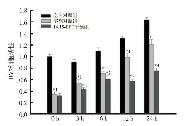

