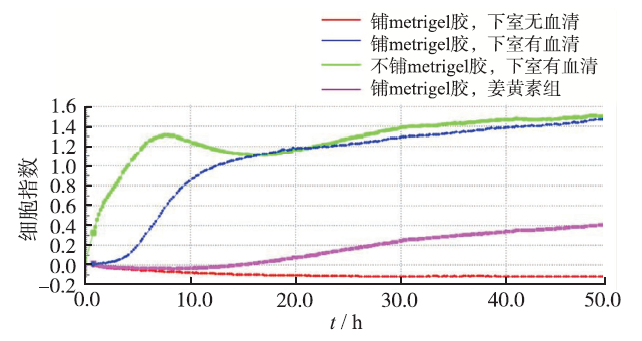
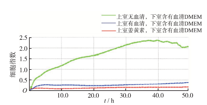
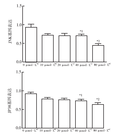
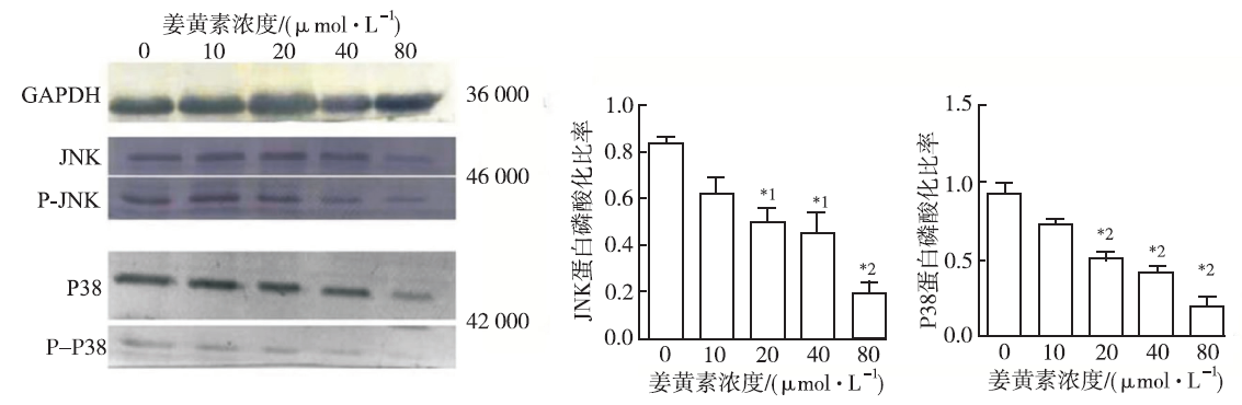
Objective To study the inhibitory effect of curcumin on the proliferation,migration and invasion of non-small cell lung cancer cell A549,and to discuss further if it is closely related to the expression of c-Jun N-terminal kinase (JNK) and relative protein p38. Methods A549 cells were cultured by conventional method,and then treated with different concentration of curcumin (10,20,40,80 μmol·L-1).The proliferation,migration and invasion of A549 cells were measured by real-time cellular analysis (RTCA).The expression levels of JNK,p-JNK,p38 and P-p38 were detected by real-time PCR and Western blotting. Results Curcumin showed an antiproliferation effect against A549 cells with IC50=40 μmol·L-1,and curcumin exhibited obviously inhibitory effect on the migration and invasion of A549 cells.Additionally,compared with control group,curcumin suppressed the expression of JNK and p38 at the gene level,and significantly inhibited the expression of JNK,P-JNK,p38 and p38 (P<0.05) at the protein level. Conclusion These results demonstrated that curcumin can inhibit the proliferation,migration and invasion of A549 cells via reducing the level of JNK,p38 phosphorylation,and blocking JNK signal transduction pathway.
Key words:
Curcumin
;
Cancer
;
lung
;
non-small cell
;
c-Jun N-terminal kinase
;
Relative protein p38
Fig.4
Comparison of gene expression and phosphory-lation of JNK and p38 among five groups of lung cancer A549 Cells(x¯±s,n=4) Compared with control group,*1P<0.05,*2P<0.01
Fig.5
Comparison of protein expression of JNK, P-JNK, p38 and P-p38 among five groups of lung cancer A549 Cells Compared with control group,*1P<0.05,*2P<0.01
BHARATB,AGGARWAL KB.Harikumar Potential thera-peutic effects of curcumin,the anti-inflammatory agent,against neurodegenerative,cardiovascular,pulmonary,metabolic,autoimmune and neoplastic diseases[J].Intern J Biochem Cell Biol,2009,41(3):40-59.
Although safe in most cases, ancient treatments are ignored because neither their active component nor their molecular targets are well defined. This is not the case, however, with curcumin, a yellow-pigment substance and component of turmeric (Curcuma longa), which was identified more than a century ago. For centuries it has been known that turmeric exhibits anti-inflammatory activity, but extensive research performed within the past two decades has shown that this activity of turmeric is due to curcumin (diferuloylmethane). This agent has been shown to regulate numerous transcription factors, cytokines, protein kinases, adhesion molecules, redox status and enzymes that have been linked to inflammation. The process of inflammation has been shown to play a major role in most chronic illnesses, including neurodegenerative, cardiovascular, pulmonary, metabolic, autoimmune and neoplastic diseases. In the current review, we provide evidence for the potential role of curcumin in the prevention and treatment of various proinflammatory chronic diseases. These features, combined with the pharmacological safety and negligible cost, render curcumin an attractive agent to explore further.
ALIN,SOHEIL AE.A review of therapeutic effects of curcumin[J].Curr Pharmac Des,2013,19(45):2032-2046.
There is a growing interest in herbal medicine. Scientific studies have demonstrated the beneficial pharmacological effects of curcumin. Curcumin is a bright yellow spice, derived from the rhizome of Curcuma longa Linn. It has been proven that curcumin is a highly pleiotropic molecule which can be a modulator of intracellular signaling pathways that control cell growth, inflammation, and apoptosis. Curcumin might be a potential candidate for the prevention and/or treatment of some diseases due to its anti-oxidant, antiinflammatory activities and an excellent safety profile. We present an updated concise review of currently available animal and clinical studies demonstrating the therapeutic effect of curcumin.
KAJIURAS,HOSOKAWAA,YOSHITAH,et al.Phase I study of docetaxel plus nedaplatin in patients with metastatic or recurrent esophageal squamous cell carcinoma after cisplatin plus 5-fluorouracil treatment[J].Am J Clin Oncol,2013,47(2):19-20.
Abstract OBJECTIVES: To date, no second-line chemotherapy regimen for esophageal squamous cell carcinoma (SCC) has been established. This clinical trial aimed to assess the optimum dose of docetaxel plus nedaplatin (cis-diammine-glycolate platinum) as second-line chemotherapy. METHODS: Patients with metastatic or recurrent esophageal SCC after treatment with cisplatin plus 5-fluorouracil received docetaxel (50 or 60 mg/m) plus nedaplatin (70 mg/m05) on day 1 every 4 weeks. The recommended dose was based on dose-limiting toxicities defined during the first cycle. RESULTS: From February 2009 to November 2011, 9 patients were enrolled in the study. Their median age was 62 years (range, 58 to 72 y). Six patients had undergone radiotherapy and 4 had undergone surgical resection of primary lesions. Dose-limiting toxicities were observed in 2 patients at dose level 1 (60 mg/m05 docetaxel, 70 mg/m05 nedaplatin) but not at dose level 0 (50 mg/m05 docetaxel, 70 mg/m nedaplatin). Thus, the maximum tolerated dose was established at dose level 1. No severe nonhematological toxicity was observed. No patient achieved complete response, but 2 (22%; 95% confidence interval, 0%-49%) achieved partial response and 3 had stable disease. Median progression-free and overall survival times were 2.1 and 9.5 months, respectively. CONCLUSIONS: Docetaxel plus nedaplatin chemotherapy seems to be a safe and feasible second-line regimen for the treatment of esophageal SCC. We recommend the administration of 50 mg/m05 docetaxel (day 1) plus 70 mg/m05 nedaplatin (day 1) every 4 weeks in a phase II study.
YUS,SHENG,KHOR TO,et al.Curcumin inhibits akt/mammalian target of rapamycin signaling through protein phosphatase-dependent mechanism[J].Mol Cancer Ther,2008,7(5):2609-2620.
[本文引用:2]
[7]
THOMASS,CHRISTIANA,MICHAEL KH.The ankyrin repeat protein diversin recruits casein kinase iepsilon to the beta-catenin degradation complex and acts in both canonical Wnt and Wnt/JNK signaling[J].Genes Dev,2002,16(8):2073-2084.
[本文引用:0]
[8]
LAN LA,YUE ZA,ZHONG HX,et al.Diversin increases the proliferation and invasion ability of non-small-cell lung cancer cells via JNK pathway[J].Cancer Letters,2014,344(2):232-238.
The expression and significance of Diversin in human tumors remains unclear. We found that Diversin was overexpressed in NSCLC, and exhibited direct correlation to poor differentiation, advanced TNM stage, lymph node metastasis and survival time. Overexpression of Diversin lead to a significant increase in proliferation and invasion of NSCLC cells, possibly through activation of JNK, cyclin B and MMP9, and the effects were blocked by JNK inhibitor. These results suggest Diversin is overexpressed in NSCLC and predict poor prognosis. Diversin may promote cell proliferation and invasion through JNK pathway.
TANG ZP,CUI QZ,DONG QZ.Ataxia-telangiectasia gr-oup D complementing gene(ATDC)upregulates matrix metalloproteinase 9(MMP-9)to promote lung cancer cell invasion by activating ERK and JNK pathways[J].Tumour Biol,2013,34(5):2835-2842.
Although the expression pattern and biological functions of ataxia-telangiectasia group D complementing gene (ATDC) had been implicated in several types of cancer, the roles and potential mechanisms of ATDC in lung cancer cell invasion are still ambiguous. In this study, we used gain- and loss-of-function analyses to explore the roles and potential mechanisms of ATDC in lung cancer cell invasion. siRNA knockdown of ATDC impaired cell invasion in A549 and H1299 cell lines, and its overexpression promoted cell invasion in HBE cell line. ATDC may contribute to the invasive ability of lung cancer cells by promoting the expression of invasion-related matrix metalloproteinase 9 (MMP-9). In addition, ATDC increased activating protein 1 (AP-1) reporter luciferase activity and the protein and mRNA levels of c-Jun and c-Fos. We further demonstrated that the roles of ATDC on cell invasion, MMP-9 upregulation, and AP-1 activation were dependent on extracellular signal-regulated protein kinase (ERK) and c-Jun N-terminal kinase (JNK) pathway activation, and ERK inhibitor U0126 or JNK inhibitor SP600125 blocked these effects of ATDC. These results suggested that ATDC upregulates MMP-9 to promote lung cancer cell invasion by activating ERK and JNK pathways.
XUY,ZHANG JJ,HANJ,et al.Curcumin inhibits tumor proliferation induced by neutrophil elastase through the upregulation of α1-antitrypsin in lung cancer[J].Mol Oncol,2012,6(4):405-417.
Lung carcinogenesis is a complex process in an unregulated inflammatory environment. Curcumin has been extensively investigated as a multi-target anti-tumor and anti-inflammation compound. In this paper, we demonstrate a novel inflammation-related mechanism for curcumin-induced inhibition of lung tumor growth. We found that neutrophil elastase, an important regulator of inflammatory processes, directly triggered tumor cell proliferation in human lung adenocarcinoma A549 cells, and curcumin could completely suppress the excess tumor proliferation induced by neutrophil elastase. α1-antitrypsin is synthesized by tumor cells and is the natural inhibitor of neutrophil elastase. We found that curcumin counteracted the decrease of α1-antitrypsin induced by neutrophil elastase by inducing the promoter activity of α1-antitrypsin and promoting its expression in A549 cells. The inhibition of neutrophil elastase-induced proliferation by curcumin was dependent on the PI3K/Akt pathway. Knockdown of α1-antitrypsin by siRNA further enhanced the tumor cell proliferation induced by neutrophil elastase and significantly blocked the anti-proliferation effect of curcumin against neutrophil elastase. Curcumin remarkably inhibited the primary tumor growth of Lewis lung carcinoma (LLC) in C57BL/6 mice. We further showed that curcumin upregulated the level of α1-antitrypsin in primary tumor tissue by promoting its local expression, and the protein level of neutrophil elastase in tumor tissue was obviously decreased in mice treated with curcumin. Overall, our results suggest that neutrophil elastase and α1-antitrypsin play important roles in modulating lung tumor proliferation in inflammatory microenvironment and curcumin inhibits neutrophil elastase-induced tumor proliferation via upregulating α1-antitrypsin expression in02vitro and in02vivo.
PITHIC,VARISAP,SUMALEE WS,et al.Curcumin sen-sitizes lung cancer cells to cisplatin-induced apoptosis through superoxide anion-mediated Bcl-2 degradation[J].Cancer Invest,2009,27(2):624-635.
[本文引用:1]
[12]
LEE HY,OH SH,HONG WK.Response of non-small ce-ll lung cancer cells to the inhibitors of phosphatidylinositol 3-kinase/Akt- and MAPK Kinase 4/c-Jun NH2-terminal kinase pathways:an effective therapeutic strategy for lung cancer[J].Clin Cancer Res,2009,11(2):6065-6074.
[本文引用:1]
Harikumar Potential thera-peutic effects of curcumin,the anti-inflammatory agent,against neurodegenerative,cardiovascular,pulmonary,metabolic,autoimmune and neoplastic diseases
Phase I study of docetaxel plus nedaplatin in patients with metastatic or recurrent esophageal squamous cell carcinoma after cisplatin plus 5-fluorouracil treatment
The ankyrin repeat protein diversin recruits casein kinase iepsilon to the beta-catenin degradation complex and acts in both canonical Wnt and Wnt/JNK signaling
2002
Diversin increases the proliferation and invasion ability of non-small-cell lung cancer cells via JNK pathway
Ataxia-telangiectasia gr-oup D complementing gene(ATDC)upregulates matrix metalloproteinase 9(MMP-9)to promote lung cancer cell invasion by activating ERK and JNK pathways
Response of non-small ce-ll lung cancer cells to the inhibitors of phosphatidylinositol 3-kinase/Akt- and MAPK Kinase 4/c-Jun NH2-terminal kinase pathways:an effective therapeutic strategy for lung cancer

 , 刘洁婷
, 刘洁婷
