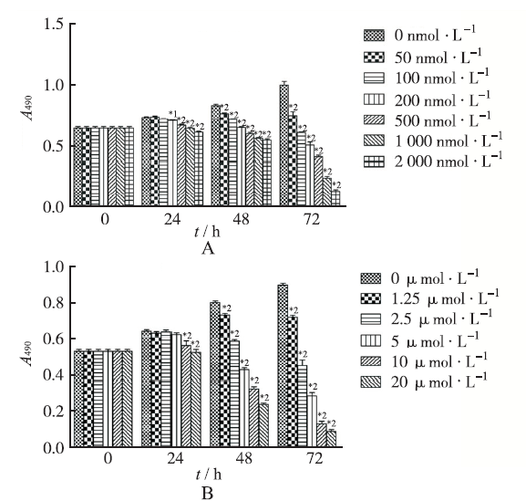
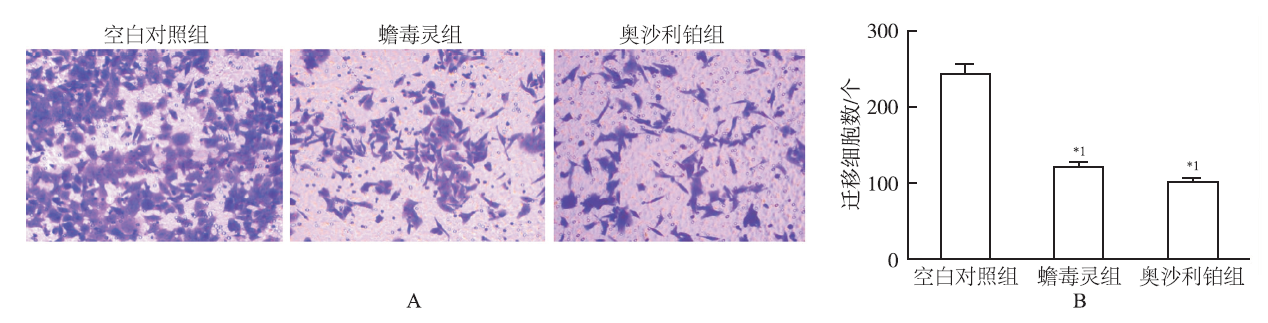
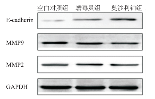
Objective To study effect of bufalin on invasion and metastasis of gastric cancer cells and related mechanism. Methods AGS human gastric cancer cell line was used for in vitro experiments.Cultured cells were treated by negative control group, bufalin group and oxaliplatin group.Cell proliferation was determined by MTT.Invasion and metastasis were observed by Wound-Healing Assay and Transwell Assay.Expression levels of E-cadherin,matrix metalloproteinases (MMP)-2,MMP-9 were detected by Western blotting. Results Bufalin was found to significantly inhibit the proliferation of AGS cells in a dose and time dependent manner.Wound-Healing Assay and Transwell Assay showed that as compared with the blank control group,bufalin inhibited invasion and metastasis of AGS cells (P<0.05),but there was no statistically significant difference between bufalin group and oxaliplatin group (P>0.05).Western blotting showed the expression of E-cadherin was increased in bufalin group as compared with the blank control group while the expression levels of MMP-9 and MMP-2 were down-regulated (P<0.05),but there was no statistically significant difference between bufalin group and oxaliplatin group (P>0.05). Conclusion Bufalin has anti-cancer activity on gastric cancer cells,and it has the ability of inhibiting cancer cell invasion and metastasis,and regulating the expression of some related gene.
Fig.1
Comparison of the inhibitory activity on AGS cells beween bufalin at different concentration(A) and oxaliplatin(B)(x¯±s,n=3) Compared with blank control group,*1P<0.05,*2P<0.01
GAIQ,SHENGX,QIN JM,et al.The effect and mechani-sm of bufalin on regulating hepatocellular carcinoma cell invasion and metastasis via Wnt/β-catenin signaling pathway[J].Int J Oncol,2016,48(1):338-348.
Hepatocellular carcinoma (HCC) is a highly malignant tumor with an extremely poor prognosis. Our preliminary study indicated that bufalin could restrain the proliferation of human hepatoma BEL-7402 cells in a time- and dose-dependent manner. In the present study, the colony formation assay, the Transwell invasion assay, the western blot analysis and the immunofluorescence method were respectively used to investigate the effect and mechanism of bufalin against HCC cell invasion and metastasis. We found that: i) bufalin had significant inhibitory effect on the cell proliferation of BEL-7402 cells; ii) bufalin markedly inhibited the migration and invasion of BEL-7402 cells; iii) bufalin could suppress the phosphorylation of GSK-3β Ser9 site in BEL-7402 cells, decrease the expression of β-catenin, cyclin02D1, metalloproteinases-7 (MMP-7) and cyclooxygenase-2 (COX-2) in the cytoplasm, and increase the expression of E-cadherin and β-catenin on the cell membrane; and iv) the expression of α-fetoprotein significantly decreased and the expression of albumin increased in BEL-7402 cells after bufalin was used. Our results indicate that: i) bufalin can regulate the expression of associated factors in Wnt/β-catenin signaling pathway of BEL-7402 cells through suppressing the phosphorylation of GSK-3β Ser9 site; ii) bufalin can strengthen intercellular E-cadherin/β-catenin complex to control epithelial-mesenchymal transition; and iii) bufalin can reverse the malignant phenotype and promote the differentiation and maturation by regulating the AFP and ALB expression in BEL-7402 cells. These are very important mechanisms of bufalin on the inhibition of the invasion and metastasis of HCC cells.
CHENY,LIM,LIZ,et al.Bufalin induces apoptosis in the U2OS human osteosarcoma cell line via triggering the mitochondrial pathway[J].Mol Med Rep,2016,13(1):817-822.
Bufalin has been shown to induce apoptosis in osteosarcoma cells; however, the underlying mechanism has not been elucidated. The purpose of the present study was to investigate whether mitochondriamediated signaling pathways trigger the process of apoptosis in the U2OS osteosarcoma cell line. Bufalin inhibited the proliferation and induced apoptosis in U2OS cells in a time- and dosedependent manner. Bufalininduced apoptosis was accompanied with a significant reduction of the mitochondrial membrane potential, release of mitochondrial cytochrome c into the cytosol, activation of caspase3, caspase9 and poly(adenosine diphosphate ribose) polymerase, as well as downregulation of B-cell lymphoma 2 (Bcl-2)/Bcl-2-associated X protein. Cyclosporin A, a specific inhibitor of the mitochondrial permeability transition pore, attenuated bufalin-induced apoptosis. In conclusion, the present study revealed that bufalin induced apoptosis in the U2OS human osteosarcoma cell line via triggering of the mitochondrial pathway.
ZHAO HY,ZHAO DL,TANG,et al.Bufalin promotes ap-optosis of gastric cancer by down-regulation of miR-298 targeting bax[J].Int J Clin Exp Med,2015,8(3):3420-3428.
[本文引用:2]
[5]
MENGZ,YANGP,SHENY,et al.Pilot study of huachansu in patients with hepatocellular carcinoma,nonsmall-cell lung cancer,or pancreatic cancer[J].Cancer,2009,115(22):5309-5318.
BACKGROUND:Huachansu, a Chinese medicine that comes from dried toad venom from the skin glands of Bufo gargarizans or B. melanostictus, has been used in the treatment of various cancers in China. The authors conducted a pilot study, using a phase 1 trial design, of huachansu in patients with advanced cancer.METHODS:Huachansu was administered intravenously for 14 days followed by 7 days off (1 cycle). Without significant adverse events or progressive disease, treatment continued beyond 2 cycles. The dose of huachansu was escalated as follows with 3 patients per cohort: 10 (level 1), 20 (level 2), 40 (level 3), 60 (level 4), and 90 (level 5) mL/m2.RESULTS:Fifteen patients (hepatocellular cancer, n = 11; nonsmall cell lung cancer, n = 2; pancreatic cancer, n = 2) were enrolled in the trial, and no dose-limiting toxicities (DLTs) were found. Eleven patients had no drug-related toxicity greater than grade 1. Six (40%) had stable disease (median duration, 6.0 months; range, 3.5-11.1 months). One of these patients (with hepatocellular cancer) had 20% regression (duration, 11 months) (dose level 1). Quality of life improved for patients with stable disease. Plasma bufalin concentration reached maximal levels at the end of the 2-hour infusion and was proportional to the amount of drug being administered (0.81-3.38 ng/mL).CONCLUSIONS:No DLT was observed with the use of huachansu at doses up to 8 higher than typically used in China. Six patients had prolonged stable disease or minor tumor shrinkage. Cancer 2009. 2009 American Cancer Society.
QIN TJ,ZHAO XH,YUNJ,et al.Efficacy and safety of gemcitabine-oxaliplatin combined with huachansu in patients with advanced gallbladder carcinoma[J].World J Gastroenterol,2008,14(33):5210-5216.
AIM:To evaluate the efficacy and safety of gemcitabine-oxaliplatin(GEMOX)cornbined with huachansu(cinobufagin)injection treatment in patients with locally advanced or metastatic gallbladder carcinoma(GBC),and to assess the quality of life(QOL)of such patients.METHODS:Twenty-five patients with locally advanced or metastatic GBC were treated with intravenous gemcitabine(1000 mg/m2)over 30 min on days 1 and 8,2 h infusion of oxaliplatin(120 mg/m2)on day 1,and 2-3 h infusion of huachansu(20 mL/m2)on days-3-11,every 3-4 wk.Treatment was continued until occurrence of unacceptable toxicity or disease progression.QOL of patients was assessed by the EORTC QLQ-C30 at baseline,at the end of the first,third and sixth chemotherapy cycles,and 1 mo after the treatment.RESULTS:Among the 25 patients with a median age of 64 years(range 42-78 years),23 were evaluable in the study.A total of 137 cycles of therapy were performed and the median cycle was 5(range 1-8)per patient.Out of the 23 patients whose response could be evaluated,8 partial responses(PR)were observed(34.8%),while 7 patients(30.4%)demonstrated a stable disease(SD).The disease control rate was 65.2%.Progression of cancer was observed in 8(34.8%)patients.The median progression-free and overall survival time was 5.8 mo(95% CI:4.5-7.1 mo)and 10.5 mo,respectively.The therapy was well tolerated,with moderate myelosuppression as the main toxicity.Anemia grade 2 was seen in 16.0%,neutropenia grade 3 in 8.0% and thrombocytopenia grade 3 in 24.0% of patients,respectively.Non-hematologic toxicity ranged from mild to moderate.No death occurred due to toxicity.The QOL of patients was improved after chemotherapy,and the scores of QOL were increased by 10 t0 20 points.CONCLUSION:GEMOX combined with huachansu(cinobufagin)injection is well tolerated,effective,thus ireproving the QOL of patients with advanced GBC.(C)2008 The WJG Press.All rights reserved.
MIAOQ,BI LL,LIX,et al.Anticancer effects of bufalin on human hepatocellular carcinoma HepG2 cells:roles of apoptosis and autophagy[J].Int J Mol Sci,2013,14(1):1370-1382.
The traditional Chinese medicine bufalin, extracted from toadamp;#8217;s skin, has been demonstrated to exert anticancer activities in various kinds of human cancers. The mechanisms of action lie in its capacity to induce apoptosis, or termed type I programmed cell death (PCD). However, type II PCD, or autophagy, participates in cancer proliferation, progression, and relapse, as well. Recent studies on autophagy seem to be controversial because of the dual roles of autophagy in cancer survival and death. In good agreement with previous studies, we found that 100 nM bufalin induced extensive HepG2 cell apoptosis. However, we also noticed bufalin triggered autophagy and enhanced Beclin-1 expression, LC3-I to LC3-II conversion, as well as decreased p62 expression and mTOR signaling activation in HepG2 cells. Blockage of autophagy by selective inhibitor 3-MA decreased apoptotic ratio in bufalin-treated HepG2 cells, suggesting a proapoptotic role of bufalin-induced autophagy. Furthermore, we investigated the underlying mechanisms of bufalin-induced autophagy. Bufalin treatment dose-dependently promoted AMPK phosphorylation while AMPK inhibition by compound C significantly attenuated bufalin-induced autophagy. Taken together, we report for the first time that bufalin induces HepG2 cells PCD, especially for autophagy, and the mechanism of action is, at least in part, AMPK-mTOR dependent.
JIANG YY,ZHANGY,LUAN JL,et al.Effects of bufalin on the proliferation of human lung cancer cells and its molecular mechanisms of action[J].Cytotechnology,2010,62(6):573-583.
Bufalin, a naturally occurring small-molecule compound from Traditional Chinese Medicine (TCM) Chansu showed inhibitory effects against human prostate, hepatocellular, endometrial and ovarian cancer cells, and leukemia cells. However, whether or not bufalin has inhibitory activity against the proliferation of human non-small cell lung cancer (NSCLC) cells is unclear. The aim of this study is to study the effects of bufalin on the proliferation of NSCLC and its molecular mechanisms of action. The cancer cell proliferation was measured by MTT assay. The apoptosis and cell cycle distribution were analyzed by flow cytometry. The protein expressions and phosphorylation in the cancer cells were detected by Western blot analysis. In the present study, we have demonstrated that bufalin suppressed the proliferation of human NSCLC A549 cell line in time- and dose-dependent manners. Bufalin induced the apoptosis and cell cycle arrest by affecting the protein expressions of Bcl-2/Bax, cytochrome c, caspase-3, PARP, p53, p21WAF1, cyclinD1, and COX-2 in A549 cells. In addition, bufalin reduced the protein levels of receptor expressions and/or phosphorylation of VEGFR1, VEGFR2, EGFR and/or c-Met in A549 cells. Furthermore, bufalin inhibited the protein expressions and phosphorylation of Akt, NF- B, p44/42 MAPK (ERK1/2) and p38 MAPK in A549 cells. Our results suggest that bufalin inhibits the human lung cancer cell proliferation via VEGFR1/VEGFR2/EGFR/c-Met-Akt/p44/42/p38-NF- B signaling pathways; bufalin may have a wide therapeutic and/or adjuvant therapeutic application in the treatment of human NSCLC.
ZHUZ,LIE,LIUY,et al.Bufalin induces the apoptosis of acute promyelocytic leukemia cells via the downregulation of survivin expression[J].Acta Haematologic,2012,128(3):144-150.
Abstract BACKGROUND AND AIMS: Bufalin is a cardiotonic steroid isolated from the Chinese toad venom preparation Chan'su and has been shown to induce leukemia cell differentiation and apoptosis under certain experimental conditions. However, the detailed mechanism by which bufalin induces the apoptosis of acute promyelocytic leukemia cells is largely unexplored. METHODS: The acute promyelocytic leukemia cell line NB4 was treated with bufalin, then the proliferation was evaluated by cell viability assay and apoptosis was detected by flow cytometry analysis. In addition, NB4 cells were treated by MEK inhibitor PD98059 in combination with bufalin, and the expression of survivin and activation of caspase-3 were detected by Western blot analysis. RESULTS: Bufalin inhibited the proliferation and induced the apoptosis of NB4 cells in a dose- and time-dependent manner. Moreover, bufalin synergized with PD98059 to inhibit the proliferation and induce the apoptosis of NB4 cells, which was associated with the downregulation of survivin expression and the upregulation of caspase-3 activation. CONCLUSIONS: Bufalin is a potential regimen to be used in combination with conventional chemotherapeutic drugs to improve acute promyelocytic leukemia therapy. Copyright 2012 S. Karger AG, Basel.
LID,QU XJ,HOU KZ,et al.PI3K/Akt is involved in bufalin-induced apoptosis in gastric cancer cells[J].Anti-Cancer Drugs,2009,20(1):59-64.
Bufalin is the active ingredient of the Chinese medicine Chan Su, and it has been reported that bufalin induces apoptosis in some human leukemia and solid cancer cell lines. The exact mechanism of bufalin-induced apoptosis is, however, still not clear. In this study, we demonstrated that bufalin inhibited the proliferation of gastric cancer MGC803 cells in a dose-dependent and time-dependent manner. At a low concentration (20 nmol/l), bufalin induced M-phase cell cycle arrest, whereas at a high concentration (80 nmol/l) it induced apoptosis in MGC803 cells. Bufalin increased the Bax/Bcl-2 ratio and activated caspase-3 during the apoptotic process of MGC803 cells. It should be noted that bufalin transiently activated the phosphatidylinositol 3-kinase (PI3K)/Akt signaling pathway and then inhibited it completely, and upregulated the Casitas B-lineage lymphoma (Cbl) family of ubiquitin ligases, upstream modulators of PI3K. A combination of bufalin and LY294002, a PI3K-specific inhibitor, enhanced apoptosis, but PD98059, an extracellular-regulated protein kinase-specific inhibitor, had no significant effect on bufalin-induced apoptosis. These results suggested that the PI3K/Akt pathway might play a key role in bufalin-induced apoptosis in gastric cancer MGC803 cells.
PATTERSON ML,ATJUBSIB SJ,KNAUPERV,et al.Specific collagenolysis by gelatinasc A,MMP-2,is determined by the hemopexin domain and not the fibronectin-like domain[J].Febs Letters,2001,503(2-3):158-162.
In view of the essential role of the hemopexin domain of the traditional interstitial collagenases, MMP-1, -8, -13 and MT1-MMP (MMP-14), in determining specific collagen cleavage we have studied the function of this domain in MMP-2, relative to that of the fibronectin-like domain that promotes gelatinolysis. Although the fibronectin-like domain promotes avid binding to collagen, our data demonstrate that the catalytic and hemopexin domains of MMP-2 are sufficient to effect the critical step in cleavage of rat type I collagen into 3/4 and 1/4 fragments. The mechanism of MMP-2 cleavage of collagen proceeds in two phases, the first resembling that of the interstitial collagenases, followed by gelatinolysis, promoted by the fibronectin-like domain.
HAGEDOM HG,BACHMEIER BE,NERLICH AG.Synthesis and degradation of basement membranes and extracellular matrix and their regulation by TGF-beta in invasive carcinomas[J].Int J Oncol,2001,18(4):669-681.
The proper structure of the extracellular matrix, in particular of the basement membrane and the adjacent interstitial matrix, are essential prerequisits for a proper function of tissues. Invasive growth in malignant tumors is associated with a destruction of various matrix structures. Due to extensive recent analyses significant advances have been made in the knowledge of the structure of the extracellular matrix, the composition of its most important constituents, their metabolism and that of matrix degrading enzymes. This information provides insight into the pathophysiology of malignant growth. Thereby, it has been shown that malignant tumor growth is associated with a loss of basement membrane (BM) material which, however, disappears not homogeneously, but affects various BM components to different degree. The loss of an intact BM as the first barrier is therefore the initial step of tumor invasion. Despite this loss there is evidence that the de novo synthesis of BM constituents in tumor and adjacent stromal cells is enhanced. Thus, it is obvious that BM material is degraded during the invasion process to significant degree. In addition, since there is a positive correlation between the amount of retained peritumoral BM and a higher degree of tumor cell differentiation the amount of retained BM material seems to represent a marker for the biological behaviour of the tumor cells. The loss of BM material is well explained by a significant expression of major matrix degrading enzymes, the matrix metalloproteinases (MMPs) both on the mRNA and protein level. Here again, there is considerable data indicating that both tumor and stroma cells are involved in the MMP synthesis. In addition to the loss of BM substances, the interstitial extracellular matrix (ECM) is disarranged. This disarrangement may comprise enhanced de novo synthesis (
ANATOLY PB,RINA IG,MARAT RG,et al.Stomach cancer:interconnection between the redox state,activity of MMP-2,MMP-9 and stage of tumor growth[J].Cancer Microenviron,2016,9(1):27-32.
Abstract High levels of reactive oxygen (ROS) and nitrogen (RNS) species can lead to the destruction of extracellular matrix facilitating tumor progression. ROS can activate matrix metalloproteinases (MMP), damage DNA and RNA. Therefore, the levels of MMP, ROS and RNS can serve as additional prognostic markers and for the estimation of the effectiveness of tumor therapy. Concerning gastric cancer, the prognostic role of MMP, its connection with the cancer staging remains controversial and correlations between the activity of MMP with the ROS and RNS levels are insufficiently confirmed. Superoxide generation rates, nitric oxide (NO) levels, concentrations of active forms of matrix metalloproteinases MMP-2 and MMP-9 in tumor and adjacent tissues of patients with stomach cancer at different disease stages were measured by electron spin resonance (ESR) including spin-trapping and polyacrylamide gel zymography. It is shown that the activity of MMP-2 and MMP-9 in tumor tissue correlate with the superoxide radicals generation rate and NO levels (r = 0.48 0.67, p < 0.05). The activity of MMP-2 and MMP-9 in tumor tissues and superoxide radical generation rates correlate positively with the stage of regional dissemination (r = 0.45 and 0.37, correspondingly, p < 0.05), but MMP-2 and MMP-9 activity inversely depends on distant metastatic degree of stomach cancer (r = 0.58; p < 0.05). Additionally, the feasibility of ESR to locally determine oxidative stress is demonstrated.
GIALELIC,THEOCHARIS AD,KARAMANOS NK.Roles of matrix metalloproteinases in cancer progression and their pharmacological targeting[J].Febs J,2011,278(1):16-27.
Matrix metalloproteinases (MMPs) consist of a multigene family of zinc-dependent extracellular matrix (ECM) remodeling endopeptidases implicated in pathological processes, such as carcinogenesis. In this regard, their activity plays a pivotal role in tumor growth and the multistep processes of invasion and metastasis, including proteolytic degradation of ECM, alteration of the cell-cell and cell-ECM interactions, migration and angiogenesis. The underlying premise of the current minireview is that MMPs are able to proteolytically process substrates in the extracellular milieu and, in so doing, promote tumor progression. However, certain members of the MMP family exert contradicting roles at different stages during cancer progression, depending among other factors on the tumor stage, tumor site, enzyme localization and substrate profile. MMPs are therefore amenable to therapeutic intervention by synthetic and natural inhibitors, providing perspectives for future studies. Multiple therapeutic agents, called matrix metalloproteinase inhibitors (MMPIs) have been developed to target MMPs, attempting to control their enzymatic activity. Even though clinical trials with these compounds do not show the expected results in most cases, the field of MMPIs is ongoing. This minireview critically evaluates the role of MMPs in relation to cancer progression, and highlights the challenges, as well as future prospects, for the design, development and efficacy of MMPIs.
EIDELMANS,DAMSKY CH,WHEELOCK MJ,et al.Expression of the cell-cell adhesion glycoprotein cell-CAM120 /80 in normal human tissues and tumors[J].Am J Pathol,1989,135(1):101-110.
Polyclonal and monoclonal antibodies raised to the 80 kd glycoprotein component of the cell to cell adhesion molecule cell-CAM 120/80 were used to map its distribution immunohistochemically in normal human tissues and in benign and malignant tumors. Cell-CAM 120/80 was found in all normal epithelial tissues, but was not expressed on neural, lymphoid, smooth, striated and cardiac muscle, connective tissue, or the germ cells in either sex. The expression of this adhesion molecule was polarized in ductal and glandular epithelia and evenly circumferential in squamous and transitional epithelia. Some organs, such as the kidney, liver and endocrine glands, showed unique organ to tissue specific patterns. Maturation-dependent loss of cell-CAM 120/80 was noticed in superficial layers of squamous epithelium and the placenta. Benign epithelial tumors expressed cell-CAM 120/80 in a manner comparable with their tissue of origin. Malignant tumors expressed cell-CAM 120/80 either in a manner similar to the tissue of their origin or assumed a less polarized phenotype. Overall, the immunoreactivity in many malignant tumors appeared weaker and the polarization was less pronounced. Thus, cell-CAM 120/80 is a universal marker of human epithelial cells, but its mode of expression differs in various anatomic sites, and may be influenced by maturation or malignant transformation of cells.
HERMISTON ML,WONG MH,GORDON JI.Forced expression of E-cadherin in the mouse intestinal epithelium slows cell migration and provides evidence for nonauto-nomous regulation of cell fate in a self-renewing system[J].Genes Dev,1996,10(8):985-996.
The adult mouse small intestinal epithelium is self-renewing. Its crypt-villus unit provides a model for studying many of the processes that occur during tissue morphogenesis such as control of proliferative status, specification of cell fate, regulation of differentiation, and induction of death. To assess the contributions of cell-cell and cell-substratum interactions to the coordinated control of these processes, 129/Sv embryonic stem (ES) cells, transfected with a recombinant DNA consisting of a fatty acid-binding protein gene (Fabp1) promoter that functions along the entire length of the crypt-villus axis linked to mouse E-cadherin, were introduced into normal C57Bl/6 (B6) blastocysts. Analyses of adult B6 129/Sv mice indicated that forced expression of E-cadherin suppresses proliferation and induces apoptosis in the crypt, and slows cell movement up the villus. The slowed migration is not accompanied by a change in distribution of terminal differentiation markers along the crypt-villus axis suggesting that differentiation is largely cell nonautonomous. To determine whether the slowed migration was a direct effect of forced expression of E-cadherin or a secondary effect of reduced crypt cell production, another Fabp promoter was used to restrict overproduction of E-cadherin to the villus epithelium of transgenic mice. Enterocytic migration was slowed, although proliferation and apoptosis were not perturbed in crypts. Augmentation of cellular E-cadherin pools was accompanied by an increase in beta-catenin levels. These findings establish that cadherins and their associated proteins modulate cellular migration, proliferation, and death programs in an adult vertebrate organ.
Forced expression of E-cadherin in the mouse intestinal epithelium slows cell migration and provides evidence for nonauto-nomous regulation of cell fate in a self-renewing system

 , 陈超, 张勇, 邢立凯, 许婕, 左青松, 蔡含, 蒋一鸣, 陈腾
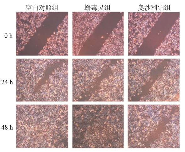
