中国科技论文统计源期刊 中文核心期刊
美国《化学文摘》《国际药学文摘》
《乌利希期刊指南》
WHO《西太平洋地区医学索引》来源期刊
日本科学技术振兴机构数据库(JST)
第七届湖北十大名刊提名奖
美国《化学文摘》《国际药学文摘》
《乌利希期刊指南》
WHO《西太平洋地区医学索引》来源期刊
日本科学技术振兴机构数据库(JST)
第七届湖北十大名刊提名奖

 , 张方方
, ZHANG Fangfang
, 张方方
, ZHANG Fangfang
目的 探讨α-细辛醚对人食管癌Eca-109细胞线粒体凋亡通路Cyt-c、Smac 及Caspase3 mRNA和蛋白表达水平的影响。方法 体外培养的人食管癌Eca-109细胞设为阴性对照组和α-细辛醚组,α-细辛醚终浓度分别为25,50,100 μg·mL-1,培养48 h后使用荧光倒置显微镜观察细胞凋亡形态改变,采用TRIzol法提取细胞总RNA,实时荧光定量聚合酶链反应检测细胞Cyt-c、Smac及Caspase3 mRNA的表达,Western blotting检测其蛋白表达,选取β-actin进行相对定量分析。结果 α-细辛醚组细胞形态发生明显凋亡改变;与阴性对照组比较,α-细辛醚组细胞Cyt-c、Smac及Caspase3 mRNA和蛋白的表达量均明显增加(
Objective To discuss the effect of α-asarone on the expression level of Cyt-c,Smac,Caspase3 mRNA and protein in human esophageal carcinoma Eca-109 cell mitochondria. Methods The Eca-109 cells were cultured
α-细辛醚注射液(山西普德药业股份有限公司,批号:201308,规格:2 mg);人食管癌细胞系 Eca-109为南京凯基生物科技发展有限公司,批号:KG189;RPMI1640培养液(Hyclone公司,批号:MyG0919);新生小牛血清(杭州四季青生物工程材料有限公司,批号:150402);实时荧光定量聚合酶链反应( real-time fluorescent quantitative polymerase chain reaction,RT-PCR)试剂盒(Takara公司,批号:DRR047S);三氯甲烷、异丙醇及无水乙醇等均为国产分析纯。小鼠抗人β-actin单克隆抗体(Santa cruz,批号:SC-81178);鼠多抗Cytochrome C(Abcam,批号:ab110325);鼠多抗Smac(Abcam,批号:ab111893);鼠多抗Caspase3(Abcam,批号:ab119794);HRP标记的羊抗小鼠IgG(北京中杉金桥生物技术有限公司,批号:ZB2305);预染蛋白参照物(PageRulerTM Prestained Protein Ladder,Fermentas,批号:SM0671);eECL Western Blot Kit高灵敏度化学发光检测试剂盒(北京康为世纪生物科技有限公司,批号:CW0049)。
1.2.1 细胞培养 Eca-109细胞用含10%胎牛血清的RPMI 1640培养液、37 ℃、5%二氧化碳培养箱中培养,实验取对数生长状态良好的细胞,2~3 d更换培养液,3~4 d传代一次。
1.2.2 免疫荧光染色检测细胞凋亡 用胰酶消化贴壁生长的药物处理48 h的Eca-109细胞及经完全培养基培养48 h的阴性对照细胞;收集细胞,磷酸盐缓冲液(PBS)洗涤细胞2次,离心1 500 r·min-1,(
1.2.3 总RNA的提取 取对数生长期细胞,收集细胞5×10⁶个,加入预冷的Trizol试剂1 mL,反复吹打细胞至透明状,静置5 min,分离核酸蛋白复合物;4 ℃,12 000 r·min-1离心5 min(
1.2.4 RNA的质量检测及cDNA的合成 按照1:50的比例,用RNase-free水稀释RNA样品,利用超微量分光光度计测定样品在260,280及320 nm下的吸光度,计算其
1.2.5 PCR引物扩增 引物序列见
1.2.6 Western blotting检测蛋白表达 按实验分组培养细胞,收获后用PBS 洗涤 2次,收集细胞,加组织细胞裂解液置冰浴裂解50 min,4 ℃离心(14 000 r·min-1)5 min后取上清液,去除细胞碎片,用BCA试剂盒测定蛋白及内参浓度,常规聚丙烯酰胺凝胶电泳,蛋白湿转法至聚偏二氟乙烯膜上,再至封闭液中室温振摇2 h,分别加入鼠多抗Cyt-c、Smac和Caspase3抗体于4 ℃孵育过夜,TBST漂洗10 min×3次,二抗为用HRP 标记的羊抗鼠IgG抗体(1:1 000),室温振摇孵育1 h,漂洗后用电化学发光法(ECL) 显色,室温孵育3 min,曝光照相,并计算灰度值。
表1 RT-PCR 引物序列
Tab.1 Primer sequence of RT-PCR
实验数据采用SPSS17.0版统计软件包进行统计学处理,计量资料以均数±标准差(
α-细辛醚处理细胞48 h后,收集细胞,根据AO/EB双染说明书对细胞进行染色,荧光倒置显微镜观察可见:阴性对照组细胞数量较多,核染色质着绿色并呈正常结构,凋亡现象很少;α-细辛醚处理组细胞出现不同程度的凋亡细胞形态,有核染色质着绿色呈固缩状或圆珠状的早期凋亡细胞;有核染色质为橘红色并呈固缩状或圆珠状的晚期凋亡细胞(
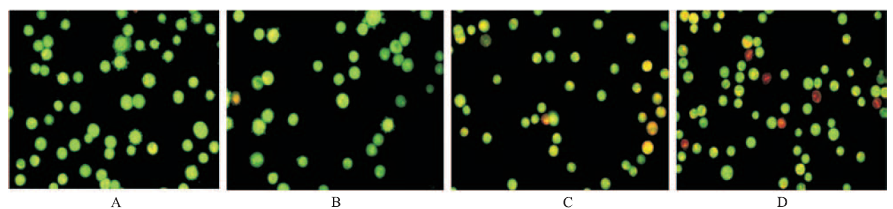
图1 人食管癌Eca-109 细胞AO/EB双染荧光染色结果(AO/EB染色,×400) A.阴性对照组;B.25 μg·mL-1α-细辛醚组;C.50 μg·mL-1α-细辛醚组;D.100 μg·mL-1α-细辛醚组
Fig.1 Results of AO/EB fluorescent double staining on Eca-109 cells(AO/EB staining,×400) A.negative control group;B.25 μg·mL-1α-asarone group;C.50 μg·mL-1α-asarone group;D.100 μg·mL-1α-asarone group
不同浓度α-细辛醚作用于人食管癌Eca-109细胞48 h后,与阴性对照组比较,α-细辛醚25,50,100 μg·mL-1组Cyt-c基因的表达水平分别为阴性对照组的1.247,1.651,3.77倍;Smac基因的表达水平分别为阴性对照组的1.323,1.65,2.133倍;Caspase3基因的表达水平分别为阴性对照组的1.612,1.726,2.568倍,见
表2 α-细辛醚干预人食管癌Eca-109细胞48 h后Cyt-c、Smac和Caspase3 mRNA的相对表达
Tab.2
Relative expression of Cyt-c、Smac and Caspase3 mRNA in Eca-109 cells treated with α-asarone for 48 h
用不同浓度α-细辛醚处理人食管癌Eca-109细胞48 h后,Cyt-c的相对表达量在α-细辛醚浓度为100 μg·mL-1时明显增加,而在25和50 μg·mL-1时表达没有明显变化;Smac的相对表达在3个浓度组都有明显增加,且呈现浓度依赖性;Caspase3的相对表达量在α-细辛醚浓度为50和100 μg·mL-1时明显增加,与阴性对照组比较差异有统计学意义(

图2 α-细辛醚不同浓度组处理人食管癌Eca-109细胞48 h后Cyt-c、Smac and Caspase3的表达
Fig.2 Expression of Cyt-c,Smac and Caspase3 in Eca-109 cells treated with different doses of α-asarone for 48 h
表3 不同浓度α-细辛醚处理Eca-109细胞48 h后Cyt-c、Smac和Caspase3 的相对表达
Tab.3
Relative expression of Cyt-c,Smac and Caspase3 in Eca-109 cells treated with different doses of α-asarone for 48 h
许多中草药及其提取物可通过诱导肿瘤细胞凋亡发挥抗肿瘤作用[7]。细胞凋亡受许多凋亡相关基因的调控。线粒体是真核细胞中重要的亚细胞结构,是合成ATP、为细胞生命活动提供能量和维持内环境稳态的重要场所。线粒体功能缺陷可能导致细胞内信号通路级联反应,启动细胞凋亡的发生。细胞色素C及Smac是在线粒体膜间最重要的两种凋亡促进因子[8]。细胞色素C是第一种被发现由线粒体释放的促细胞凋亡蛋白[9-10],其主要通过两个途径诱导细胞凋亡:一是Caspase途径,当细胞受到刺激时,Cyt-c释放入细胞质,与凋亡蛋白酶激活因子Apaf-1结合,启动Caspase-9形成全酶,然后裂解,导致pro-Caspase-9活化,激活下游的 pro-Caspase3进入内源性和外源性凋亡途径的最后通路最终导致细胞死亡;二是Caspase非依赖途径,该途径目前还不完全清楚,可能与bax有关。Smac是2000年DU等[11]发现一种新型线粒体膜间隙蛋白,在细胞发生凋亡时依赖Cyt-c的释放入细胞质,继而也从线粒体释放入细胞质,通过解除凋亡抑制蛋白对Caspase3,7,9的作用而诱导凋亡,尤其是Caspase3在凋亡级联反应中处于核心地位[12-13]。Caspase3 是细胞凋亡过程中激活的关键酶,也是细胞凋亡的主要效应分子[14]。
本研究结果发现不同浓度α-细辛醚可诱导人食管癌 Eca-109 细胞凋亡。荧光倒置显微镜观察细胞形态发生改变,提示细胞发生凋亡,RT-PCR和Western blotting检测发现药物处理组细胞特别是α-细辛醚为100 μg·mL-1时,线粒体Cyt-c、Smac表达明显增加,从线粒体膜间隙向细胞质内释放,经线粒体Caspase依赖凋亡通路诱导食管癌细胞凋亡,细胞色素C释放进入细胞质中,作用于Caspase家族,形成Caspase级联反应。Cyt-c、Smac mRNA及蛋白的高表达,引起Caspase 3 mRNA和蛋白的高表达,从而诱导细胞发生凋亡。从本研究结果也看出,α-细辛醚为25 μg·mL-1时,其诱导食管癌细胞凋亡的作用不是很明显,Cyt-c和Smac的表达虽有所增加,但差异不明显,而当α-细辛醚逐渐加大至终浓度100 μg·mL-1,其诱导凋亡的形态改变越来越明显,具有浓度依赖性;且Cyt-c、Smac和Caspase3的表达在终浓度100 μg·mL-1时与阴性对照组比较差异均有统计学意义。
综上所述,α-细辛醚处理组细胞Cyt-c、Smac和Caspase3基因的表达增加,提示α-细辛醚可能通过上
调线粒体凋亡通路相关蛋白Cyt-c、Smac和Caspase3的表达诱导食管癌Eca-109细胞的凋亡,但其作用机制尚不明确,有待研究。
The authors have declared that no competing interests exist.
| [1] |
目的:探讨α-细辛醚对人食管癌Eca-109细胞凋亡的影响. 方法:体外培养人食管癌细胞株(Eca-109),分别给以不同浓度的α-细辛醚对Eca-109细胞进行干预.DAPI染色倒置荧光显微镜观察细胞形态 变化,MTT检测细胞增殖率,流式细胞仪Annexin V-PI双染检测细胞凋亡率的变化.结果:不同浓度、不同作用时间的α-细辛醚组细胞增殖抑制率比较,差异有显著性(P<0.05);细胞凋亡指数与阴性 对照组比较,差异具有显著性(P<0.05).结论 ·α-细辛醚对人食管癌Eca-109细胞凋亡有促进作用.
[本文引用:1]
|
| [2] |
α-Asarone is a main component of Acorus gramineus widely known as an oriental traditional medicinal stuff. A. gramineus has been known to have a variety of medicinal efficacies such as anti-gastric ulcer and anti-allergic activities, inhibition of histamine release and antioxidant effect. However, its effect on angiogenesis remains unclear. The aim of this study was to investigate the effect of α-asarone on induction of angiogenesis through modulation of matrix metalloproteinase (MMP). First of all, MTT assay was performed to evaluate the effect of α-asarone on cell viability using MTT assay, and then tube formation assay with human umbilical vein endothelial cells (HUVEC) in vitro and rat aorta ring assay ex vivo were carried out to elucidate its effect on angiogenesis. Treatment with α-asarone below 602μM showed no cytotoxicity in human fibrosarcoma cells (HT1080) and HUVEC. It was observed that α-asarone not only promotes tube formation of HUVEC but also induces angiogenesis of rat aorta. In addition, the effects of α-asarone on the expressions of protein and gene were evaluated using western blot analysis and RT-PCR assay. α-Asarone increased the expression levels of MMP-2 and MMP-9 stimulated by phenazine methosulfate (PMS) and phorbol 12-myristate 13-acetate (PMA) in HT1080. Especially, the expression level of antioxidant enzyme such as glutathione reductase was increased in the presence of α-asarone. Therefore, above findings suggest that α-asarone may play an important role in pathological diseases related to MMP and angiogenesis.
[本文引用:1]
|
| [3] |
|
| [4] |
|
| [5] |
|
| [6] |
为研究姜黄挥发油(turmeric volatile oil,TVO)对皮肤鳞癌(CSCC)A431(以下简称A431)细胞增殖及凋亡的影响并探讨其机制,该实验采用不同质量浓度(5~80 mg·L-1)TVO体外作用于A431细胞,利用细胞增殖/毒性检测试剂盒(celcounting kit-8,CCK-8)法检测细胞增殖活性;倒置显微镜观察细胞形态变化;吖啶橙(AO)/溴化乙锭(EB)双染色荧光显微镜观察A431细胞凋亡情况;流式细胞仪检测细胞凋亡;Western blot法检测半胱氨酸天冬氨酸蛋白酶-3(caspase-3)、半胱氨酸天冬氨酸蛋白酶-9(caspase-9)表达量。结果显示,随TVO质量浓度的增加对A431细胞生长具有显著的抑制增殖作用,呈剂量依赖关系,各组间差异有统计学意义(P0.05)。与对照组比较,不同质量浓度TVO组均出现细胞皱缩及细胞破碎现象,细胞凋亡率增加,并呈剂量依赖性,caspase-3,caspase-9的相对表达量上调,各组间差异有统计学意义(P0.05),提示了TVO能抑制皮肤鳞癌A431细胞增殖,并诱导其凋亡,其机制可能与上调caspase-3,caspase-9的表达有关。
[本文引用:1]
|
| [7] |
目的 研究龙葵碱对人乳腺癌MCF-7细胞凋亡的影响并探讨相关机制。方法 采用四甲基偶氮唑蓝(MTT)法观察不同浓度龙葵碱对人乳腺癌MCF-7细胞的生长抑制作用;4,6-二脒基-2-苯基吲哚(DAPI)染色荧光显微镜进行细胞凋亡核形态学观察;DNA琼脂糖凝胶电泳进行DNA片段化分析,FITC- Annexin V/PI荧光标记流式细胞术检测细胞凋亡率,荧光显微镜结合Fluo-8/Am法、流式细胞术和分光光度比色法分别检测胞内Ca<sup>2+</sup>浓度、线粒体膜电位(△ψm)和caspase-3,caspase-8活性变化。结果 四甲基偶氮唑蓝法结果显示,龙葵碱对人乳腺癌MCF-7细胞有生长抑制作用。10.0 mmol·L<sup>-1</sup>龙葵碱处理2 d,4,6-二脒基-2-苯基吲哚染色可见核浓缩及边缘现象,DNA电泳出现特征性的凋亡条带。10.0 mmol·L<sup>-1</sup>龙葵碱处理1,2,3 d的细胞凋亡率分别为(20.9±7.3)%、(42.6±8.8)% 和(74.9±12.8)%;细胞内Ca<sup>2+</sup>荧光强度分别为35.6±2.9、52.3±5.6和27.2±2.2;线粒体膜电位(△ψm)值分别下降7.7%、33.2%和46.9%;caspase-8活性在1 d达最高(1.85±0.09)U·μg <sup>-1</sup>,caspase-3活性则在2 d达最高(2.18±0.09)U·μg <sup>-1</sup>,与对照组相比均有统计学显著性差异(<i>P</i><0.05)。结论 龙葵碱可诱导人乳腺癌MCF-7细胞凋亡,其诱导凋亡的机制可能与胞内Ca<sup>2+</sup>浓度升高、线粒体膜电位降低和caspase-3、8活化有关。
[本文引用:1]
|
| [8] |
Smac/DIABLO is a mitochondrial protein that is released along with cytochrome c during apoptosis and promotes cytochrome c-dependent caspase activation by neutralizing inhibitor of apoptosis proteins (IAPs). We provide evidence that Smac/DIABLO functions at the levels of both the Apaf-1-caspase-9 apoptosome and effector caspases. The N terminus of Smac/DIABLO is absolutely required for its ability to interact with the baculovirus IAP repeat (BIR3) of XIAP and to promote cytochrome c-dependent caspase activation. However, it is less critical for its ability to interact with BIR1/BIR2 of XIAP and to promote the activity of the effector caspases. Consistent with the ability of Smac/DIABLO to function at the level of the effector caspases, expression of a cytosolic Smac/DIABLO in Type II cells allowed TRAIL to bypass Bcl-xL inhibition of death receptor-induced apoptosis. Combined, these data suggest that Smac/DIABLO plays a critical role in neutralizing IAP inhibition of the effector caspases in the death receptor pathway of Type II cells.
DOI:10.1074/jbc.C000533200
PMID:10950947
[本文引用:1]
|
| [9] |
The aim of this study was to construct an expression vector carrying the /radiation dual62sensitive chimeric response element (HRE)/early response 1 (621) promoter in order to overexpress the therapeutic second 62derived activator of (). Using this expression vector, the present study aimed to explore the molecular mechanism underlying radiotherapy62induced A549 and apoptosis under . The plasmids, pcDNA3.16262(pE62) and pcDNA3.162HRE/62(pH/E62), were constructed and transfected into A549 using the liposome method. CoCl2 was used to chemically simulate , followed by the administration of 2 Gy X62ray irradiation. An MTT assay was performed to detect and an 62apoptosis detection kit was used to detect apoptosis. Quantitative polymerase chain reaction and western blot analyses were used for the detection of mRNA and expression, respectively. with the pE62and pH/E62plasmids in combination with radiation and/or was observed to enhance the expression of . Furthermore, overexpression was found to enhance the radiation62induced inhibition of and promotion of cycle arrest and apoptosis. The c/629/623 pathway was identified to be involved in this regulation of apoptosis. Plasmid in combination with X62ray irradiation was found to markedly induce under . In conclusion, the /radiation dual62sensitive chimeric HRE/621 promoter was observed to enhance the expression of the therapeutic , as well as enhance the radiation62induced inhibition of and promotion of cycle arrest and apoptosis under . This apoptosis was found to involve the mitochondrial pathway.
[本文引用:1]
|
| [10] |
|
| [11] |
We report here the identification of a novel protein, Smac, which promotes caspase activation in the cytochrome c/Apaf-1/caspase-9 pathway. Smac promotes caspase-9 activation by binding to i nhibitor of a poptosis p roteins, IAPs, and removing their inhibitory activity. Smac is normally a mitochondrial protein but is released into the cytosol when cells undergo apoptosis. Mitochondrial import and cleavage of its signal peptide are required for Smac to gain its apoptotic activity. Overexpression of Smac increases cells' sensitivity to apoptotic stimuli. Smac is the second mitochondrial protein, along with cytochrome c, that promotes apoptosis by activating caspases.
[本文引用:1]
|
| [12] |
STUDY DESIGN.: A biochemical and radiologic comparison of 4 disc injury models to produce disc degeneration in the rabbit was carried out in 2 experiments. OBJECTIVES.: To develop a reliable animal model of intervertebral disc degeneration. SUMMARY OF BACKGROUND DATA.: In order to study various interventions for retarding or preventing disc degeneration, a reliable animal model of disc degeneration is needed. METHODS.: First experiment: 7 New Zealand white rabbits (1 year old, 3.5-4.5 kg body weight) were used to test 4 different disc injury models; intradiscal injection of Camptothecin (an apoptotic agent) using a 23-gauge needle at L2-L3, nucleus aspiration using a 21-gauge needle at L3-L4, 3 anulus punctures using a 21-gauge needle at L4-L5, and 1 anulus puncture using a 18-gauge needle at L5-L6. The L1-L2 level was used as a control. Rabbits were killed 12 weeks later. Lumbar spinal magnetic resonance images were assessed using 4 grades of disc degeneration. The water content of the nucleus was measured. Dimethylmethylene blue (DMMB) assay was used to measure the sulfated-glycosaminoglycan content. Second experiment: the 21-gauge 3-puncture and the 18-gauge 1-puncture models, thought most effective in producing disc degeneration in the first experiment, were again used in a second study. Six rabbits were killed 8 weeks later, the water and sulfated-glycosaminoglycan contents being measured as in the first experiment. RESULTS.: In the first experiment, the water content in the aspiration and puncture models was significantly decreased. Only the sulfated-glycosaminoglycan content in the aspiration model showed a significant decrease as compared to the control. Disc heights and magnetic resonance grades documented significant degeneration occurring in the aspiration and puncture models. In the second experiment, the water content showed a significant decrease in the 21-gauge 3-puncture model, whereas neither of the results for the sulfated-glycosaminoglycancontent
[本文引用:1]
|
| [13] |
|
| [14] |
目的 探讨山竹果皮中总氧杂蒽酮提取纯化物对人鼻咽癌CNE2细胞增殖及凋亡的影响及其作用机制.方法 将人鼻咽癌CNE2细胞随机分成阴性对照组和不同浓度山竹果皮中总氧杂蒽酮提取纯化物组.阴性对照组不加药物,正常培养;总氧杂蒽酮提取纯化物组分别以200,400,600,800 μmol·L-1总氧杂蒽酮作用24,48,72 h.采用噻唑蓝(MTT)法检测不同浓度总氧杂蒽酮提取纯化物对CNE2细胞增殖的影响,Annexin-V/PI双重染色、碘化丙啶单染进行流式细胞术检测总氧杂蒽酮提取纯化物对CNE2细胞周期和凋亡的影响.Caspase-3试剂盒检测总氧杂蒽酮提取纯化物对CNE2细胞Caspase-3酶活化的影响.结果 总氧杂蒽酮提取纯化物随着浓度增加,可显著抑制人鼻咽癌CNE2细胞的增殖活性,浓度为371.536 7 μmol·L-1时可诱导人鼻咽癌CNE2细胞出现明显早期凋亡,并且随着药物作用时间的增加(24,48,72 h),凋亡早期癌细胞的比例显著上升(分别为0.03%,10.54%,26.47%)(P<0.05);总氧杂蒽酮提取纯化物使CNE2细胞G1期细胞比例大幅升高,同时S期细胞比例明显下降;对Caspase-3具有激活作用,Caspase-3酶的活性与药物浓度成正比.结论 山竹果皮中总氧杂蒽酮提取纯化物对人鼻咽癌CNE2细胞增殖有显著抑制作用和促凋亡作用,其作用机制可能与抑制鼻咽癌细胞增殖活性、抑制细胞进入S期和激活Caspase-3有关.
[本文引用:1]
|
