Objective To investigate the possible mechanism of berberine on the cardiovascular in diabetic rats by observing the effect of berberine on the blood glucose, blood lipid and factors involved in cardiovascular injury. Methods The diabetic model was induced by intraperitoneal injection of streptozotocin(STZ). The 43 rats of the model were randomly divided into model control group (n=9), metformin group (n=8) and berberine low, medium, high dose groups (50,100,150 mg·kg-1·d-1,n=9,9,8). Normal rats (n=8) were set as normal control group. After 12 weeks of gavage administration, rats’ heart was taken and heart mass index(HMI) was calculated;concentration of TG, TC, HDL-C, LDL-C, NO was examined by kit;rats’ serum concentration of blood TXB2, 6-keto-PGFα and ET was determined by radioimmunoassay;the expression of ICAM-1and VCAM-1 in thoracic aorta tissue was tested by real-time quantitative PCR (RT-PCR). Results Compared with the model control group, levels of BG, HMI, TG, TC, LDL-C were significantly decreased(P<0.01)in berberine medium, high dose groups and metformin group;the serum concentration of NO and 6-keto-PGFα was increased(P<0.01) while ET and TXB2 was decreased(P<0.01);the expression of ICAM-1and VCAM-1 in thoracic aorta tissue was decreased obviously(P<0.01). Conclusion Berberine has protective effects on the cardiovascular in diabetic rats by means of regulating blood lipid, recovering blood vessel’s dynamic balance of systolic and diastolic, playing a role in anti- atherosclerosis.
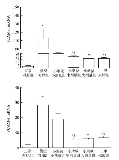
Fig.1
Relative mRNA expression of ICAM-1 and VCAM-1 detected by real-time quantitative PCR Compared with normal control group,*1P<0.01;Compared with model control group,*2P<0.01
STHEHOUWER C DA,LAMBERTJ,DONKER A TM,et al.Endothelial dysfunction and pathogenesis of diabetic patients [J].Cardiovasc Res,1997,34(1):55.
To review, from the clinical perspective, the contribution of dysfunction of the vascular endothelium to the pathogenesis of diabetic micro- and macroangiopathy.Available data indicate that endothelial dysfunction in diabetes complicated by micro- or macroalbuminuria (renal microangiopathy) is generalised. The close linkage between microalbuminuria and endothelial dysfunction is an attractive explanation for the fact that microalbuminuria is a risk marker for atherosclerotic cardiovascular disease in diabetes. Endothelial dysfunction precedes the occurrence of even early diabetic microangiopathy. However, it is not clear whether endothelial dysfunction is a feature of the diabetic state per se or whether additional factors are required to induce endothelial dysfunction given the presence of diabetes. Convincing data from animal and in vitro models exist to indicate that endothelial dysfunction in diabetes may be related to hyperglycaemic pseudohypoxia, activation of protein kinase C, increased expression of transforming growth factor-beta and vascular endothelial growth factor, non-enzymatic glycation, oxidative stress, activation of the coagulation cascade, increased expression of tumour necrosis factor-alpha, and high levels of insulin and insulin precursor molecules. However, the importance of these proposed mechanisms have not yet been extensively assessed in diabetes in man.Endothelial dysfunction plays a key role in the pathogenesis of diabetic angiopathy in man. The biochemical basis of endothelial dysfunction in diabetic man, however, has yet to be fully elucidated.
PUTHANVEETILP,WANA,RODRIGUESB.Lipoprotein lipase and angiopoietin-like 4-Cardiomyocyte secretory proteins that regulate metabolism during diabetic heart disease[J].Crit Rev Clin Lab Sci,2015,52(3):138-149.
Abstract Abstract Cardiac diseases have been extensively studied following diabetes and altered metabolism has been implicated in its initiation. In this context, there is a shift from glucose utilization to predominantly fatty acid metabolism. We have focused on the micro- and macro-environments that the heart uses to provide fatty acids to the cardiomyocyte. Specifically, we will discuss the cross talk between endothelial cells, smooth muscles and cardiomyocytes, and their respective secretory products that allows for this shift in metabolism. These changes will then be linked to alterations in the cardiovascular system and the augmented heart disease observed during diabetes. Traditionally, the heart was only thought of as an organ that supplies oxygen and nutrients to the body through its function as a pump. However, the heart as an endocrine organ has also been suggested. Secreted products from the cardiomyocytes include the natriuretic peptides atrial natriuretic peptide (ANP) and brain natriuretic peptide (BNP). Both have been shown to have vasodilatory, diuretic and antihypertensive effects. These peptides have been extensively studied and their deficiency is considered to be a major cause for the initiation of cardiovascular and cardiometabolic disorders. Another secretory enzyme, lipoprotein lipase (LPL), has been implicated in diabetic heart disease. LPL is a triglyceride-hydrolyzing enzyme that is synthesized within the cardiomyocyte and secreted towards the lumen under various conditions. For example, moderate or short-term hyperglycemia stimulates the release of LPL from the cardiomyocytes towards the endothelial cells. This process allows LPL to contact lipoprotein triglycerides, initiating their break down, with the product of lipolysis (free fatty acids, FA) translocating towards the cardiomyocytes for energy consumption. This mechanism compensates for the lack of glucose availability following diabetes. Under prolonged, chronic conditions of hyperglycemia, there is a need to inhibit this mechanism to avoid the excess delivery of FA to the cardiomyocytes, an effect that is known to induce cardiac cell death. Thus, LPL inhibition is made possible by a FA-induced activation of PPAR 尾/未, which augments angiopoietin-like 4 (Angptl4), an inhibitor of LPL activity. In the current review, we will focus on the mediators and conditions that regulate LPL and Angptl4 secretion from the cardiomyocyte, which are critical for maintaining cardiac metabolic homeostasis.
DWORACKAM,KRAYZAGORSKAE,WESOLOWSKAA,et al.Circulating monocyte chemotactic protein 1 (MCP-1),vascular cell adhesion molecule 1 (VCAM-1) and angiogenin in type 2 diabetic patients treated with statins in low doses [J].Eur J Pharmacol,2014,740:474-479.
Statins are known as agents promoting a biphasic dose-dependent effect on angiogenesis under experimental conditions. Dysregulation of angiogenesis plays an important role in the development of atherosclerosis and it may be affected by metabolic factors. The aim of this research was to explain how low doses of statins modify serum concentrations of pro-angiogenic factors MCP-1 and angiogenin in type 2 diabetic patients. Measurements of metabolic control parameters were performed in 30 patients with type 2 diabetes treated with low doses of statin, and in 34 statin-free patients with type 2 diabetes. The serum levels of MCP-1 and VCAM-1 in statin-treated patients were lower than those of the statin-free group. ANCOVA results revealed that these effects were dependent only on the use of statins. In type 2 diabetic subjects, overall positive correlation was found between total cholesterol or LDL serum concentration and MCP-1 serum level. The angiogenin concentration in the serum did not show differences and was comparable in both groups. The angiogenin serum level correlated negatively with HDL, LDL and with HbA 1 c. Multivariate regression analysis indicated that angiogenin serum levels in type 2 diabetic patients were determined mainly by HbA 1 c, HDL-cholesterol and diabetes duration. It has been shown that statins used in low doses in type 2 diabetic subjects decrease MCP-1 and VCAM-1serum levels, most likely due to the statins-related effect on the lipid profile, while angiogenin serum levels in this group are determined rather by the current metabolic control.
EL-SAYED SS,ZAKARIA MN,ABDEL-GHANY RH,et al.Cystathionine-γ lyase-derived hydrogen sulfide mediates the cardiovascular protective effects of moxonidine in diabetic rats[J].Eur J Pharmacol,2016,783:73-84.
Blunted cystathionine lyase (CSE) activity (reduced endogenous H 2 S-level) is implicated in hypertension and myocardial dysfunction in diabetes. Here, we tested the hypothesis that CSE derived H 2 S mediates the cardiovascular protection conferred by the imidazoline I 1 receptor agonist moxonidine in a diabetic rat model. We utilized streptozotocin (STZ; 55/mg/kg i.p) to induce diabetes in male Wistar rats. Four weeks later, STZ-treated rats received vehicle, moxonidine (2 or 6/mg/kg; gavage), CSE inhibitor DL-propargylglycine, (37.5/mg/kg i.p) or DL-propargylglycine with moxonidine (6/mg/kg) for 3 weeks. Moxonidine improved the glycemic state, and reversed myocardial hypertrophy, hypertension and baroreflex dysfunction in STZ-treated rats. Ex vivo studies revealed that STZ caused reductions in CSE expression/activity, H 2 S and nitric oxide (NO) levels and serum adiponectin and elevations in myocardial imidazoline I 1 receptor expression, p38 and extracellular signal-regulated kinase, ERK1/2, phosphorylation and lipid peroxidation (expressed as malondialdehyde). Moxonidine reversed these biochemical responses, and suppressed the expression of death associated protein kinase-3. Finally, pharmacologic CSE inhibition (DL-propargylglycine) abrogated the favorable cardiovascular, glycemic and biochemical responses elicited by moxonidine. These findings present the first evidence for a mechanistic role for CSE derived H 2 S in the glycemic control and in the favorable cardiovascular effects conferred by imidazoline I 1 receptor activation (moxonidine) in a diabetic rat model.
FARAHK,ADILAP,SAVITAS,et al.Improvement in myocardial function by terminalia arjuna in streptozotocin-induced diabetic rats:possible mechanisms[J].J Cardio Pharm Therap,2013,18(5):481-489.
WANGC,LIJ,LVX,et al.Ameliorative effect of berberine on endothelial dysfunction in diabetic rats induced by high-fat diet and streptozotocin[J].Eur J Pharmacol,2009,12(620):131-137.
Abstract Berberine can improve insulin resistance, lower blood glucose, and regulate lipid metabolism disorders which cause endothelial dysfunction, leading to vascular complications of type 2 diabetes mellitus. The aim of the present study was to investigate the effects of berberine on endothelial dysfunction of aortas in type 2 diabetes mellitus rats and its mechanism. Wistar rats were randomly divided into four groups: diabetic rats, control rats, diabetic rats treated with berberine (100 mg/kg), and control rats treated with berberine. The serum fasting blood glucose, insulin, total cholesterol, triglyceride and nitric oxide (NO) levels were tested. Acetylcholine-induced endothelium-dependent relaxation and sodium nitroprusside induced endothelium-independent relaxation were measured in aortas for estimating endothelial function. The expression of endothelial nitric oxide synthase (eNOS) mRNA was measured by RT-PCR, and the protein expressions of eNOS and NADPH oxidase (NOX4) were analyzed by western blot. The results showed that berberine significantly decreased fasting blood glucose, and triglyceride levels in diabetic rats. Berberine also improved endothelium-dependent vasorelaxation impaired in aorta. The expressions of eNOS mRNA and protein were significantly increased, while NOX4 protein expression was decreased in aortas from diabetic rats with berberine treatment. Moreover, serum NO levels were elevated after berberine treatment. In conclusion, berberine restores diabetic endothelial dysfunction through enhanced NO bioavailability by up-regulating eNOS expression and down-regulating expression of NADPH oxidase.
VAN DER VALK F M,VAN WIJK DF,STRORES ES.Novel anti-inflammatory strategies in atherosclerosis[ J ].Curr Opin Lipidol,2012,23(6):532-539.
Inflammation has been widely acknowledged to contribute throughout all stages of atherogenesis. However, these recent advances in our understanding have not been translated into clinical practice in which the mainstay of treatment is still lipid-targeted therapy. This review provides an overview of promising anti-inflammatory therapies in atherosclerosis, and discusses potential drawbacks and clinical benefits.Immunosuppressive drugs are likely to beneficially affect atherogenesis. Several novel anti-inflammatory targets have been scrutinized, of which some have reached clinical development stage, such as cytokine targets interleukin-1 and interleukin-6, CCR2 antagonist, selective phospholipase, and leukotriene inhibitors. Novel imaging modalities such as MRI and PET-computed tomography provide valuable surrogate inflammatory endpoints for risk stratification and testing anti-inflammatory agents in cardiovascular randomized trials.Anti-inflammatory therapies hold great promise in cardiovascular prevention regimens; however, atherosclerosis is a chronic disease, and systemic long-term use of anti-inflammatory agents carries the risk of complications arising from immunosuppression. In order to successfully add immunosuppressive drugs to our routine armament, we need to identify high-risk patients who benefit from anti-inflammatory treatment, increase our insight into the inflammatory pathogenesis of atherogenesis, and find safe and effective compounds capable of directly suppressing plaque inflammation.
JIANGY,JIANGLL,MAIMAITIREXIATI X M,et al.Irbesartan attenuates TNF-α-induced ICAM-1,VCAM-1,and E-selectin expression through suppression of NF-κB pathway in HUVECs[J].Eur Rev Med Pharmacol Sci,2015,19(17):3295-3302.
It is widely recognized that atherosclerosis is a chronic inflammatory disease. Intracellular adhesion molecule-1 (ICAM-1), vascular cellular adhesion molecule-1 (VCAM-1), and E-selectin play vital roles in inflammatory processes. ICAM-1, VCAM-1, and E-selectin expression is regulated by nuclear factor (NF)-κB signaling. It has been reported that irbesartan can decrease expression of atrial fibrillation-Induced atrial adhesion molecule and reduce secretion of inflammation associated cytokines from cultured human carotid atheroma. In this study, we examined whether irbesartan prevents TNF-α-induced ICAM-1, VCAM-1, and E-selectin expression in human umbilical vein endothelial cells (HUVECs). HUVECs were cultured. The expression of ICAM-1, VCAM-1 and MCP-1 was measured by real-time quantitative PCR and ELISA. The expression of NF-κB and p-IκB-α was measured by Western blot. It indicated that in HUVECs irbesartan inhibited expression and secretion of TNFα-induced ICAM-1, VCAM-1, and E-selectin. Furthermore, irbesartan inhibited TNF-α-induced IκB-α phosphorylation and NF-κB P65 nuclear translocation substantially. In conclusion, irbesartan attenuates TNFα-induced ICAM-1, VCAM-1, and E-selectin expression by way of suppressing the NF-κB pathways in HUVECs. Irbesartan might postpone the progression of inflammatory diseases, including atherosclerosis. Irbesartan attenuates TNFα-induced ICAM-1, VCAM-1 and MCP-1 expression through the suppression of NF-κB pathways. These results suggest irbesartan would be of great benefit to delaying the progression of inflammatory diseases, including atherosclerosis.
Circulating monocyte chemotactic protein 1 (MCP-1),vascular cell adhesion molecule 1 (VCAM-1) and angiogenin in type 2 diabetic patients treated with statins in low doses [J]

