Objective To investigate the effect of fisetin on myocardial ischemia reperfusion (I/R) injury in isolated rats and its mechanism. Methods The myocardial I/R injury model was established through Langendorff system. A total of 32 SPF grade SD rats were randomly divided into 4 groups (n=8): normal control group, model control group, low dose fisetin group (0.5 μmol·L-1) and high dose fisetin group (5 μmol·L-1). The myocardial function was measured by TTC staining. The content of CK-MB was measured by enzyme-linked immunosorbent assay (ELISA). The levels of SOD, MDA, GSH, GSSG, TNF-α and IL-6 in the myocardium were measured by ELISA. The changes of myocardial tissue were observed by HE staining. The degree of apoptosis was detected by TUNEL staining. Results Compared with the normal control group, the cardiac function parameters of the model control group were decreased significantly, serious infarction and necrosis developed in myocardium, and the leaking amount of CK-MB was significantly increased. The contents of MDA, GSSG, IL-6 and TNF-α in myocardium were significantly increased, and the activities of SOD and GSH significantly decreased. Severe apoptosis appeared in the cardiomyocytes of the model control group. Compared with the model control group, the pretreatment of fisetin could significantly reverse the changes of myocardial injury induced by I/R, and the protective effect of high dose of fisetin was more obvious. Conclusion Fisetin can significantly alleviate I/R-induced myocardial injury and exerts a dose-dependent effect. The protective effect of fisetin may be related to its anti-oxidation, anti-inflammatory and anti-apoptotic effects.
漆黄素(Solarbio公司,含量98%,批号:SF8050-20),水合氯醛(成都科龙化工试剂厂,批号:2015090601),苏木精-伊红染试剂盒(Solarbio公司,批号:2016030601),氯化三苯基四氮唑(triphenyltetrazolium chloride,TTC,Sigma公司,超氧化物歧化酶(SOD,批号:20160822)、丙二醛(MDA,批号:20160816)、还原型谷胱甘肽(GSH,批号:20161124)、氧化型谷胱甘肽(oxidized glutathione,GSSG,批号:20161108)、肌酸激酶同工酶(creatine kinase-MB,CK-MB,批号:20160816)、白细胞介素-6(IL-6,批号:20160826)、肿瘤坏死因子-α(TNF-α,批号:20160805)、In Situ Cell Death Detection Kit POD试剂盒(批号:20170314)购自Tsz公司,4-0线,其他试剂均为国产分析纯。
1.3 实验仪器
Langendorff体外心脏灌流系统、BL420S生物机能实验系统、恒温水浴器(成都泰盟软件有限公司),超低温冰箱(海尔有限公司),恒温箱(上海博讯实业有限公司),数码相机(佳能公司,型号:Canon EOS 600D),电子分析天平(赛多利斯公司,型号:SQP型,感量:1 mg),酶标仪(Thermo公司,型号:infinite 200Pro),切片机(Leica公司,型号:RM2235),显微镜(Leica公司),离心机(Leica公司)。
Fig.1
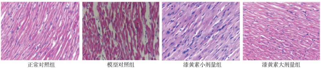
Rate of myocardial infarct in four groups of rats(x¯±s,n=8) Compared with normal control group,*1P<0.01;Compared with model control group,*2P<0.01
Fig.3
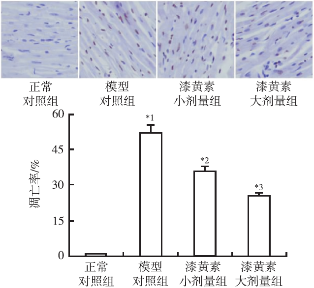
Apoptosis of myocardium in four groups of rats (×400) Compared with normal control group,*1P<0.01;Compared with model control group,*2P<0.05,*3P<0.01
CHOU RH,HSIEH SC,YU YL,et al.Fisetin inhibits migration and invasion of human cervical cancer cells by down- regulating urokinase plasminogen activator expression through suppressing the p38 MAPK- dependent NF-kappaB signaling pathway[J].PLoS One,2013,8(8):e71983.
ZHOUM,LIUL,WANGW,et al.Role of licochalcone C in cardioprotection against ischemia/reperfusion injury of isolated rat heart via antioxidant,anti-inflammatory,and anti-apoptotic activities[J].Life Sci,2015,132:27-33.
This study aimed to evaluate the protective effect of licochalcone C against myocardial ischemia/reperfusion injury in rats. Left ventricular developed pressure (LVDP) and its maximum up/down rate (dp/dtmax) were recorded as myocardial function. Levels of creatine kinase (CK), lactate dehydrogenase (LDH), malondialdehyde (MDA), superoxide dismutase (SOD), glutathione/glutathione disulfide (GSH/GSSG) ratio, and tumor necrosis factor-alpha (TNF-伪) were determined by using enzyme-linked immunosorbent assay. Cell morphology was observed and mitochondrial damage was assessed by HE coloration and transmission electron microscopy, respectively. Cardiomyocyte apoptosis was determined by using terminal deoxynucleotidyl transferased UTP nick-end labeling (TUNEL). Pretreatment with licochalcone C significantly improved the recovery of LVDP and dp/dtmax, and increased the levels of SOD and GSH/GSSG ratio. However, pretreatment with licochalcone C not only decreased the TUNEL-positive cell ratio and morphological changes, but also weaken the mitochondrial injury and the levels of CK, LDH, MDA, and TNF-伪. These results suggested an important function of licochalcone C extracted from traditional Chinese medicine in the cardioprotection via antioxidant, anti-inflammatory, and anti-apoptotic activities.
HANJ,WANGD,YUB,et al.Cardioprotection against ischemia/reperfusion by licochalcone B in isolated rat hearts[J].Oxid Med Cell Longev,2014,2014:134862.
Abstract The generation of reactive oxygen species (ROS) is a major cause of heart injury induced by ischemia-reperfusion. The left ventricular developed pressure (LVDP) and the maximum up/down rate of left ventricular pressure (00±dp/dt(max)) were documented by a physiological recorder. Myocardial infarct size was estimated macroscopically using 2,3,5-triphenyltetrazolium chloride staining. Coronary effluent was analyzed for lactate dehydrogenase (LDH) and creatine kinase (CK) release to assess the degree of cardiac injury. The levels of C-reactive protein (CRP), interleukin-8 (IL-8), tumor necrosis factor-02± (TNF-02±), and interleukin-6 (IL-6) were analyzed to determine the inflammation status of the myocardial tissue. Cardiomyocyte apoptosis analysis was performed using the In Situ Cell Death Detection Kit, POD. Accordingly, licochalcone B pretreatment improved the heart rate (HR), increased LVDP, and decreased CK and LDH levels in coronary flow. SOD level and GSH/GSSG ratio increased, whereas the levels of MDA, TNF-02±, and CRP and activities of IL-8 and IL-6 decreased in licochalcone B-treated groups. The infarct size and cell apoptosis in hearts from licochalcone B-treated group were lower than those in hearts from the I/R control group. Therefore, the cardioprotective effects of licochalcone B may be attributed to its antioxidant, antiapoptotic, and anti-inflammatory activities.
MAHERP,DARGUSCHR,EHREN J L,et a1.Fisetin lowers methylglyoxal dependent protein glycation and limits the complications of diabetes[J].PLoS One,2011,6(6):e21226.
Abstract The elevated glycation of macromolecules by the reactive dicarbonyl and -oxoaldehyde methylglyoxal (MG) has been associated with diabetes and its complications. We have identified a rare flavone, fisetin, which increases the level and activity of glyoxalase 1, the enzyme required for the removal of MG, as well as the synthesis of its essential co-factor, glutathione. It is shown that fisetin reduces two major complications of diabetes in Akita mice, a model of type 1 diabetes. Although fisetin had no effect on the elevation of blood sugar, it reduced kidney hypertrophy and albuminuria and maintained normal levels of locomotion in the open field test. This correlated with a reduction in proteins glycated by MG in the blood, kidney and brain of fisetin-treated animals along with an increase in glyoxalase 1 enzyme activity and an elevation in the expression of the rate-limiting enzyme for the synthesis of glutathione, a co-factor for glyoxalase 1. The expression of the receptor for advanced glycation end products (RAGE), serum amyloid A and serum C-reactive protein, markers of protein oxidation, glycation and inflammation, were also increased in diabetic Akita mice and reduced by fisetin. It is concluded that fisetin lowers the elevation of MG-protein glycation that is associated with diabetes and ameliorates multiple complications of the disease. Therefore, fisetin or a synthetic derivative may have potential therapeutic use for the treatment of diabetic complications.
DE VRIES DK,KORTEKAAS KA,TSIKASD,et al.Oxidative damage in clinical ischemia/reperfusion injury:a reappraisal[J].Antioxid Redox Signal,2013,19(6):535-545.
Ischemia/reperfusion (I/R) injury is a common clinical problem. Although the pathophysiological mechanisms underlying I/R injury are unclear, oxidative damage is considered a key factor in the initiation of I/R injury. Findings from preclinical studies consistently show that quenching reactive oxygen and nitrogen species (RONS), thus limiting oxidative damage, alleviates I/R injury. Results from clinical intervention studies on the other hand are largely inconclusive. In this study, we systematically evaluated the release of established biomarkers of oxidative and nitrosative damage during planned I/R of the kidney and heart in a wide range of clinical conditions.Sequential arteriovenous concentration differences allowed specific measurements over the reperfused organ in time. None of the biomarkers of oxidative and nitrosative damage (i.e., malondialdehyde, 15(S)-8-iso-prostaglandin F2伪, nitrite, nitrate, and nitrotyrosine) were released upon reperfusion. Cumulative urinary measurements confirmed plasma findings. As of these negative findings, we tested for oxidative stress during I/R and found activation of the nuclear factor erythroid 2-related factor 2 (Nrf2), the master regulator of oxidative stress signaling.This comprehensive, clinical study evaluates the role of RONS in I/R injury in two different human organs (kidney and heart). Results show oxidative stress, but do not provide evidence for oxidative damage during early reperfusion, thereby challenging the prevailing paradigm on RONS-mediated I/R injury.Findings from this study suggest that the contribution of oxidative damage to human I/R may be less than commonly thought and propose a re-evaluation of the mechanism of I/R.
WOZNIAKD,DRYSA,MATKOWSKIA.Antiradical and antioxidant activity of flavones from scutellariae baicalensis radix[J].Nat Prod Res,2015,29(16):1567-1570.
We evaluated the antioxidant properties of four main flavones from : baicalein, wogonin and their glucuronides – baicalin and wogonoside. We used three assays: free radical scavenging with 2,2′-diphenylpicrylhydrazyl radical, transition metal ions reducing power by phosphomolybdenum assay and inhibition of the hydroxyl radical-induced peroxidation of linoleic acid assay. All flavones have antioxidant capacity, which differs depending on the structure and mechanisms of activity. In all tests, only baicalein – the aglycone with three adjacent hydroxyl groups – exhibited consistent antioxidant effect. Wogonin protected linoleic acid against oxidation. Baicalin displayed less potent antioxidant properties whereas wogonoside did not have significant antioxidant activity.
SYKIOTIS GB,BOHMANN D.Stress-activated cap'n'collar transcription factors in aging and human disease[J].Sci Signal,2010,3(112):re3.
Cap'n'collar (Cnc) transcription factors are conserved in metazoans and have important developmental and homeostatic functions. The vertebrate Nrf1, Nrf2, and Nrf3; the Caenorhabditis elegans SKN-1; and the Drosophila CncC comprise a subgroup of Cnc factors that mediate adaptive responses to cellular stress. The most studied stress-activated Cnc factor is Nrf2, which orchestrates the transcriptional response of cells to oxidative stressors and electrophilic xenobiotics. In rodent models, signaling by Nrf2 defends against oxidative stress and aging-associated disorders, such as neurodegeneration, respiratory diseases, and cancer. In humans, polymorphisms that decrease Nrf2 abundance have been associated with various pathologies of the skin, respiratory system, and digestive tract. In addition to preventing disease in rodents and humans, Cnc factors have life-span-extending and anti-aging functions in invertebrates. However, despite the pro-longevity and antioxidant roles of stress-activated Cnc factors, their activity paradoxically declines in aging model organisms and in humans suffering from progressive respiratory disease or neurodegeneration. We review the roles and regulation of stress-activated Cnc factors across species, present all reported instances in which their activity is paradoxically decreased in aging and disease, and discuss the possibility that the pharmacological restoration of Nrf2 signaling may be useful in the prevention and treatment of age-related diseases.
ANF,YANG GD,TIAN JM,et al.Antioxidant effects of the orientin and vitexin in Trollius chinensis Bunge in D-galactose-aged mice[J].Neural Regen Res,2012,7(33):2565-2575.
Total flavonoids are the main pharmaceutical components of Trollius chinensis Bunge, and orientin and vitexin are the monomer components of total flavonoids in Trollius chinensis Bunge. In this study, an aged mouse model was established through intraperitoneal injection of D-galactose for 8 weeks, followed by treatment with 40, 20, or 10 mg/kg orientin, vitexin, or a positive control (vitamin E) via intragastric administration for an additional 8 weeks. Orientin, vitexin, and vitamin E improved the general medical status of the aging mice and significantly increased their brain weights. They also produced an obvious rise in total antioxidant capacity, superoxide dismutase, catalase, and glutathione peroxidase levels in the serum, and the levels of superoxide dismutase, catalase and glutathione peroxidase, Na+-K+-ATP enzyme, and Ca2+-Mg2+-ATP enzyme in the liver, brain and kidneys. In addition, they significantly reduced malondialdehyde levels in the liver, brain and kidney and lipofuscin levels in the brain. They also significantly improved the neuronal ultrastructure. The 40 mg/kg dose of orientin and vitexin had the same antioxidant capacity as vitamin E. These experimental findings indicate that orientin and vitexin engender anti-aging effects through their antioxidant capacities.
GOTTLIEB RA.Cell death pathways in acute ischemia/reperfusion injury[J].J Cardiov Pharmacol Therap,2011,16(3/4):233-238.
Abstract The consequence of myocardial ischemia is energetic stress, while reperfusion is accompanied by abrupt ionic shifts and considerable oxidative stress. Cells die by apoptotic and necrotic pathways. After the acute injury, the healing myocardium is subject to biomechanical stress and inflammation, which can trigger a smaller but more sustained wave of cell death, as well as changes in the metabolic and functional characteristics of surviving cells. The goal of cardioprotection is to prevent cell death during the acute injury as well as to modulate the detrimental processes that ensue during remodeling. This review will focus on acute injury, and the central premise is that mitochondria are the key determinant of cardiomyocyte fate.
The present study examined whether atorvastatin, when used for pharmacological postconditioning, attenuated myocardial ischemia-reperfusion (I/R) injury in a manner similar to ischemic postconditioning (I-PostC), that is, by inhibition of endoplasmic reticulum (ER) stress-related apoptosis. In the present study, markers for myocardial injury, infarct area, and hemodynamics, and indicators of ER stress and apoptosis were compared in ischemic and atorvastatin-induced postconditioning as a means of evaluating the protective effect of atorvastatin postconditioning in I/R injury and whether, as in I-PostC, inhibition of ER stress is involved. Both ischemic and atorvastatin-mediated postconditioning significantly decreased indications of cardiac damage and reduced serum concentrations of markers for myocardial injury, reduced the infarct area seen at the end of reperfusion, and improved left ventricular systolic function. We found that high-dose atorvastatin- and I-PostC significantly downregulated expression of glucose-regulating protein 78 and calreticulin (CRT; ER stress markers), expression of C/EBP homologous protein (CHOP), and caspase 12 (markers for ER stress-related apoptosis), and Bax (downstream molecule of CHOP), in the myocardial area at risk. Atorvastatin and I-PostC have similar cardioprotective effects in I/R injury and inhibit the ER stress-related apoptotic pathway.
SINGH SS,KANG PM.Mechanisms and inhibitors of apoptosis in cardiovascular diseases[J].Curr Pharm Des,2011,17(18):1783-1793.
Apoptosis or progress of programmed cell death is a tightly regulated process which plays an important role in various cardiovascular diseases particularly in myocardial infarction, reperfusion injury, and heart failure. Over the past two decades, investigations of several pathways have broadened our understanding of programmed cell death. Many anti-apoptotic interventions have targeted ischemia- reperfusion, however only a limited number have been considered at the chronic stage of heart failure. Endogenous inhibitors, caspase inhibitors, PARP-1 inhibitors, as well as various other agents have been implicated as anti-apoptotic interventions. This review summarizes the apoptotic pathways associated with heart failure, discusses the current anti-apoptotic interventions available and reviews the clinical implications. <br/> <br/> <br/>
Fisetin inhibits migration and invasion of human cervical cancer cells by down- regulating urokinase plasminogen activator expression through suppressing the p38 MAPK- dependent NF-kappaB signaling pathway
Role of licochalcone C in cardioprotection against ischemia/reperfusion injury of isolated rat heart via antioxidant,anti-inflammatory,and anti-apoptotic activities

 , 郝雯瑾, 李德芳, 王博, 韩吉春, 郑秋生
