中国科技论文统计源期刊 中文核心期刊
美国《化学文摘》《国际药学文摘》
《乌利希期刊指南》
WHO《西太平洋地区医学索引》来源期刊
日本科学技术振兴机构数据库(JST)
第七届湖北十大名刊提名奖
美国《化学文摘》《国际药学文摘》
《乌利希期刊指南》
WHO《西太平洋地区医学索引》来源期刊
日本科学技术振兴机构数据库(JST)
第七届湖北十大名刊提名奖

 , 罗志强, 李昱, LUO Zhiqiang, LI Yu
多孔二氧化硅微球作为药物载体具有较大的内外比表面积及孔体积,可调控孔尺寸,良好的胶体稳定性和生物相容性,容易功能化修饰等特性,在纳米药物制剂领域受到的广泛关注。该文首先对实心等级多孔、核壳结构、中空球结构的二氧化硅微球的制备策略进行综述;其次探讨多孔二氧化硅微球用于可控药物释放体系的各种响应手段;最后对多孔二氧化硅微球在药物控释方面的应用进行概述。
With high internal and external surface area and pore volume,adjustable pore size,colloidal stability,favorable biocompatibility,diverse surface chemistry,scientists in nanomedicine field pay intensive attention on porous silica spheres.In this review,at first,the fabrication strategy of porous silica spheres with different structure,such as hierarchical solid sphere,core shell spheres and porous hollow spheres was discussed;then explore the various responsive strategy used for controllable drug delivery system;and lastly review the application of porous silica carriers in controlled drug delivery.
在这篇综述中,笔者讨论了介孔二氧化硅微球的制备响应性刺激手段及其在药物控释中的应用。介孔二氧化硅通用的介孔结构与孔结构使得其在智能载药的研究中处于优势地位。首先介孔二氧化硅的孔尺寸与形貌可调,可以装载各种物质。其次,介孔二氧化硅微球的表面改性可以实现药物载体的可控释放。多功能化的介孔二氧化硅在响应性药物释放或者癌症治疗中能提供潜在应用。研究者在介孔二氧化硅载药系统已经获得的结果有较大进展,有望在未来的生物医学应用中发挥较大作用。基于介孔二氧化硅生物载药系统在生物医学实际研究中的发展过程见
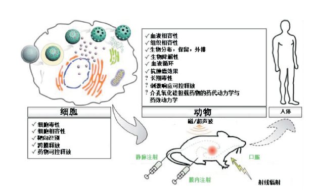
图18 多功能化多孔二氧化硅可控载药体系在生物医学应用中的研究路线
Fig.18 Research route for the biomedicine applications of multifunctional porous silica controlled drug delivery system
然而,介孔二氧化硅微球在治疗人类疾病的实际应用方面仍面临着巨大挑战。例如,具有新颖性、多功能化、高效、规律性释药的介孔二氧化硅的设计,并通过成像手段能够在生物体内被实时检测。虽然已经有些关于多功能化介孔二氧化硅的研究,但是在功能化方面的创新仍然很有限。此外,介孔二氧化硅在临床诊断与治疗方面的细胞毒性仍需进一步研究。
(全文完)
The authors have declared that no competing interests exist.
| [1] |
Cancer is among the most serious diseases characterized by uncontrollable cell growth and spread of abnormal cells. Cancerous cells form tumors that negatively impact the functions of the body, inducing serious malfunctioning leading to fatalities in most cases. Up to now, the effective diagnosis and treatments of cancer have remained a big challenge. Nanotechnology is an emerging field encompassing science, engineering and medicine, which has attracted great attention for cancer therapy in recent years. Among the numerous nanomaterials, mesoporous silica nanoparticles (MSNs) have attracted great attention and are being considered as promising biomedical materials for the development of cancer therapies because of their size tunability, surface functionality, optically transparent properties, low toxicity and high drug loading efficiency. In this review, we first outline the properties and structure of different configurations of MSNs and their subsequent application in the field of cancer theranostics. Thereafter, the potential of MSNs as multifunctional delivery platforms for therapeutic agents and their significance in next generation cancer therapy is discussed.
[本文引用:0]
|
| [2] |
Although ordered mesoporous silica materials have been studied for almost 20 years, their utilization within life science applications is relatively new and unexplored. An increasing number of researchers are transcending their respective fields in order to bridge the knowledge gap between materials chemistry and biotechnology, and to exploit the potential of mesoporous materials. Their intricate porosity with order in the nanoscale translates into high surface areas above 1000 m(2)/g, high selectivity for the encapsulation of biorelevant molecules as well as controlled surface chemistry. Their uses in pharmaceutics to improve drug formulation, drug bioavailability, mitigate drug toxicity and in cellular targeting, through controlled drug delivery strategies, have been shown. The incorporation of a high concentration of fluorescent and nuclear markers within their pores, whilst retaining good diffusion through their porous matrix, has shown them to be ideal candidates for sensing devices, in immunoassays such as flow cytometry and for their use in novel theranostic applications. This article aims to bring to the forefront some of the most important properties of mesoporous materials, which prove advantageous for their use in nanomedical applications and to highlight some of the potential areas into which the field may now emerge.
[本文引用:0]
|
| [3] |
Abstract In the past decade, mesoporous silica nanoparticles (MSNs) have attracted more and more attention for their potential biomedical applications. With their tailored mesoporous structure and high surface area, MSNs as drug delivery systems (DDSs) show significant advantages over traditional drug nanocarriers. In this review, we overview the recent progress in the synthesis of MSNs for drug delivery applications. First, we provide an overview of synthesis strategies for fabricating ordered MSNs and hollow/rattle-type MSNs. Then, the in vitro and in vivo biocompatibility and biotranslocation of MSNs are discussed in relation to their chemophysical properties including particle size, surface properties, shape, and structure. The review also highlights the significant achievements in drug delivery using mesoporous silica nanoparticles and their multifunctional counterparts as drug carriers. In particular, the biological barriers for nano-based targeted cancer therapy and MSN-based targeting strategies are discussed. We conclude with our personal perspectives on the directions in which future work in this field might be focused.
[本文引用:0]
|
| [4] |
Abstract A new application of MCM-41 mesoporous materials has been developed. Two kinds of surfactants, C16TAB and C12TAB, have been employed to get different pore sizes. The samples were disk-shaped conformed before and after charging with ibuprofen, an anti-inflammatory drug. In all the cases the weight percent ratio of drug/MCM-41 was 30%. The drug release plots show a different behavior depending on the method for charging the drug in the material but not on the employed surfactant.
DOI:10.1021/cm0011559
URL
[本文引用:0]
|
| [5] |
A system of chemical reactions has been developed which permits the controlled growth of spherical silica particles of uniform size by means of hydrolysis of alkyl silicates and subsequent condensation of silicic acid in alcoholic solutions. Ammonia is used as a morphological catalyst. Particle sizes obtained in suspension range from less than 0.05 μ to 2 μ in diameter.
[本文引用:0]
|
| [6] |
This paper reviews the progress made recently in synthesis and applications of spherical silica micro/nanomaterials with multilevel (hierarchical) structures. The spherical silica micro/nanomaterials with hierarchical structures are classified into four main structural categories that include (1) hollow mesoporous spheres, (2) core-in-(hollow porous shell) spheres, (3) hollow spheres with multiple porous shells and (4) hierarchically porous spheres. Due to the complex structures and being focused on spherical silica micro/nanomaterials, some novel methods based on the combination of two routine methods or two surfactants, and some special synthetic strategies are proposed to produce the spherical silica micro/nanomaterials with hierarchical structures. Compared with the same-sized solid, porous or hollow silica spheres, these fantastic spherical silica micro/nanomaterials with hierarchical structures exhibit enhanced properties which may enable them to be used in broad and promising applications as ideal scaffolds (carriers) for biological, medical, and catalytic applications.
[本文引用:0]
|
| [7] |
|
| [8] |
We describe a "surface-protected etching" strategy that allows convenient conversion of sol-gel derived silica into porous structures. Poly(vinyl pyrrolidone) is used to protect the near surface layer, and NaOH is used to selectively etch the interior of the silica spheres. Etching initially yields porous structures and eventually removes the core to leave behind hollow silica spheres with porous shells. This strategy is useful for constructing core-shell systems where active nanomaterials are embedded in silica shell for enhanced stability against aggregation. We experimentally demonstrate use of the surface-protected etching approach to create openings on silica shells; these openings allow dissolved chemical species to reach embedded catalytic particles to be chemically transformed while the porous shells continue to act as effective barriers against aggregation and loss of activity of the core particles. We also show that, by controlling the extent of etching, it is possible to control the permeation rate of the chemical species through the shells.
[本文引用:0]
|
| [9] |
DOI:10.1021/cm102829m
URL
[本文引用:0]
|
| [10] |
Fibrous nanosilica: A new family of high-surface-area silica nanospheres (KCC-1) have been prepared (see picture). KCC-1 features excellent physical properties, including high surface area, unprecedented fibrous surface morphology, high thermal (up to 95009000900°C) and hydrothermal stabilities, and high mechanical stability.
[本文引用:0]
|
| [11] |
DOI:10.1021/la100196j
URL
[本文引用:0]
|
| [12] |
DOI:10.1021/la200014w
URL
[本文引用:0]
|
| [13] |
Silica powder containing organized pores was prepared by a spray drying method. Silica and polystyrene latex (PSL) nanoparticles colloids were mixed and atomized to form micrometer-sized droplets. Nitrogen carrier gas was used to carry the resulting droplets into a vertical reactor that contained two heating zones:65 200 °C and 450 °C, which were used, respectively, to evaporate the dispersing medium (water) and to decompose the PSL particles to give a porous silica powder. The pores on the surface of the powders were found to be arranged into a hexagonal packing, indicating that a self-organization process occurred spontaneously during evaporation of the solvent. The pore size was controlled by changing the size of the PSL particles. By adding an additional zone (third zone) maintained at high temperatures, the produced powders could be in-situ annealed. A comparison of the average volume of the powder before and after annealing (at 1500 °C) indicated that the porosity of the powder was about 70%. Togethe...
DOI:10.1021/nl015662g
URL
[本文引用:0]
|
| [14] |
A spray drying method was used to produce a silica powder that contained ordered mesopores. A colloidal mixture of silica nanoparticles and polystyrene latex nanoparticles was mixed and sprayed as droplets into a vertical reactor that contained two temperature zones. The solvent in the droplets was evaporated at the front part of the reactor to produce a powder composite consisting of silica and PS nanoparticles. The PS nanoparticles in the powder were evaporated in the back portion of the reactor to produce a silica powder consisting of mesopores. The mesopores was observed to be arranged into a hexagonal packing, indicating the self-organization process occurred spontaneously during the solvent evaporation. The entire process was completed in only several seconds, which is contrary to currently available methods that require several hours or up to several days to complete this self-organization process.
[本文引用:0]
|
| [15] |
|
| [16] |
|
| [17] |
|
| [18] |
DOI:10.1021/nn1015117
URL
[本文引用:0]
|
| [19] |
We demonstrate a water-based etching strategy for converting solid silica shells into porous ones with controllable permeability. It overcomes the challenges of the alkaline-based surface-protected etching process that we previously developed for the production of porous and hollow silica nanostructures. Mild etching around the boiling point of water partially breaks the imperfectly condensed silica network and forms soluble monosilicic acid, eventually producing mesoscale pores in the silica structures. With the surface protection from poly(vinyl pyrrolidone) (PVP), it is possible to maintain the overall shape of the silica structures while at the same time to create porosity inside. By using bulky PVP molecules which only protect the near-surface region, we are able to completely remove the interior silica and produce hollow particles. Because the etching is mild and controllable, this process is particularly useful for treating small silica particles or core-shell particles with very thin silica shells for which the alkaline-based etching method has been difficult to control. We demonstrated the precise control of the permeation of the chemical species through the porous silica shells by using a model reaction which involves the etching of Ag encapsulated inside Ag@SiO(2) by a halocarbon. It is expected that the water-based surface-protected etching method can be conveniently extended to the production of various porous silica shells containing functional materials whose diffusion to outside and/or reaction with outside species can be easily controlled.
[本文引用:0]
|
| [20] |
|
| [21] |
DOI:10.1021/nn901398j
URL
[本文引用:0]
|
| [22] |
Abstract Rattle-type silica particles with metal cores, applicable to catalysts and metal/inorganic composite coating materials, were prepared by the pre-shell/post-core method that can control the size of metal cores inside silica capsules and exchange from metal cores into different ones with a metal displacement reaction.
[本文引用:0]
|
| [23] |
|
| [24] |
A simple, mild, and effective self-templated approach has been developed to directly convert solid SiO2 microspheres into hollow structures. The reaction involves initial partial dissolution of silica cores in a NaBH4 solution and subsequent shell formation due to the redeposition of the silicate species back onto the colloid surfaces. The increasing concentration of NaBO2 as the result of the slow decomposition of NaBH4 in water is found to be responsible for the regrowth of the silica shell. This method allows the production of hollow silica spheres with sizes ranging from 6570 nanometers to several micrometers, largely determined by the size of the starting silica colloids. The solid-to-hollow transformation mechanism is investigated in detail by transmission electron microscopy (TEM), scanning electron microscopy (SEM), Fourier Transform Infrared (FTIR) spectrometry, X-ray absorption spectroscopy (XAS), N2 adsorption61desorption, and X-ray diffraction (XRD). We also study the reaction conditions that al...
DOI:10.1021/jp810360a
URL
[本文引用:0]
|
| [25] |
Abstract From the inside out: Silica colloids can be spontaneously transformed from solid spheres to hollow structures in aqueous solutions of NaBH4 (see picture). The high pH value and gradual decomposition of NaBH4 facilitate the formation of hollow structures first by partial dissolution of silica cores and then by regrowth of the silicate species on the colloid surfaces to form shells. (Figure Presented).
[本文引用:0]
|
| [26] |
This paper explores the capability of the urface-protected etching process for the creation of rattle-type SiO 2 @void@SiO 2 colloidal structures featuring a mesoporous silica shell and a mesoporous movable silica core. The surface-protected etching process involves stabilization of the particle surface using a polymer ligand, and then selective etching of the interior to form hollow structures. In this paper, this strategy has been extended to the formation of rattle-like structures by etching SiO 2 @SiO 2 core shell particles which are synthesized by a two-step sol gel process. The key is to introduce a protecting polymer of polyvinylpyrrolidone (PVP) to the surface of both core and shell in order to tailor their relative stability against chemical etching. Upon reacting with NaOH, the outer layer silica becomes a hollow shell as only the surface layer is protected by PVP and the interior is removed, while the core remains its original size thanks to the protection of PVP on its surface. This process can be carried out at room temperature without the need of additional templates or complicated heterogeneous coating procedures. The etching process also results in the rattle-type colloids having mesoscale pores with two distinct average sizes. In our demonstration of a model drug delivery process, such mesoporous structures show an interesting two-step elution profile which is believed to be related to the unique porous rattle structures.
[本文引用:0]
|
| [27] |
DOI:10.1021/ja8059039
URL
[本文引用:0]
|
| [28] |
Hollow nanomaterials have shown great potential in many fields, including as drug carriers, building blocks of photonic crystals, and nanometer-scale reaction vessels. Recently, a new kind of hollow structure, the "nanorattle", has attracted more and more attention. In many applications, such as drug delivery systems, confined nanoreactors, and catalysts, nanorattles show considerable advantages.
[本文引用:0]
|
| [29] |
Hollow silica is a special type of novel inorganic material with one or more cavities inside. In addition to the excellent properties as with its solid counterparts, hollow silica exhibits unique characteristics, such as low density, high specific surface and good adsorption performance. Researchers have developed many routes to prepare mono-dispersed hollow silica with regular morphology. However, most studies focused on hollow silica spheres, ignoring the structural superiority of other hollow structures. Template synthesis is highly prominent due to its flexibility and versatility. What's more, it is suitable for the preparation of hollow silica with various morphologies. In this article, the research progress of template synthesis was firstly provided. Then different morphologies of hollow silica were introduced, including hollow spheres, hollow tubes, hollow cubes, etc. To better demonstrate the advantages and potential value of hollow silica materials, their performance in diverse applications were discussed. Finally, some perspectives on the future research and development of hollow silica were put forward.
[本文引用:0]
|
| [30] |
|
| [31] |
|
| [32] |
DOI:10.1021/ic301371q
URL
[本文引用:0]
|
| [33] |
Hollow silica nanospheres with their size distribution ranging from 350 nm to 450 nm are synthesized by using polystyrene (PS) templates in the present study. On the basis of PS templates, silica, the hydrolyzate of TEOS(tetraethyl orthosilicate) under moist alkaline condition at ambient temperatures and atmospheric pressures, it is set to be coated on the surface of the PS spheres. Since the size of PS sphere core can be easily controlled, it is expected to serve various needs of different sized hollow silica nanospheres in industrial applications. It is proposed that the PS cores be removed by either thermal pyrogenation or solvent dissolved. Morphology of the hollow silica nanospheres is characterized by scanning electron microscopy (SEM).
[本文引用:0]
|
| [34] |
DOI:10.1021/jp710990b
URL
[本文引用:0]
|
| [35] |
In this paper, we report a novel method for the fabrication of small monodisperse hollow silica spheres. In this approach, when silica shells were coated on polystyrene particles by the sol-gel method, the polystyrene cores were dissolved subsequently, even synchronously, in the same medium to form monodisperse hollow spheres. Neither additional dissolution nor a calcination process was needed to remove the polystyrene cores. Transmission electron microscopy, scanning electron microscopy, and porosity measurements were used to characterize the monodisperse hollow silica spheres.
[本文引用:0]
|
| [36] |
|
| [37] |
DOI:10.1021/la800366j
URL
[本文引用:0]
|
| [38] |
Raspberry-like superhydrophobic hollow silica particles were prepared through a sacrificial polymer template method. The St枚ber method was adopted to coat silica onto the surface of cationic polymethylmethacrylate(PMMA) particles by electrostatic interaction. The surface of the PMMA-silica composite particles exhibited raspberry-like morphology with high surface roughness. Hollow silica particles were then obtained by calcination to selectively remove the PMMA core. Subsequent modification with nonafluorohexyltriethoxysilane (NFH-silane) conferred superhydrophobicity on the hollow silica particles. The surface property of this particles were investigated by measuring their water contact angle, and the results showed that such perfluorinated raspberry-like hollow particles had unique superhydrophobic.
[本文引用:0]
|
| [39] |
|
| [40] |
Three kinds of different ZnO colloid particles (flowerlike particles, nanoribbons and microspheres) and one kind of ZnO film have been coated with silica via a simple sol–gel method in the St02ber system and ZnO/silica core–shell microparticles or films have been obtained. The thickness of silica shell can be controlled by adjusting the concentration of TEOS added into the system. If the ZnO core is etched off by HCl, corresponding, hollow silica particles or film will be generated.
[本文引用:0]
|
| [41] |
In this work, hollow silica colloids with different shapes, such as pseudocubes, ellipsoids, capsules, and peanuts, have been synthesized through the following process: silica coating on the surface of hematite colloidal particles with different shapes (pseudocubes, ellipsoids, capsules, and peanuts) and the sequential acid dissolution of the hematite cores. The as-obtained hollow silica colloids with different shapes have uniform sizes, shapes, and shells.
[本文引用:0]
|
| [42] |
Hollow inorganic particles have attracted great interest because of their unique physicochemical properties. In this study, hollow silica microparticles were prepared using a rod-shaped gram-negative bacterium, Escherichia coli (KP7600), as a biological template. Silica nanoparticles were generated in addition to coated biological templates when the reaction rate was increased, so control of reaction rate is important for coating silica smoothly onto the bacterial surface. Silica coating was also carried out using the fixed cells (with and without internal water) using glutaraldehyde as templates. When the fixed cells without internal water were used as templates, no rod-shaped particles were observed after calcination of the synthesized particles. By contrast, silica hollow particles were formed using the fixed cells with internal water as templates. This means that the internal water inside biological cells acts as an initiator for hydrolysis of tetraethyl orthosilicate (TEOS) and results in the formation of smooth silica shell surface and indicates that the use of dry cultured bacteria templates is not required. Thus, there is a significant benefit in using gram-negative bacteria as templates for producing hollow silica microparticles, compared with the method using dried gram-positive bacteria templates.
[本文引用:0]
|
| [43] |
Silica needle-shaped nano-hollow structure with high specific surface area was prepared by a sol–gel approach using needle-shaped calcium carbonate (CaCO 3 ) nanoparticles as novel template and sodium silicate as the silica source, followed by washing in acidic solution to remove the CaCO 3 sacrificial template. The sample was characterized by transmission electron microscope (TEM), scanning electron microscope (SEM), energy disperse spectroscopy (EDS), X-ray diffraction (XRD), Fourier transform infrared (FTIR) and BET. The results show that regular uniform silica nano-hollow tubes are obtained: the length of the hollow tubes ranges from 200nm to 300nm with a inside diameter about 20nm, the thickness of the tubes is around 10nm, the specific surface area of the sample is up to 321.4m 2 /g, the average pore diameter is 63.8303 and the total pore volume is 0.5128cm 3 /g.
[本文引用:0]
|
| [44] |
Abstract Ordered mesostructured porous silicas that are also macroscopically structured were created by control of the interface on two different length scales simultaneously. Micellar arrays controlled the nanometer-scale assembly, and at the static boundary between an aqueous phase and an organic phase, control was achieved on the micrometer to centimeter scale. Acid-prepared mesostructures of silica were made with the p6, Pm3n, and the P63/mmc structures in the form of porous fibers 50 to 1000 micrometers in length, hollow spheres with diameters of 1 to 100 micrometers, and thin sheets up to 10 centimeters in diameter and about 10 to 500 micrometers in thickness. These results might have implications for technical applications, such as slow drug-release systems or membranes, and in biomineralization, where many processes are also interface-controlled.
[本文引用:0]
|
| [45] |
DOI:10.1021/cm8024236
URL
[本文引用:0]
|
| [46] |
DOI:10.1021/la101225j
URL
[本文引用:0]
|
| [47] |
DOI:10.1021/cm801411y
URL
[本文引用:0]
|
| [48] |
|
| [49] |
|
| [50] |
Hollow microspheres with ordered mesoporous walls are synthesised under ambient conditions by a simple procedure involving dilution and neutralisation of an aqueous tetraethoxysilane/reaction mixture.
[本文引用:0]
|
| [51] |
DOI:10.1021/la062542l
URL
[本文引用:0]
|
| [52] |
Hollow mesoporous silica spheres were synthesized by a sol–gel/emulsion (oil-in-water/ethanol) approach, in which cetyltrimethylammonium bromide (CTAB) surfactant was employed to stabilize and direct the hydrolysis of oil droplets of tetraethoxysilane (TEOS). The diameters of the hollow spheres can be tuned in the range from 210 to 720 nm by varying the ratio of ethanol-to-water and their shell thickness can be mediated by changing the concentration of CTAB used in the system. BET surface areas of the hollow silica spheres are determined to be in the range of 924–1766 m 2 g 611 and their pore sizes are around 3.10 nm as determined by BJH method.
[本文引用:0]
|
| [53] |
DOI:10.1021/ja028650l
URL
[本文引用:0]
|
| [54] |
DOI:10.1021/ja901831u
URL
[本文引用:0]
|
| [55] |
|
| [56] |
DOI:10.1021/ja104501a
URL
[本文引用:0]
|
| [57] |
This paper proposes a new nanoscopic molecular movable gate-like functional hybrid system consisting of nanoscopic MCM-41-based material functionalized onto pore outlets with a saccharide derivative capable of interacting with boronic acid functionalized gold nanoparticles (AuNPs) acting as nanoscopic caps. The gating mechanism involves the reversible reaction between polyalcohols and boronic acids to form boronate esters. Functionalized AuNPs thus act as a suitable nanoscopic cap via the reversible formation of the corresponding boroester bonds with the saccharide derivative anchored on the external surface of the mesoporous silica-based solid. The developed platform operates in aqueous solution and can be triggered by two simple external stimuli such as pH changes or light. The hydrolysis of the boroester bond takes place at pH 3, which results in rapid delivery of the safranine cargo from the pore voids into the aqueous solution. However, at pH 5 the pores are capped with nanoparticles and the delivery is strongly inhibited. The kinetics of the delivery was studied at pH = 3, assuming a simple diffusion process and that the kinetics of guest release from the pore voids of the hybrid material can be explained by the Higuchi model. It is possible to deliver the cargo in small portions by carrying out on-off aperture cycles via changing the pH from 3 to 5. AuNPs also open the possibility of employing light as a suitable stimulus for release procedures using the AuNPs' capacity for raising their temperature locally by absorption of laser light. The plasmonic heating using a Nd:YAG laser at 1064 nm results in the cleavage of the boronic ester linkage that anchors the nanoparticles to the surface of the mesoporous silica-based material, allowing the release of the entrapped guests. Further studies also demonstrated that it is possible to fine-tune the amount of cargo delivered by simply controlling the laser irradiation opening the possibility to designing laser-induced pulsatile release supports.
[本文引用:0]
|
| [58] |
|
| [59] |
|
| [60] |
|
| [61] |
A novel pH-controlled delivery system has been developed based on hollow mesoporous silica spheres using pH-sensitive polyelectrolyte multilayer coated on the spheres as a switch to store and release gentamicin molecules (a model drug). The polyelectrolyte layers with an average thickness of 12 nm was coated on hollow mesoporous silica spheres through a layer-by-layer technique. Gentamicin molecules were successfully stored in these spheres by means of adjusting the gentamicin solution from pH 2 to pH 8. The storage capacity can reach 614.8 mg/g (34.11%) at an initial gentamicin concentration of 60 mg/ml. The controlled release of gentamicin molecules from this system has been achieved by simply changing the pH value in the release media. Therefore, this type of material is of potentials for the controlled drug release applications.
[本文引用:0]
|
| [62] |
Aqueous solutions of poly(N-isopropyl acrylamide) show a lower critical solution temperature. The thermodynamic properties of the system have been evaluated from the phase diagram and the heat absorbed during phase separation and the phenomenon is ascribed to be primarily due to an entropy effect. From viscosity, sedimentation, and light-scattering studies of solutions close to conditions of phase separation, it appears that aggregation due to formation of nonpolar and intermolecular hydrogen bonds is important. In addition, a weakening of the ordering effect of the water-amide hydrogen bonds as the temperature is raised contributes to the stability of the two-phase system.
[本文引用:0]
|
| [63] |
Poly(N-isopropylacrylamide) : experiment, theory and application SCHILD H. G. Prog. Polym. Sci. 17, 163-249, 1992
[本文引用:0]
|
| [64] |
|
| [65] |
|
| [66] |
|
| [67] |
|
| [68] |
ABSTRACT Inorganic photochemistry is one of the most rapidly growing fields of chemistry. It evolved from simple molecular systems and reached its "adolescence" via convergence with supramolecular chemistry, heterogeneous catalysis, and biochemistry. New trends in inorganic photochemistry explore such diverse areas as medicine, environmental sciences, materials technology, and technology related to new energy sources. This review highlights the recent advances and main frontiers in bioinorganic photochemistry, the interdisciplinary field which intersects inorganic photochemistry with biological, medical, and environmental sciences. It is emphasized that the understanding of the photochemistry and photophysics of metal compounds has matured to the point that solutions to various practical problems of the modern world are accessible.
|
| [70] |
|
| [71] |
|
| [72] |
DOI:10.1021/ja107184z
URL
[本文引用:0]
|
| [73] |
DOI:10.1021/ja209793b
URL
[本文引用:0]
|
| [74] |
|
| [75] |
|
| [76] |
|
| [77] |
|
| [78] |
DOI:10.1021/ja046572r
URL
[本文引用:0]
|
| [79] |
|
| [80] |
|
| [81] |
|
| [82] |
|
| [83] |
Bernardos A, Aznar E, Marcos MD, Martínez-Má09ez R, Sancenón F, Soto J, Barat JM, Amorós P.
[本文引用:0]
|
| [84] |
DOI:10.1021/nn101499d
URL
[本文引用:0]
|
| [85] |
Mesoporous silica nanoparticles, capable of storing a payload of small molecules and releasing it following specific catalytic activation by an esterase, have been designed and fabricated. The storage and release of the payload is controlled by the presence of [2]rotaxanes, which consist of tri(ethylene glycol) chains threaded by -cyclodextrin tori, located on the surfaces of the nanoparticles and terminated by a large stoppering group. These modified silica nanoparticles are capable of encapsulating guest molecules when the [2]rotaxanes are present. The bulky stoppers, which serve to hold the tori in place, are stable under physiological conditions but are cleaved by the catalytic action of an enzyme, causing dethreading of the tori and release of the guest molecules from the pores of the nanoparticles. These snap-top covered silica nanocontainers (SCSNs) are prepared by a modular synthetic method, in which the stoppering unit, incorporated in the final step of the synthesis, may be changed at will to target the response of the system to any of a number of hydrolytic enzymes. Here, the design, synthesis, and operation of model SCSNs that open in the presence of porcine liver esterase (PLE) are reported. The empty pores of the silica nanoparticles were loaded with luminescent dye molecules (rhodamine B), and stoppering units that incorporate adamantyl ester moieties were then attached in the presence of 伪-cyclodextrin using the copper-catalyzed azide lkyne cycloaddition (CuAAC), closing the SCSNs. The release of rhodamine-B from the pores of the SCSN, following PLE-mediated hydrolysis of the stoppers, was monitored using fluorescence spectroscopy.
[本文引用:0]
|
| [86] |
|
| [87] |
DOI:10.1021/ja9061085
URL
[本文引用:0]
|
| [88] |
Abstract Stimuli-responsive gate mechanisms offer potential for the controlled passage of payload molecules from a porous carrier vehicle on-demand. We describe a method for the enzyme-mediated release of macromolecular guest molecules from inorganic silica particles coated with a bioactive peptide shell, synthesized precisely by Fmoc chemistry. Specific enzymatic hydrolysis of the peptide shell removes the bulky peptide-terminated Fmoc groups, permitting the selective release of previously entrapped guest molecules.
[本文引用:0]
|
| [89] |
|
| [90] |
|
| [91] |
DOI:10.1021/cm402592t
URL
[本文引用:0]
|
| [92] |
|
| [93] |
ABSTRACT The preparation of fluorescent mesoporous silica nanoparticles (FMSN) as a delivery system for hydrophobic anticancer drugs were discussed. The FMSNs were prepared by using a base-catalyzed sol-gel method at high temperature with a modification of established procedures. It was found that for efficient cellular uptake of the particles that the FMSNs should remain dispersed and do not aggregate in the buffer solution. The uptake of the nanoparticles by various cancer-cell lines was also revealed by using fluorescence and confocal microscopy. The results show that these FMSNs can be successfully used for the delivery of the hydrophobic anticancer drug camptothecin (CPT).
[本文引用:0]
|
| [94] |
DOI:10.1021/jz101483u
URL
[本文引用:0]
|
| [95] |
DOI:10.1002/jps.21638
URL
[本文引用:0]
|
| [96] |
It is known that the energy of the amorphous state of itraconazole loaded in ordered mesoporous materials is high relative to that of the crystalline state and is responsible for enhanced solubility and dissolution rate. We investigated the effects of particle size (0.7–5 μm), mesostructure (2D p 6 mm , cubic Ia -3 d and cubic Fm -3 m ) and pore size (2.2–15.4 nm) of mesoporous silicas on the release performance of itraconazole. Results indicated that the release performance was not influenced by the particle sizes tested here, that the release performance increased with increasing pore diameter due to the lower probability of drug molecules colliding to recrystallize in large pores, and that the release performance was decreased in the cage-type pore structure ( Fm -3 m ) compared to that in the cylindrical pore structures ( p 6 mm and Ia -3 d ) because of the small entrance to the cagelike pores that retards the drug release.
[本文引用:0]
|
| [97] |
|
| [98] |
|
| [99] |
Protein-based nanomedicine platforms for drug delivery comprise naturally self-assembled protein subunits of the same protein or a combination of proteins making up a complete system. They are ideal for drug-delivery platforms due to their biocompatibility and biodegradability coupled with low toxicity. A variety of proteins have been used and characterized for drug-delivery systems, including the ferritin/apoferritin protein cage, plant-derived viral capsids, the small Heat shock protein (sHsp) cage, albumin, soy and whey protein, collagen, and gelatin. There are many different types and shapes that have been prepared to deliver drug molecules using protein-based platforms, including various protein cages, microspheres, nanoparticles, hydrogels, films, minirods, and minipellets. The protein cage is the most newly developed biomaterial for drug delivery and therapeutic applications. The uniform size, multifunctionality, and biodegradability push it to the frontier of drug delivery. In this Review, the recent strategic development of drug delivery is discussed with emphasis on polymer-based, especially protein-based, nanomedicine platforms for drug delivery. The advantages and disadvantages are also discussed for each type of protein-based drug-delivery system.
[本文引用:0]
|
| [100] |
|
| [101] |
An MCM-41-type mesoporous silica nanoparticle (MSN) material with a large average pore diameter (5.4 nm) is synthesized and characterized. The in vitro uptake and release profiles of cytochrome c by the MSN were investigated. The enzymatic activity of the released protein was quantitatively analyzed and compared with that of the native cytochrome c in physiological buffer solutions. We found that the enzymes released from the MSNs are still functional and highly active in catalyzing the oxidation of 2,2'-azino-bis(3-ethylbenzthiazoline-6-sulfonate) (ABTS) by hydrogen peroxide. In contrast to the fact that cytochrome c is a cell-membrane-impermeable protein, we discovered that the cytochrome c-encapsulated MSNs could be internalized by live human cervical cancer cells (HeLa) and the protein could be released into the cytoplasm. We envision that these MSNs with large pores could serve as a transmembrane delivery vehicle for controlled release of membrane-impermeable proteins in live cells, which may lead to many important biotechnological applications including therapeutics and metabolic manipulation of cells.
[本文引用:0]
|
| [102] |
|
| [103] |
|
| [104] |
Mesoporous silica nanoparticles (MSNs) are highly attractive supports for the design of controlled delivery systems able to act as containers for the encapsulation of therapeutic agents overcoming common issues such as poor water solubility and poor stability of some drugs and also enhancing their bioavailability. In this context, we describe herein the development of polyglutamic acid (PGA)-capped MSNs able to selectively deliver rhodamine B and doxorubicin. PGA-capped MSNs remained closed in an aqueous environment yet are able to deliver the cargo in the presence of pronase due to the hydrolysis of the peptide bonds in PGA. The solids prepared released less than 20% of the cargo in one day, whereas they were able to reach 90% of the maximum release of the entrapped guest in ca. 5 h in the presence of pronase. Studies of the PGA-capped nanoparticles with SK-BR-3 breast cancer cells was also tested. Rhodamine-loaded nanoparticles were not toxic, while doxorubicin-loaded nanoparticles were able to kill eff...
[本文引用:0]
|
| [105] |
|
| [106] |
Abstract Surface-functionalized silica nanoparticles can deliver DNA and drugs into animal cells and tissues. However, their use in plants is limited by the cell wall present in plant cells. Here we show a honeycomb mesoporous silica nanoparticle (MSN) system with 3-nm pores that can transport DNA and chemicals into isolated plant cells and intact leaves. We loaded the MSN with the gene and its chemical inducer and capped the ends with gold nanoparticles to keep the molecules from leaching out. Uncapping the gold nanoparticles released the chemicals and triggered gene expression in the plants under controlled-release conditions. Further developments such as pore enlargement and multifunctionalization of these MSNs may offer new possibilities in target-specific delivery of proteins, nucleotides and chemicals in plant biotechnology.
[本文引用:0]
|
| [107] |
DOI:10.1021/nn900918w
URL
[本文引用:0]
|
| [108] |
DOI:10.1021/jp807956r
URL
[本文引用:0]
|
| [109] |
DOI:10.1021/nn103130q
URL
[本文引用:0]
|
| [110] |
The interaction between DNA and mesopores is one of the basic concerns when mesoporous silica nanoparticle (MSN) is used as a DNA carrier. In this work, we have synthesized a type of mesoporous silica nanoparticle that has a Fe(3)O(4) inner core and mesoporous silica shell. This magnetic mesoporous silica nanoparticle (denoted as M-MSN) offers us a convenient platform to manipulate the DNA adsorption and desorption processes as it can be easily separated from solution by applying a magnetic field. The DNA adsorption behavior is studied as a function of time in chaotropic salt solution. The maximum amount of adsorbed DNA is determined as high as 121.6 mg/g. We have also developed a method to separate the DNA adsorbed onto the external surface and into the mesopores by simply changing temperature windows. The desorption results suggest that, within the whole adsorbed DNA molecules, about 89.5% has been taken up by M-MSN mesopores. Through the dynamic light scattering experiment, we have found that the hydrodynamic size for M-MSN with DNA in its mesopores is higher than the naked M-MSN. Finally, the preliminary result of the adsorption mechanism study suggests that the DNA adsorption into mesopores may generate more intermolecular hydrogen bonds than those formed on the external surface.
[本文引用:0]
|
| [111] |
|
| [112] |
DOI:10.1002/smll.v6:3
URL
[本文引用:0]
|
| [113] |
DOI:10.1021/jp203454g
URL
[本文引用:0]
|
| [114] |
DOI:10.1038/nmat2992
URL
[本文引用:0]
|
| [115] |
|
| [116] |
|
