Effect of Hydromorphone Post-treatment on the Reperfusion Arrhythmia and the Expression of Myocardial Cx43 During Ischemia-Reperfusion in Isolated Rat Hearts
易菁1,, 段宏伟2, 高鸿1,, 曾庆繁1, 王子君3, 王贵龙3, 刘艳秋1
1.贵州医科大学附属医院麻醉科,贵阳 550004
2.复旦大学附属浦东医院麻醉科,上海 201301
3.贵州医科大学麻醉学院,贵阳 550004
YI Jing1,, DUAN Hongwei2, GAO Hong1,, ZENG Qingfan1, WANG Zijun3, WANG Guilong3, LIU Yanqiu1
1. Department of Anesthesiology, the Affiliated Hospital of Guizhou Medical University, Guiyang 550004,China
2.Department of Anesthesiology, the Affiliated Pudong Hospital of Fudan University, Shanghai 201301,China
3.School of Anesthesiology, Guizhou Medical University, Guiyang 550004, China
Objective To investigate the effect of hydromorphone postconditioning on the reperfusion arrhythmia and the expression of myocardial connexin 43 (Cx43) during ischemia-reperfusion in isolated rat hearts and its mechanism. Methods The SD rats (n=24) were randomly divided into three groups: control group (group C,n=18),ischemia-reperfusion group(group I/R,n=8),hydromorphone post-treatment group(group HM,n=8).The langendorff heart or isolated perfused heart assay was established. The heart rate (HR) and ECG during the whole experiment period were recorded . And the reperfusion arrhythmia during the period of reperfusion were detected by ECG. The expression of Akt and Cx43 protein were detected by Western blotting technique. Results Although the HR at different time points and the reperfusion arrhythmia score of three groups were not statistically significant(P>0.05), the duration of reperfusion arrhythmia of group HM was significantly shorter than that of group IR, and the incidence of ventricular tachycardia and ventricular fibrillation in group HM were also less than that in group IR (P<0.05). When Akt expression level in group IR was compared with group C, there was no significant differences(P>0.05), but Akt expression level in group HM was obviously increased; Cx43 expression level in group IR and group HM were both significantly reduced. While compared with the group IR, Cx43 expression level in group HM was significantly increased(P<0.05). Conclusion The mechanism by which hydromorphone post-treatment inhibits reperfusion arrhythmia induced by myocardial IR is associated with up-regulated expression of myocardial Cx43 after activation of Akt signaling pathway during ischemia-reperfusion in isolated rat hearts.
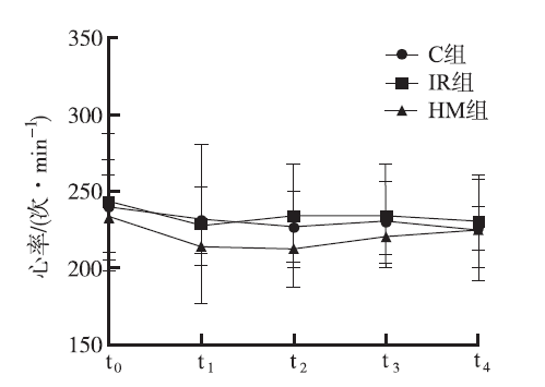
图1
3组大鼠各时点心率比较($\bar{x}\pm{s}$,n=8) t0:平衡灌注20 min;t1、t2、t3、t4:再灌注10,25,45,60 min
Fig.1
Comparison of the heart rate among three groups of rats at different time points($\bar{x}\pm{s}$,n=8) t0:balance perfusion for 20 min;t1、t2、t3、t4:reperfusion of 10,25,45,60 min
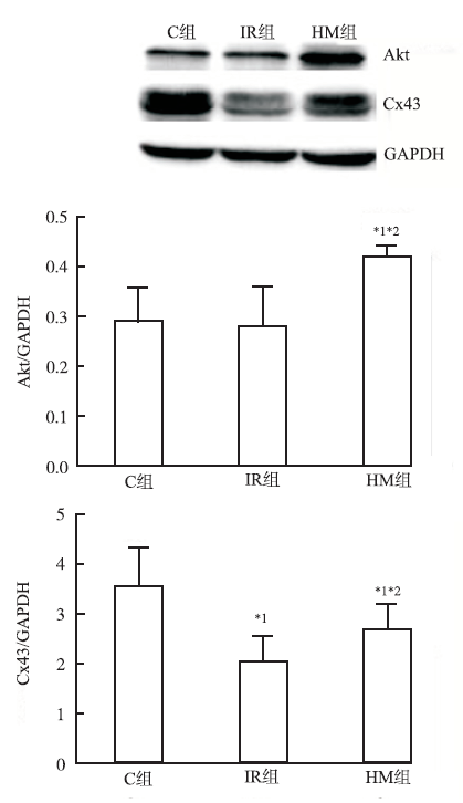
Fig.2
Comparison of relative protein expression of Akt and Cx43 in isolated hearts among three groups of rats Compared with group C, *1P<0.05;Compared with group IR, *2P<0.05
TSEG,YEO JM.Conduction abnormalities and ventricular arrhythmogenesis:the roles of sodium channels and gap junctions[J].IJC Heart Vascul,2015,9(12):75-82.
Ventricular arrhythmias arise from disruptions in the normal orderly sequence of electrical activation and recovery of the heart. They can be categorized into disorders affecting predominantly cellular depolarization or repolarization, or those involving action potential (AP) conduction. This article briefly discusses the factors causing conduction abnormalities in the form of unidirectional conduction block and reduced conduction velocity (CV). It then examines the roles that sodium channels and gap junctions play in AP conduction. Finally, it synthesizes experimental results to illustrate molecular mechanisms of how abnormalities in these proteins contribute to such conduction abnormalities and hence ventricular arrhythmogenesis, in acquired pathologies such as acute ischaemia and heart failure, as well as inherited arrhythmic syndromes.
DESPLANTEZT.Cardiac Cx43,Cx40 and Cx45 coassemb-ling:involvement of connexins epitopes in formation of hemichannels and Gap junction channels[J].BMC Cell Biol,2017,18(Suppl 1):3.
This review comes after the International Gap Junction Conference (IGJC 2015) and describes the current knowledge on the function of the specific motifs of connexins in the regulation of the formation of gap junction channels. Moreover the review is complemented by a summarized description of the distinct contribution of gap junction channels in the electrical coupling. Complementary biochemical and functional characterization on cell models and primary cells have improved our understanding on the oligomerization of connexins and the formation and the electrical properties of gap junction channels. Studies mostly focused cardiac connexins Cx43 and Cx40 expressed in myocytes, while Cx45 and Cx30.2 have been less investigated, for which main findings are reviewed to highlight their critical contribution in the formation of gap junction channels for ensuring the orchestrated electrical impulse propagation and coordination of atrial and ventricular contraction and heart function, whereas connexin dysfunction and remodeling are pro-arrhythmic factors. Common and specific motifs of residues identified in different domain of each type of connexin determine the connexin homo- and hetero-oligomerization and the channels formation, which leads to specific electrical properties. These motifs and the resulting formation of gap junction channels are keys to ensure the tissue homeostasis and function in each connexin expression pattern in various tissues of multicellular organisms. Altogether, the findings to date have significantly improved our understanding on the function of the different connexin expression patterns in healthy and diseased tissues, and promise further investigations on the contribution in the different types of connexin.
MICHELAP,VELIAV,ALDOP,et al.Role of connexin 43 in cardiovascular diseases[J].Eur J Pharm,2015,768(12):71-76.
Gap junctions (GJs) channels provide the basis for intercellular communication in the cardiovascular system for maintenance of the normal cardiac rhythm, regulation of vascular tone and endothelial function as well as metabolic interchange between the cells. They allow the transfer of small molecules and may enable slow calcium wave spreading, transfer of “death” or of “survival” signals. In the cardiomyocytes the most abundant isoform is Connexin 43 (Cx43). Alterations in Cx43 expression and distribution were observed in myocardium disease; i.e. in hypertrophic cardiomyopathy, heart failure and ischemia. Recent reports suggest the presence of Cx43 in the mitochondria as well, at least in the inner mitochondrial membrane, where it plays a central role in ischemic preconditioning. In this review, the current knowledge on the relationship between the remodeling of cardiac gap junctions and cardiac diseases are summarized.
SCHULZR,GÖRGEP M,GÖRBEA,et al.Connexin 43 is an emerging therapeutic target in ischemia/reperfusion injury,cardioprotection and neuroprotection[J].Pharmacol Therap,2015,153(9):90-106.
Connexins are widely distributed proteins in the body that are crucially important for heart and brain functions. Six connexin subunits form a connexon or hemichannel in the plasma membrane. Interactions between two hemichannels in a head-to-head arrangement result in the formation of a gap junction channel. Gap junctions are necessary to coordinate cell function by passing electrical current flow between heart and nerve cells or by allowing exchange of chemical signals and energy substrates. Apart from its localization at the sarcolemma of cardiomyocytes and brain cells, connexins are also found in the mitochondria where they are involved in the regulation of mitochondrial matrix ion fluxes and respiration. Connexin expression is affected by age and gender as well as several pathophysiological alterations such as hypertension, hypertrophy, diabetes, hypercholesterolemia, ischemia, post-myocardial infarction remodeling or heart failure, and post-translationally connexins are modified by phosphorylation/de-phosphorylation and nitros(yl)ation which can modulate channel activity. Using knockout/knockin technology as well as pharmacological approaches, one of the connexins, namely connexin 43, has been identified to be important for cardiac and brain ischemia/reperfusion injuries as well as protection from it. Therefore, the current review will focus on the importance of connexin 43 for irreversible injury of heart and brain tissues following ischemia/reperfusion and will highlight the importance of connexin 43 as an emerging therapeutic target in cardio- and neuroprotection.
BATRAN,RIQUELME MA,BURRAS,et al.Direct regu-lation of osteocytic connexin 43 hemichannels through AKT kinase activated by mechanical stimulation[J].J Biolog Chem,2014,289(15):10582-10591.
Background: Opening of Cx43 hemichannels by mechanical stress releases factors important for bone remodeling; however, the regulatory mechanism is unknown. Result: Upon mechanical stimulation, AKT phosphorylates integrin 5 and Cx43, increases interaction, and opens hemichannels. Conclusion: Phosphorylation of Cx43 and 5 by AKT is critical for hemichannel opening. Significance: This is the first report demonstrating the functional importance of AKT in regulation of Cx43 hemichannels. Connexin (Cx) 43 hemichannels in osteocytes are thought to play a critical role in releasing bone modulators in response to mechanical loading, a process important for bone formation and remodeling. However, the underlying mechanism that regulates the opening of mechanosensitive hemichannels is largely unknown. We have recently shown that Cx43 and integrin 5 interact directly with each other, and activation of PI3K appears to be required for Cx43 hemichannel opening by mechanical stimulation. Here, we show that mechanical loading through fluid flow shear stress (FFSS) increased the level of active AKT, a downstream effector of PI3K, which is correlated with the opening of hemichannels. Both Cx43 and integrin 5 are directly phosphorylated by AKT. Inhibition of AKT activation significantly reduced FFSS-induced opening of hemichannels and disrupted the interaction between Cx43 and integrin 5. Moreover, AKT phosphorylation on Cx43 and integrin 5 enhanced their interaction. In contrast to the C terminus of wild-type Cx43, overexpression of the C-terminal mutant containing S373A, a consensus site previously shown to be phosphorylated by AKT, failed to bind with 5 and hence could not inhibit hemichannel opening. Together, our results suggest that AKT activated by FFSS directly phosphorylates Cx43 and integrin 5, and Ser-373 of Cx43 plays a predominant role in mediating the interaction between these two proteins and Cx43 hemichannel opening, a crucial step to mediate the anabolic function of mechanical loading in the bone.
DUNN CA,SUV,LAU AF,et al.Activation of Akt,not connexin 43 protein ubiquitination,regulates gap junction stability[J].J Biolog Chem2012,287(4):2600-2607.
The pore-forming gap junctional protein connexin 43 (Cx43) has a short (1-3 h) half-life in cells in tissue culture and in whole tissues. Although critical for cellular function in all tissues, the process of gap junction turnover is not well understood because treatment of cells with a proteasomal inhibitor results in larger gap junctions but little change in total Cx43 protein whereas lysosomal inhibitors increase total, mostly nonjunctional Cx43. To better understand turnover and identify potential sites of Cx43 ubiquitination, we prepared constructs of Cx43 with different lysines converted to arginines. However, when transfected into cells, a mutant version of Cx43 with all lysines converted to arginines behaved similarly to wild type in the presence of proteasomal and lysosomal inhibitors, indicating that ubiquitination of Cx43 did not appear to be playing a role in gap junction stability. Through the use of inhibitors and dominant negative constructs, we found that Akt (protein kinase B) activity controlled gap junction stability and was necessary to form larger stable gap junctions. Akt activation was increased upon proteasomal inhibition and resulted in phosphorylation of Cx43 at Akt phosphorylation consensus sites. Thus, we conclude that Cx43 ubiquitination is not necessary for the regulation of Cx43 turnover; rather, Akt activity, probably through direct phosphorylation of Cx43, controls gap junction stability. This linkage of a kinase involved in controlling cell survival and growth to gap junction stability may mechanistically explain how gap junctions and Akt play similar regulatory roles.
DOUM,WUH,ZHUH,et al.Remifentanil preconditioning protects rat cardiomyocytes against hypoxia-reoxygenation injury via delta-opioid receptor mediated activation of PI3K/Akt and ERK pathways[J].Eur J Pharm,2016,789(8):395-401.
MASLOV LN,KHALIULINI,OELTGEN PR,et al.Pros-pects for creation of cardioprotective and antiarrhythmic drugs based on opioid receptor agonists[J].Med Res Rev,2016,36(5):871-923.
Abstract It has now been demonstrated that the μ, δ1, δ2, and κ1 opioid receptor (OR) agonists represent the most promising group of opioids for the creation of drugs enhancing cardiac tolerance to the detrimental effects of ischemia/reperfusion (I/R). Opioids are able to prevent necrosis and apoptosis of cardiomyocytes during I/R and improve cardiac contractility in the reperfusion period. The OR agonists exert an infarct-reducing effect with prophylactic administration and prevent reperfusion-induced cardiomyocyte death when ischemic injury of heart has already occurred; that is, opioids can mimic preconditioning and postconditioning phenomena. Furthermore, opioids are also effective in preventing ischemia-induced arrhythmias.
JELEAZCOVC,IHMSENH,SAARI TI,et al.Patientcon-trolled analgesia with target-controlled infusion of hydromorphone in postoperative pain therapy[J].Anesthesiology,2016,124(1):56-68.
Patient-controlled analgesia (PCA) is a common method for postoperative pain therapy, but it is characterized by large variation of plasma concentrations. PCA with target-controlled infusion (TCI-PCA) may be an alternative. In a previous analysis, the authors developed a pharmacokinetic model for hydromorphone. In this secondary analysis, the authors investigated the feasibility and efficacy of TCI-PCA for postoperative pain therapy with hydromorphone. Fifty adult patients undergoing cardiac surgery were enrolled in this study. Postoperatively, hydromorphone was applied intravenously during three sequential periods: (1) as TCI with plasma target concentrations of 1 to 265ng/ml until extubation; (2) as TCI-PCA with plasma target concentrations between 0.8 and 1065ng/ml during the following 6 to 865h; and (3) thereafter as PCA with a bolus dose of 0.265mg until the next morning. During TCI-PCA, pain was regularly assessed using the 11-point numerical rating scale (NRS). A pharmacokinetic/pharmacodynamic model was developed using ordinal logistic regression based on measured plasma concentrations. Data of 43 patients aged 40 to 81 yr were analyzed. The hydromorphone dose during TCI-PCA was 0.2665mg/h (0.07 to 0.9365mg/h). The maximum plasma target concentration during TCI-PCA was 2.365ng/ml (0.9 to 7.065ng/ml). The NRS score under deep inspiration was less than 5 in 83% of the ratings. Nausea was present in 30%, vomiting in 9%, and respiratory insufficiency in 5% of the patients. The EC50 of hydromorphone for NRS of 4 or less was 4.165ng/ml (0.6 to 12.865ng/ml). TCI-PCA with hydromorphone offered satisfactory postoperative pain therapy with moderate side effects.
CURTIS MJ,WALKER MJ.Quantification of arrhythmias using scoring systems:an examination of seven scores in an in vivo model of regional myocardial ischaemia[J].Cardiovasc Res,1988,22(9):656-665.
Abstract Arrhythmia scores have been used in recent years to facilitate the analysis of arrhythmias, particularly in relation to regional myocardial ischaemia. The recent Lambeth Conventions recommended caution in the use of arrhythmia scores since their use may be misleading. In the present study seven scoring systems were examined in an attempt to validate the use of arrhythmia scores. A strong positive correlation was present between all seven scores. Furthermore, the scores all correlated with the incidences of ventricular fibrillation, ventricular tachycardia, and ventricular premature beats in early myocardial ischaemia. All seven scores successfully detected statistically significant reductions in the incidence of ventricular fibrillation resulting from the administration of two drugs. Some of the scores occasionally showed statistically significant reductions when effects on the raw arrhythmia data were not statistically significant. In this respect, parametric statistical analysis of arrhythmia scores may be a more sensitive method of quantifying arrhythmias than non-parametric analysis of binomially distributed raw data such as the incidence of ventricular fibrillation (in accordance with the power of such tests) indicating that the scores have precision. However, none of the scores incorrectly showed a statistically significant reduction when the raw data expressed a statistically significant or non-significant increase, indicating that the scores have accuracy. In conclusion, it is possible to design many arrhythmia scores that show changes in arrhythmia severity when more conventional analyses show only non-statistically significant trends. When used in conjunction with raw arrhythmia data, comprehensive drug dose ranges, and appropriate parametric statistical tests, arrhythmia scores facilitate the quantification of arrhythmias. It is recommended that arrhythmia scores should be used only for quantifying group data and model building and not for prognostic purposes in individuals.
DE HERTS,MOERMANA.Myocardial injury and protec-tion related to cardiopulmonary bypass[J].Best Practice Res Clin Anaesth,2015,29(2):137-149.
During cardiac surgery with cardiopulmonary bypass, the heart is isolated from the circulation. This inevitably induces myocardial ischemia. In addition to this ischemic insult, an additional hit will occur upon reperfusion, which may worsen the extent of tissue damage and organ dysfunction. Over the years, several strategies have been developed that aim to attenuate and/or modulate the extent of this ischemia eperfusion injury related to the episode of cardiopulmonary bypass. This article reviews the pathophysiology of myocardial injury related to cardiopulmonary bypass and summarizes potential therapeutic strategies that may modulate the extent of this myocardial injury.
DE JONG JS S G,MARSMAN RF,HENRIQUES J PS,et al.Prognosis among survivors of primary ventricular fibrillation in the percutaneous coronary intervention era[J].Am Heart J,2009,158(3):467-472.
Sudden cardiac death (SCD) constitutes one of the most prevalent modes of death and is mainly caused by primary ventricular fibrillation (VF), that is, VF in the acute setting of a first acute myocardial infarction (MI). Current guidelines for secondary prevention of SCD are based on data from the thrombolysis era. We analyzed follow-up data of a large group of primary VF survivors to determine prognosis and risk of SCD in patients who received contemporary MI treatment. Patients in this study were included in the ongoing Dutch multicenter primary VF study between December 1999 and April 2007. Primary VF was defined as VF during the first ST-elevation myocardial infarction (STEMI). Patients surviving the first 30 days were analyzed in this study. Data on mortality, cause of death, hospitalization, and implantable cardioverter-defibrillator (ICD) implantation were retrieved from national databases. In addition, data on left ventricular ejection fraction and medication use during follow-up were retrieved. In total, 341 primary VF patients (cases) and 292 STEMI patients without VF (controls) were included in the study. Demographic and infarct characteristics were comparable between both groups. The median follow-up was 3.33 years for cases and 3.69 for controls ( P = .02). The left ventricular ejection fraction post-STEMI was 45.1% versus 46.5% ( P = .342). During follow-up, 19 cases died versus 24 controls. Cox regression analysis showed no significant difference in survival between cases and controls (relative risk 0.59, 95% CI 0.15-2.30). Implantable cardioverter-defibrillators were implanted in 22 cases and 2 controls ( P < .001), but only 2 cases and 1 control patient received appropriate ICD shocks. -Blocker use during follow-up was 84.4% in cases versus 76.2% in controls ( P = .049). Of cases, 2.5% were rehospitalized for acute MI versus 10.1% of controls ( P < .001). The numbers of admissions for acute coronary syndromes and chest pain were not different between groups. In conclusion, patients who survive the first month after primary VF have a similar prognosis as patients with a STEMI without VF. This is the first study to address this question in the modern era of reperfusion therapy. Implantable cardioverter-defibrillator treatment in primary VF patients without residual ischemia or other risk factors can be safely withheld.
OSMANCIK PP,STROSP,HERMAND.Inhospital arrhy-thmias in patients with acute myocardial infarction - the relation to the reperfusion strategy and their prognostic impact[J].Acute Card Care,2008,10(1):15-25.
MIURAT,YANOT,NAITOHK,et al.Opioid receptor acti-vation before ischemia reduces gap junction permeability in ischemic myocardium by PKC -mediated phosphorylation of connexin 43[J].AJP:Heart Circul Physiol,2007,293(3):H1425-H1431.
TUI,YEN HD,CHENGH,et al.Baicalein protects chic-ken embryonic cardiomyocyte against hypoxia-reoxygenation injury via μ- and δ- but not κ-opioid receptor signaling[J].Eur J Pharm,2008,588(2/3):251-258.
Remifentanil preconditioning protects rat cardiomyocytes against hypoxia-reoxygenation injury via delta-opioid receptor mediated activation of PI3K/Akt and ERK pathways

 , 段宏伟
, 段宏伟