中国科技论文统计源期刊 中文核心期刊
美国《化学文摘》《国际药学文摘》
《乌利希期刊指南》
WHO《西太平洋地区医学索引》来源期刊
日本科学技术振兴机构数据库(JST)
第七届湖北十大名刊提名奖
美国《化学文摘》《国际药学文摘》
《乌利希期刊指南》
WHO《西太平洋地区医学索引》来源期刊
日本科学技术振兴机构数据库(JST)
第七届湖北十大名刊提名奖

 , 张洪
, ZHANG Hong
, 张洪
, ZHANG Hong
目的 观察药根碱对糖尿病大鼠血管蛋白激酶B/一磷酸腺苷活化蛋白激酶/内皮型一氧化氮合酶(Akt/AMPK/eNOS)信号通路的影响及对糖尿病大鼠可能的保护作用机制。方法 将60只雄性Wistar大鼠随机分为正常对照组、模型对照组、药根碱小剂量组和大剂量组,除正常对照组外,其他组通过诱导胰岛素抵抗后空腹腹腔注射链脲佐菌素制作2型糖尿病模型,正常对照组和模型对照组大鼠每日灌胃5%羧甲基纤维素钠溶液,药根碱大剂量组和小剂量组每日分别给予药根碱100,50 mg·kg-1,连续8周。检测各组大鼠体质量、血糖及血清胰岛素水平,酶联免疫吸附测定(ELISA)法比较各组大鼠血清中炎症因子白细胞介素-1β(IL-1β)、肿瘤坏死因子-α(TNF-α)水平,Western blotting法检测各组大鼠血管中eNOS蛋白及Akt/AMPK通路蛋白的表达。结果 与正常对照组比较,模型对照组大鼠体质量下降,血糖升高,血清胰岛素水平显著性升高,差异有统计学意义(
Objective To observe the influence of jatrorrhizine on the Akt/AMPK/eNOS signaling pathways and potential protective function in blood vessel of diabetes rats. Methods Male Wistar rats (
药根碱(jatrorrhizine)是从防己科(Menispermaceae)植物青牛胆[
SPF级Wistar大鼠,雄性,体质量80~100 g,均购自北京华阜康生物科技股份有限公司,实验动物生产许可证号:SCXK(京)2014-0004,合格证号:11401300029610),控制环境温度在(22±2)℃ 、相对湿度为40%,昼夜12 h节律循环和自由饮水进食。
链脲佐菌素(streptozotocin,STZ)购自美国Sigma公司(批号:S0130);白细胞介素(IL)-1β试剂盒(批号:R013)、肿瘤坏死因子-α(TNF-α)试剂盒(批号:R019)购自南京建成生物工程研究所;胰岛素放射免疫检测试剂盒购自北京北方生物试剂研究所(批号:F01);抗eNOS、蛋白激酶B(protein kinase B,Akt)、pAkt、一磷酸腺苷活化蛋白激酶(Adenosine 5’-monophosphate(AMP)-activated protein kinase,AMPK)、pAMPK抗体购自美国CST公司(批号分别为9570S,9271,9272,2531,2532);抗β-actin和二抗购自武汉谷歌生物科技有限公司(批号分别为GB13001-3,GB23303)。高糖高脂饲料(含胆固醇1.5%、胆酸钠0.25%、猪油10%、蔗糖5%以及基础饲料83.25%)购自北京科澳协力饲料有限公司,正常饲料购自武汉万千佳兴生物科技有限公司。
BT-25型电子天平(德国Sartorius公司,感量:0.01 mg);3-18K高速冷冻离心机(德国Sigma公司);DFM-96型放射免疫γ计数器(合肥众成机电技术开发有限公司);血糖试纸、安稳型血糖测试仪(深圳市三诺电子有限公司);snergy2型多功能酶标仪(美国Bio Tek公司);Powerpac基础型Western blotting电泳仪、转膜仪均购自美国Bio Rad公司;G:BOX型显影仪(英国SYNGENE公司)。
大鼠适应性喂养1周后,按照体质量排序后随机分为4组,分别为正常对照组(10只)、模型对照组(20只)、药根碱大剂量组(15只)、药根碱小剂量组(15只)。其中正常对照组给予正常饲料,模型对照组和药根碱大、小剂量组喂养高糖高脂饲料。4周后,行糖耐量实验检测是否形成胰岛素抵抗模型。随后饥饿8 h,大鼠尾静脉注射STZ 23 mg·kg-1,3 d后大鼠尾静脉取血测随机血糖,血糖高于16.7 mmol·L-1为糖尿病模型,并于2周后重复测定,若稳定在血糖高值,则视为模型成功;若血糖低于16.7 mmol·L-1则重复注射STZ,直至成为稳定的糖尿病模型。正常对照组和模型对照组大鼠每日灌胃5%羧甲纤维素钠溶液,药根碱大剂量组和小剂量组每日分别给予药根碱100,50 mg·kg-1,用5%羧甲纤维素钠混溶后灌胃。继续原饲料喂养8周,每2周取大鼠尾静脉血检测随机血糖1次,每天注意观察大鼠摄食、饮水等情况,并每周记录大鼠体质量变化。实验结束时,正常对照组、模型对照组、药根碱大剂量组和小剂量组大鼠各剩余10,12,8,11只。
大鼠高糖高脂喂养4周后,禁食过夜,腹腔注射40%葡萄糖2 g·kg-1。分别于注射后0,15,30,60,90,120 min尾静脉取血,用血糖测试仪严格按照说明书测其血糖值。
末次给药并检测血糖24 h后,采用戊巴比妥钠麻醉大鼠,并取腹主动脉血,自然放置30 min后1 500×
取“1.6”项中血清,严格按照说明书采用酶联免疫吸附测定(ELISA)方法对血清中TNF-α和IL-1β进行检测。
取胸主动脉血管30 mg,剥离干净周围附着组织,加入RIPA组织裂解液300 μL,匀浆后于4 ℃ 13 000 r·min-1离心15 min。以BCA法对上清液中蛋白进行定量,加入5×上样缓冲液煮沸后晾至室温,置于-20 ℃ 冰箱保存。制备十二烷基硫酸钠-聚丙烯酰胺凝胶电泳(sodium dodecyl sulfate polyacrylamide gel electrophoresis,SDS-PAGE),上样量为每泳道20~50 μg,转膜后脱脂牛奶室温封闭1 h,加入一抗4 ℃孵育过夜,TBST洗3次后,加入辣根过氧化物酶标记的二抗室温孵育1 h,最后再用TBST洗3次,使用化学发光(enhanced chemiluminescence,ECL)显影液避光孵育约3 min后放入自动显影仪显色。以β-actin为内参对目的条带进行分析。
采用SPSS 16.0版统计软件进行分析,计量资料以均数±标准差(
在注射STZ前,利用高糖高脂饲料喂养4周已成功诱导出大鼠糖耐量异常,与正常对照组比较,高脂饲料喂养的大鼠在腹腔注射葡萄糖后,血糖恢复到正常的速度明显减慢,说明已发生胰岛素抵抗,见
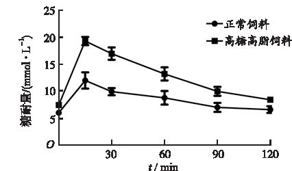
图1
高脂饲养诱导胰岛素抵抗大鼠的糖耐量变化(
Fig.1
Variation of glucose tolerance in high fat-induced diabetes rats with insulin resistance(
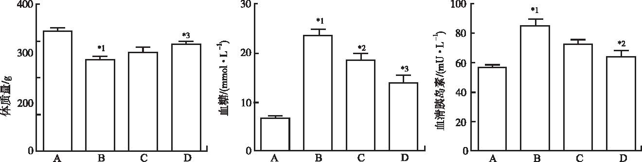
图2
4组大鼠血糖,体质量及血清胰岛素水平比较(
A.正常对照组;B.模型对照组;C.药根碱小剂量组;D.药根碱大剂量组;与正常对照组比较,*1
Fig.2
Comparison of blood glucose, body weight and serum insulin among four groups of rats(
A.normal control group;B.model control group;C.low-dose jatrorrhizine group;D.high-dose jatrorrhizine group;compared with normal control group,*1
正常对照组血清中IL-1β为(92.3±4.3) pg·mL-1,模型对照组为(152.4± 20.0) pg·mL-1,IL-1β显著升高,与正常对照组比较差异有统计学意义(
Western blotting实验结果见
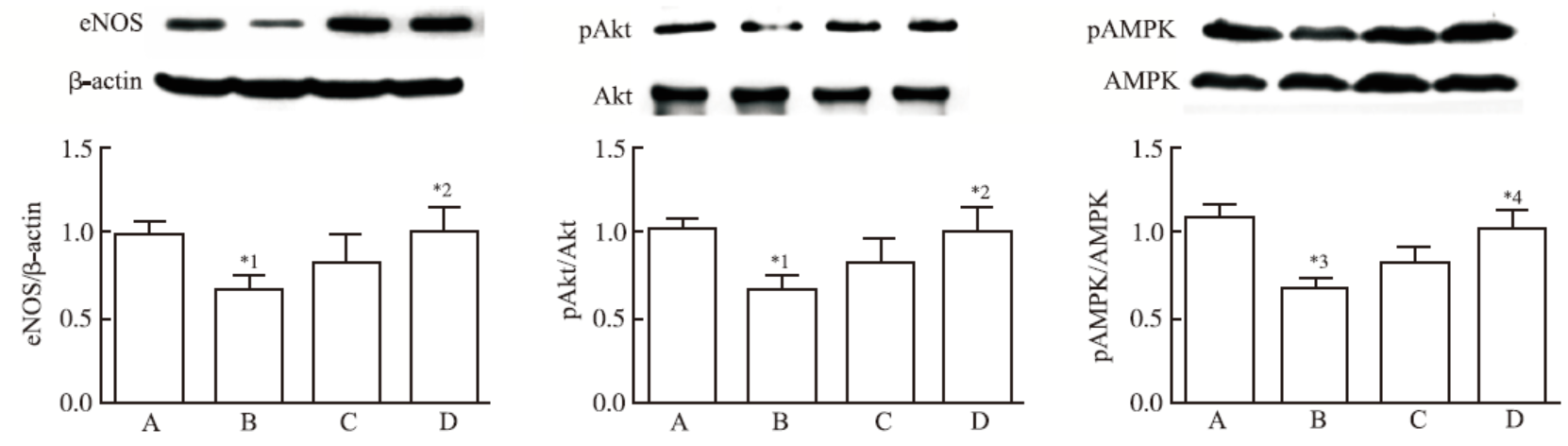
图3
4组大鼠血管eNOS信号通路比较(
A.正常对照组;B.模型对照组;C.药根碱小剂量组;D.药根碱大剂量组;与正常对照组比较,*1
Fig.3
Comparison of vascular eNOS signaling pathway among four groups of rats(
A.normal control group;B.model control group;C.low-dose jatrorrhizine group;D.high-dose jatrorrhizine group;compared with normal control group,*1
根据国际糖尿病联盟(international diabetes federation,IDF)在2015年世界糖尿病大会上发布数据,世界范围内患有糖尿病成年人4.15亿人。中国糖尿病发生例数最多,随着糖尿病发病率的日益增长,糖尿病相关心血管疾病并发症威胁人类健康。这些并发症包括肾小球、视网膜、周围神经等微血管病变,还有大血管病变,如动脉粥样硬化、心肌缺血、卒中和末端血管病变等,从而导致失明、肾衰竭、心肌梗死,甚至截肢等严重后果[6]。糖尿病并发症多,究其原因与长期受到高血糖刺激引起血管内皮功能紊乱密切相关[7-8],血管内皮细胞在体内具有多种生物功能,如分泌、合成、代谢和免疫等,不仅与血管张力通透性和凝血相关,还可促进炎症因子,如TNF-α、IL-6、IL-1β等分泌,而在炎症反应中起重要作用,从而损伤内皮细胞,导致内皮功能紊乱。其中以一氧化氮(NO)生物利用度受损为主的内皮功能紊乱是血管疾病重要发病机制。
NO是由血管内皮细胞分泌的eNOS调控生成,具有重要的生理功能。一氧化氮合酶在生物体内已发现有3种亚型,分别是神经细胞分泌nNOS,诱导型iNOS和主要由血管内皮细胞分泌eNOS,各有不同生理功能。eNOS主要作用通过
AMPK是体内能量代谢的关键调节激酶,在控制葡萄糖转运,脂肪酸氧化和脂质合成等过程发挥重要作用[10],活化的AMPK不管在体外还是在体内都证明能激活eNOS[11-12]。Akt、AMPK蛋白水平的正常表达在维持体内糖代谢内平衡上非常重要,表达失衡通常会导致肥胖和糖尿病的发生。所以这个通路的蛋白被认为可能成为下一个治疗糖尿病的靶向分子,如二甲双胍能激活AMPK活性,也证明具有激活Akt的作用[13]。并且以这两个蛋白为靶点的药物已经实验性治疗肿瘤[13-14]。
黄连主要活性成分为小檗碱和药根碱。小檗碱对糖尿病肾病、内皮损伤、脑病等并发症的缓解与治疗作用明显[15-16],并且毒副作用小。小檗碱的水溶性较差,口服吸收较少。研究表明,药根碱与小檗碱一样具有相似的药理活性,所以药根碱具有潜在的治疗血糖异常的作用。WU等[17]研究发现药根碱安全性远大于小檗碱,具有良好的调节血脂效果。
本实验结果显示,糖尿病大鼠血清IL-1β、TNF-α水平升高,血管组织Akt、AMPK、eNOS表达也明显下降,经药根碱治疗后,蛋白水平有所改善,提示药根碱可能通过Akt/AMPK/eNOS信号通路恢复血管内皮功能紊乱,使eNOS表达增加,炎症反应减轻,从而对糖尿病大鼠产生保护作用。
The authors have declared that no competing interests exist.
| [1] |
|
| [2] |
Malignant melanoma is the most aggressive form of skin cancer. Although various antimelanoma approaches have been used in the clinics to treat the disease over the last three decades, none of the drugs significantly prolonged the survival of metastatic melanoma patients; hence, effective drugs against metastatic melanoma are highly desired. In this study, we explored an antimetastatic melanoma agent derived from traditional Chinese medicinal herbs and found that jatrorrhizine hydrochloride (JH), an active component of the traditional Chinese medicinal herb Coptis chinensis, inhibited the proliferation and neovascularization of C8161 human metastatic melanoma cells. JH suppressed C8161 cell proliferation in a dose-dependent manner, with a half-maximal inhibitory concentration of 47.4卤1.6 渭mol/l; however, it did not induce significant cellular apoptosis at doses up to 320 渭mol/l. Mechanistic studies showed that JH-induced C8161 cell cycle arrest at the G0/G1 transition, which was accompanied by overexpression of the cell cycle-suppressive genes p21 and p27 at higher doses. Moreover, JH reduced C8161 cell-mediated neovascularization in vitro and in vivo and impeded the expression of the gene for VE-cadherin, a key protein in tumor vasculogenic mimicry and angiogenesis. Taken together, the effective inhibitory effects of JH on metastatic melanoma cell proliferation and neovascularization with low toxicity suggest that JH is a potential new antimelanoma drug candidate.
[本文引用:1]
|
| [3] |
目的:观察大黄及其分别与枳实、黄连、牡丹、桃仁和甘遂配伍后对小鼠足肿胀的抗炎作用,研究大黄药对的配伍规律。方法小鼠分别灌胃给予15,30 g?kg-1大黄及大黄枳实、大黄黄连、大黄牡丹、大黄桃仁及大黄甘遂提取物,早晚各1次,连续7 d,采用角叉菜胶致小鼠足肿胀法观察其抗炎作用;采用经典急性毒性实验法比较大黄及其5个药对的急性毒性。结果与模型对照组比较,大黄及其5个药对提取物均能抑制角叉菜胶引起的小鼠足肿胀炎症反应,并可降低小鼠炎症组织丙二醛(MDA)和一氧化氮(NO)含量,提高其超氧化物歧化酶(SOD)活力(P<0.05)。急性毒性实验中各组小鼠无法测出半数致死量(LD50),测得最大给药量按含生药量计算大黄、大黄枳实、大黄黄连、大黄牡丹、大黄桃仁及大黄甘遂分别为145.33,142.30,117.53,103.45,113.09,182.36 g?kg-1,相当于人临床日用量的581,569,470,418,452及729倍。结论大黄各药对配伍后均具有抗炎作用,大黄甘遂抗炎作用最优,其次为大黄黄连和大黄牡丹。大黄经过配伍后毒副作用降低。
[本文引用:0]
|
| [4] |
目的:比较研究黄连粉末及煎煮液中的盐酸小檗碱(BER)和盐酸药根碱(JAT)在2型糖尿病大鼠药动学变化,揭示古方应用黄连治疗消渴症选择丸剂的科学内涵。方法:将黄连粉末及其煎煮液灌胃于2型糖尿病大鼠,应用RP-HPLC测定血浆中小檗碱、JAT的血药浓度,DAS2.0药动学软件计算药动学参数。结果:BER、JAT的药动学过程均为二室开放模型;与煎煮液组比较,黄连粉末口服给药组BER、JAT AUC均显著增加,V1/F显著减小,CL/F显著降低,Tmax提前,t1/2Ka及t1/2α缩短,t1/2β延长。结论:与煎煮液比较,黄连粉末效应物质生物利用度更高,吸收更快,消除更慢,提示黄连用于治疗2型糖尿病选择粉末形态如丸剂入药优于煎剂入药。
[本文引用:1]
|
| [5] |
Jatrorrhizine, a protoberberine alkaloid derived from Coptis chinensis, is currently under investigation as a natural gastric prokinetic drug candidate. In vitro and in vivo studies were conducted to characterize its pharmacokinetics and metabolism. After intravenous administration, the plasma concentration kinetics and major metabolites in rats were investigated. The metabolic kinetics, key cytochrome P450 enzymes and UDP-glucuronosyltransferase isoforms (UGTs) of jatrorrhizine were studied in rat liver microsomes (RLMs). After intravenous administration, plasma jatrorrhizine concentrations showed a biphasic decline, dose-independent clearance and half-life of terminal elimination phase, and a relatively large distribution volume. The metabolic pathway for the conversion of jatrorrhizine was important for its elimination. In addition, the demethylated and glucuronidated products were found to be the major metabolites in rats. The enzyme kinetics for both demethylation and glucuronidation were fitted to the hyperbolic Michaelis-Menten equation in RLMs. CYP3A1/2 and CYP2D2 were mainly responsible for demethylation, and UGT 1A1 and 1A3 were responsible for glucuronidation in RLMs. The metabolic properties of jatrorrhizine suggest multiple metabolic pathways. These results will contribute to promote further research and development of jatrorrhizine. Copyright 漏 2012 John Wiley & Sons, Ltd.
[本文引用:1]
|
| [6] |
DOI:10.1038/414813a
URL
[本文引用:1]
|
| [7] |
Abstract Although the etiology of vascular dysfunction in diabetes has been extensively investigated in both humans as well as animal models of human diabetes, the relative importance of the cellular pathways involved is still not fully understood. In this review, we focus on reviewing the literature that provides insights into how an acute exposure to hyperglycemia results in a dysregulation of endothelial nitric oxide synthase function, the subsequent downstream effects of endothelial nitric oxide synthase dysregulation, and the development of endothelial dysfunction. Copyright (c) 2010 American Society of Hypertension. Published by Elsevier Inc. All rights reserved.
[本文引用:1]
|
| [8] |
Hyperglycemia and insulin resistance are key players in the development of atherosclerosis and its complications. A large body of evidence suggest that metabolic abnormalities cause overproduction of reactive oxygen species (ROS). In turn, ROS, via endothelial dysfunction and inflammation, play a major role in precipitating diabetic vascular disease. A better understanding of ROS-generating pathways may provide the basis to develop novel therapeutic strategies against vascular complications in this setting. Part I of this review will focus on the most current advances in the pathophysiological mechanisms of vascular disease: (i) emerging role of endothelium in obesity-induced insulin resistance; (ii) hyperglycemia-dependent microRNAs deregulation and impairment of vascular repair capacities; (iii) alterations of coagulation, platelet reactivity, and microparticle release; (iv) epigenetic-driven transcription of ROS-generating and proinflammatory genes. Taken together these novel insights point to the development of mechanism-based therapeutic strategies as a promising option to prevent cardiovascular complications in diabetes.
[本文引用:1]
|
| [9] |
Functional consequences of impaired endothelial nitric oxide synthase (eNOS) activity causing organ-specific abnormalities on a diabetic setting are not completely understood. In this study, we extensively characterized a diabetic mouse model (lepr(db/db)) in which eNOS expression is genetically disrupted (eNOS-/-). The eNOS-/-/ lepr(db/db) double-knockout (DKO) mice developed obesity, hyperglycemia, hyperinsulinemia and hypertension. Analysis of tissues from DKO mice showed large islets in the pancreas and fat droplets in hepatocytes. Interestingly, the aorta was normal and atherogenic lesions were not observed. Abnormalities in the aorta including poor re-endothelialization and increased medial wall thickness were evident only in response to deliberate injury. In contrast, significant glomerular capillary damage in the kidney was identified, with DKO mice demonstrating a robust diabetic nephropathy similar to human disease. The vascular and renal impairments in DKO mice were pronounced despite lower fasting plasma glucose levels compared to lepr(db/db) mice, indicating that eNOS is a critical determinant of hyperglycemia-induced organ-specific complications and their severity in diabetes. Results provide the first evidence that absence of eNOS in diabetes has a greater deleterious effect on the renal microvasculature than on the larger aortic vessel. The DKO model may suggest novel therapeutic strategies to prevent both vascular and renal complications of diabetes.
[本文引用:1]
|
| [10] |
In cells expressing only the Glut1 isoform of glucose transporters, we have shown that glucose transport is markedly stimulated in response to hypoxia or inhibition of oxidative phosphorylation, conditions that would be expected to cause a stimulation of AMP-activated protein kinase (AMPK) activity. In the present study we tested the hypothesis that the stimulation of AMPK activity might be accompanied by an enhancement of Glut1-mediated glucose transport. Exposure of Clone 9 cells, 3T3-L1 preadipocytes, and C2C12 myoblasts (cells that express only the Glut1 isoform) to 5-aminoimidazole-4-carboxamide ribonucleoside (AICAR), an adenosine analog that stimulates AMPK activity, resulted in a marked increase in the rate of glucose transport (ranging from four- to sixfold) that was accompanied by activation of AMPK. This stimulation of AMPK activity was associated with an increase in the phosphorylation of threonine 172 on the activation loop of its alpha subunit, with the predominant change being in the alpha-a isoform. Exposure of Clone 9 cells to 5-iodotubercidin, an inhibitor of adenosine kinase, abolished the accumulation of AICAR-5'-monophosphate (ZMP), stimulation of AMPK, and the enhancement of glucose transport in response to AICAR. There was no significant increase in the content of Glut1 in plasma membranes of Clone 9 cells exposed to AICAR, We conclude that stimulation of AMPK activity is associated with enhancement of Glut1-mediated glucose transport, and that the glucose! transport response is mediated by activation of Glut1 transporters preexisting in the plasma membrane. (C) 2000 Academic Press. [References: 30]
[本文引用:1]
|
| [11] |
Abstract AIM: To investigate the effects of bezafibrate on the proliferation and differentiation of osteoblastic MC3T3-E1 cells, and to determine the signaling pathway underlying the effects. METHODS: MC3T3-E1 cells, a mouse osteoblastic cell line, were used. Cell viability and proliferation were examined using MTT assay and colorimetric BrdU incorporation assay, respectively. NO production was evaluated using the Griess reagent. The mRNA expression of ALP, collagen I, osteocalcin, BMP-2, and Runx-2 was measured using real-time PCR. Western blot analysis was used to detect the expression of AMPK and eNOS proteins. RESULTS: Bezafibrate increased the viability and proliferation of MC3T3-E1 cells in a dose- and time-dependent manner. Bezafibrate (100 0204mol/L) significantly enhanced osteoblastic mineralization and expression of the differentiation markers ALP, collagen I and osteocalcin. Bezafibrate (100 0204mol/L) increased phosphorylation of AMPK and eNOS, which led to an increase of NO production by 4.08-fold, and upregulating BMP-2 and Runx-2 mRNA expression. These effects could be blocked by AMPK inhibitor compound C (5 0204mol/L), or the PPAR0205 inhibitor GSK0660 (0.5 0204mol/L), but not by the PPAR02± inhibitor MK886 (10 0204mol/L). Furthermore, GSK0660, compound C, or N(G)-nitro-L-arginine methyl ester hydrochloride (L-NAME, 1 mmol/L) could reverse the stimulatory effects of bezafibrate (100 0204mol/L) on osteoblast proliferation and differentiation, whereas MK886 only inhibited bezafibrate-induced osteoblast proliferation. CONCLUSION: Bezafibrate stimulates proliferation and differentiation of MC3T3-E1 cells, mainly via a PPAR0205-dependent mechanism. The drug might be beneficial for osteoporosis by promoting bone formation.
[本文引用:1]
|
| [12] |
Endothelial dysfunction is a critical factor during the initiation of cardiovascular complications in diabetes. Berberine can ameliorate endothelial dysfunction induced by diabetes. However, the underlying mechanisms remain unclear. The aim of this study was to investigate the protective effect and mechanism of berberine on palmitate-induced endothelial dysfunction in human umbilical vein endothelial cells (HUVECs). The cell viability of HUVECs was determined by MTT assays. Nitric oxide (NO) level and production of reactive oxygen species (ROS) were determined in supernatants or in the cultured HUVECs. The mRNA level of endothelial nitric oxide synthase (eNOS) was measured by RT-PCR, and the protein levels of eNOS, p-eNOS, Akt, p-Akt, AMPK, p-AMPK, and NADPH oxidase (NOX4) were analyzed. The results demonstrated that berberine significantly elevated NO levels and reduced the production of ROS. The expressions of eNOS were significantly increased, while NOX4 protein expression was decreased in berberine-treated HUVECs. Moreover, berberine upregulated the protein expression of AMPK and p-AMPK in palmitate-treated HUVECs, but had no effect on the levels of Akt. Therefore, berberine ameliorates palmitate-induced endothelial dysfunction by upregulating eNOS expression and downregulating expression of NOX4. This regulatory effect of berberine may be related to the activation of AMPK.
[本文引用:1]
|
| [13] |
Metformin is an antidiabetic drug with anticancer properties, which mainly acts through induction of AMP-activated protein kinase (AMPK). In the present study we investigated the influence of metformin on the in vitro anticancer activity of the well-known chemotherapeutic agent cisplatin. Cell viability was determined by MTT and LDH release assay, oxidative stress and apoptosis (caspase activation, DNA fragmentation, and phosphatidylserine exposure) were assessed by flow cytometry, while activation of AMPK and Akt was analyzed by immunoblotting. Although metformin reduced the number of tumour cells when applied alone, it surprisingly antagonized the cytotoxicity of cisplatin towards U251 human glioma, C6 rat glioma, SHSY5Y human neuroblastoma, L929 mouse fibrosarcoma and HL-60 human leukemia cell lines. Only in B16 mouse melanoma cells metformin augmented the cytotoxicity of cisplatin. In U251 glioma cells metformin suppressed cisplatin-induced apoptotic cell death through inhibition of oxidative stress and caspase activation. The observed cytoprotection was apparently AMPK-independent, as metformin did not further increase cisplatin-induced AMPK activation in U251 cells and other pharmacological AMPK activators failed to block cisplatin-mediated apoptosis. On the other hand, metformin induced Akt activation in cisplatin-treated cells and Akt inhibitor 10-DEBC hydrochloride or phosphoinositide 3-kinase/Akt inhibitor LY294002 abolished metformin-mediated antioxidant and antiapoptotic effects. In conclusion, the antidiabetic drug metformin reduces cisplatin in vitro anticancer activity through AMPK-independent upregulation of Akt survival pathway. These data warrant caution when considering metformin for treatment of diabetic cancer patients receiving cisplatin or as a potential adjuvant in cisplatin-based chemotherapeutic regimens.
[本文引用:2]
|
| [14] |
The aberrant regulation of phosphatidylinositide 3-kinases (PI3-K)/Akt, AMP-activated protein kinase (AMPK) and mammalian target of rapamycin (m-TOR) signaling pathways in cancer has prompted significant interest in the suppression of these pathways to treat cancer. Caffeic acid (CA) has been reported to possess important anti-inflammatory actions. However, the molecular mechanisms by which CA derivatives including caffeic acid phenethyl ester (CAPE) and caffeic acid phenylpropyl ester (CAPPE), exert inhibitory effects on the proliferation of human colorectal cancer (CRC) cells have yet to be elucidated. CAPE and CAPPE were evaluated for their ability to modulate these signaling pathways and suppress the proliferation of CRC cells both in vitro and in vivo. Anti-cancer effects of these CA derivatives were measured by using proliferation assays, cell cycle analysis, western blotting assay, reporter gene assay and immunohistochemical (IHC) staining assays both in vitro and in vivo. This study demonstrates that CAPE and CAPPE exhibit a dose-dependent inhibition of proliferation and survival of CRC cells through the induction of G0/G1 cell cycle arrest and augmentation of apoptotic pathways. Consumption of CAPE and CAPPE significantly inhibited the growth of colorectal tumors in a mouse xenograft model. The mechanisms of action included a modulation of PI3-K/Akt, AMPK and m-TOR signaling cascades both in vitro and in vivo. In conclusion, the results demonstrate novel anti-cancer mechanisms of CA derivatives against the growth of human CRC cells. CA derivatives are potent anti-cancer agents that augment AMPK activation and promote apoptosis in human CRC cells. The structure of CA derivatives can be used for the rational design of novel inhibitors that target human CRC cells.
[本文引用:1]
|
| [15] |
|
| [16] |
The incidence of is increasing rapidly worldwide, and the of novel anti-diabetic drugs is emerging. However, most anti-diabetic drugs cannot be used in patients with hepatic dysfunction, , and , which makes pharmacological therapy of complicated. Despite continued introduction of novel agents, the search for an ideal drug that is useful as both a and to reduce -related complications remains elusive. is an extract that has shown promise as a in the management of in animal and studies. Mechanistic studies have revealed beneficial effects of on -related complications. Although there have been few clinical reports of the anti-diabetic effects of , little documentation of adverse effects in positions it as a potential candidate drug to treat . In the present review, the anti-diabetic mechanism of , its effect on -related complications, and its recent use in clinical studies is highlighted. In addition, we summarize the different treatments for in adults and children.
[本文引用:1]
|
| [17] |
In conclusion, jatrorrhizine was a safe and potential antihypercholesterolemic agent from RC which could improve the utilization and excretion of cholesterol by up-regulating the mRNA and protein expression of LDLR and CYP7A1.
[本文引用:1]
|
