Objective To investigate the influence of curcumin and its analogue H8 on glucose and lipid metabolism disorder in db/db mice. Methods The type 2 diabetes mouse model (db/db mice) was intragastrically administrated with curcumin and analogue H8 for 8 weeks.The blood biochemical indexes were measured.The expression of PEPCK and G6Pase mRNA was detected by real-time PCR in liver tissues.The expression of PEPCK and G6Pase protein was detected by Western blotting. Results Curcumin analogue H8 reduced blood glucose and lipids in db/db mice (P<0.01) and improved liver function related enzymes significantly.The levels of PEPCK and G6Pase mRNA in db/db mice were significantly decreased (P<0.01) and the expression levels of PEPCK and G6Pase protein were significantly decreased (P<0.01). Conclusion Curcumin analogue H8 improves the glucose and lipid metabolism disorder in db/db mice,and it is related to inhibiting the expression of PEPCK and G6Pase gene and protein.
Fig.1
Comparsion of the mRNA expression of PEPCK and G6Pase in livers among six groups of db/db mice A. expression of PEPCK mRNA; B.expression of G6Pase mRNA;compared with Con group,*1P<0.01;compared with Dia group,*2P<0.01 ,*3P<0.05
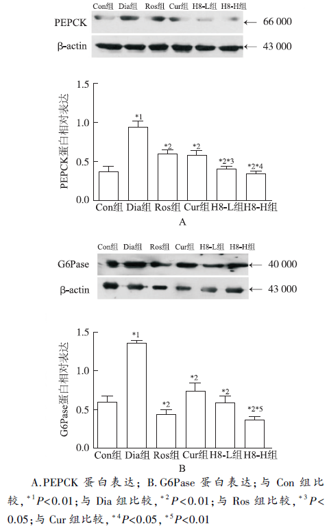
Fig.2
Comparison of the protein expression of PEPCK and G6Pase in livers among six groups of db/db mice A.expression of PEPCK protein;B.expression of G6Pase protein;compared with Con group,*1P<0.01;compared with Dia group,*2P<0.01;compared with Ros group,*3P<0.05;compared with Cur group,*4P<0.05,*5P<0.01
GUPTA SC,KISMALIG,AGGARWAL BB.Curcumin,a component of turmeric: from farm to pharmacy[J].Biofactors,2013,39(1):2-13.
Curcumin, an active polyphenol of the golden spice turmeric, is a highly pleiotropic molecule with the potential to modulate the biological activity of a number of signaling molecules. Traditionally, this polyphenol has been used in Asian countries to treat such human ailments as acne, psoriasis, dermatitis, and rash. Recent studies have indicated that curcumin can target newly identified signaling pathways including those associated with microRNA, cancer stem cells, and autophagy. Extensive research from preclinical and clinical studies has delineated the molecular basis for the pharmaceutical uses of this polyphenol against cancer, pulmonary diseases, neurological diseases, liver diseases, metabolic diseases, autoimmune diseases, cardiovascular diseases, and numerous other chronic diseases. Multiple studies have indicated the safety and efficacy of curcumin in numerous animals including rodents, monkeys, horses, rabbits, and cats and have provided a solid basis for evaluating its safety and efficacy in humans. To date, more than 65 human clinical trials of curcumin, which included more than 1000 patients, have been completed, and as many as 35 clinical trials are underway. Curcumin is now used as a supplement in several countries including the United States, India, Japan, Korea, Thailand, China, Turkey, South Africa, Nepal, and Pakistan. In this review, we provide evidence for the pharmaceutical uses of curcumin for various diseases. 2013 BioFactors, 39(1):2-13, 2013
AGGARWAL BB,GUPTA SC,SUNGB,et al.Curcumin:an orally bioavailable blocker of TNF and other pro-inflammatory biomarkers[J].Br J Pharmacol,2013,169(8) :1672-1692.
TNFs are major mediators of inflammation and inflammation-related diseases, hence, the United States Food and Drug Administration (FDA) has approved the use of blockers of the cytokine, TNF-, for the treatment of osteoarthritis, inflammatory bowel disease, psoriasis and ankylosis. These drugs include the chimeric TNF antibody (infliximab), humanized TNF- antibody (Humira) and soluble TNF receptor-II (Enbrel) and are associated with a total cumulative market value of more than $20 billion65a year. As well as being expensive ($1565000–2065000 per person per year), these drugs have to be injected and have enough adverse effects to be given a black label warning by the FDA. In the current report, we describe an alternative, curcumin (diferuloylmethane), a component of turmeric (Curcuma longa) that is very inexpensive, orally bioavailable and highly safe in humans, yet can block TNF- action and production in in vitro models, in animal models and in humans. In addition, we provide evidence for curcumin's activities against all of the diseases for which TNF blockers are currently being used. Mechanisms by which curcumin inhibits the production and the cell signalling pathways activated by this cytokine are also discussed. With health-care costs and safety being major issues today, this golden spice may help provide the solution.Linked ArticlesThis article is part of a themed section on Emerging Therapeutic Aspects in Oncology. To view the other articles in this section visit http://dx.doi.org/10.1111/bph.2013.169.issue-8
YUANX,LIH,BAIH,et al.Synthesis of novel curcumin analogues for inhibition of 11 beta-hydroxysteroid dehydrogenase type 1 with antidiabetic properties[J].Eur J Med Chem,2014,77:223-230.
In the present study, a series of mono-carbonyl analogues of curcumin were designed and synthesized by deleting the reactive beta-diketone moiety, which is responsible for the pharmacokinetic limitation of curcumin. We demonstrated that 4 of 9 curcumin analogues were selective inhibitors of human and rodent 11β-HSD1. The level of this inhibitor was 4–20 times more than that of curcumin. Curcumin analogues weakly inhibited 11β-HSD2, and further analyses revealed that these compounds were highly selective, favoring 11β-HSD1. These 4 curcumin analogues are potential therapeutic agents for type-2 diabetes by targeting 11β-HSD1. The compound 8 displays anti-diabetic properties in diabetic mice induced by streptozocin and high-fat-diet (STZHFD).
KIMT,DAVISJ,ZHANG AJ,et al.Curcumin activates A-MPK and suppresses gluconeogenic gene expression in hepatoma cells[J].Biochem Biophys Res Commun,2009,388(2):377-382.
Curcumin, the bioactive component of curry spice turmeric, and its related structures possess potent anti-oxidant and anti-inflammatory properties. Several lines of evidence suggest that curcumin may play a beneficial role in animal models of diabetes, both by lowering blood glucose levels and by ameliorating the long-term complications of diabetes. However, current understanding of the mechanism of curcumin action is rudimentary and is limited to its anti-oxidant and anti-inflammatory effects. In this study we examine potential anti-diabetic mechanisms of curcumin, curcumin C3 complex 庐; , and tetrahydrocurcuminoids (THC). Curcuminoids did not exert a direct effect on receptor tyrosine kinase activity, 2-deoxy glucose uptake in L6-GLUT4myc cells, or intestinal glucose metabolism measured by DPP4/伪-glucosidase inhibitory activity. We demonstrate that curcuminoids effectively suppressed dexamethasone-induced phosphoenol pyruvate carboxy kinase (PEPCK) and glucose6-phosphatase (G6Pase) in H4IIE rat hepatoma and Hep3B human hepatoma cells. Furthermore, curcuminoids increased the phosphorylation of AMP-activated protein kinase (AMPK) and its downstream target acetyl-CoA carboxylase (ACC) in H4IIE and Hep3B cells with 400 times (curcumin) to 100,000 times (THC) the potency of metformin. These results suggest that AMPK mediated suppression of hepatic gluconeogenesis may be a potential mechanism mediating glucose-lowering effects of curcuminoids.
FENGY,HUANG SL,DOUW,et al.A natural product,sel-ectively inhibits 11beta-hydroxysteroid dehydrogenase type 1 and ameliorates metabolic disorder in diet-induced obese mice[J].Br J Pharmacol,2010,161(1):113-126.
Abstract Top of page Abstract Introduction Methods Results Discussion Acknowledgements Conflict of interest References Supporting Information BACKGROUND AND PURPOSE 11β-Hydroxysteroid dehydrogenase type 1 (11β-HSD1) is an attractive therapeutic target of type 2 diabetes and metabolic syndrome. Emodin, a natural product and active ingredient of various Chinese herbs, has been demonstrated to possess multiple biological activities. Here, we investigated the effects of emodin on 11β-HSD1 and its ability to ameliorate metabolic disorders in diet-induced obese (DIO) mice. EXPERIMENTAL APPROACH Scintillation proximity assay was performed to evaluate inhibition of emodin against recombinant human and mouse 11β-HSDs. The ability of emodin to inhibit prednisone- or dexamethasone-induced insulin resistance was investigated in C57BL/6J mice and its effect on metabolic abnormalities was observed in DIO mice. KEY RESULTS Emodin is a potent and selective 11β-HSD1 inhibitor with the IC 50 of 186 and 86nM for human and mouse 11β-HSD1, respectively. Single oral administration of emodin inhibited 11β-HSD1 activity of liver and fat significantly in mice. Emodin reversed prednisone-induced insulin resistance in mice, whereas it did not affect dexamethasone-induced insulin resistance, which confirmed its inhibitory effect on 11β-HSD1 in vivo . In DIO mice, oral administration of emodin improved insulin sensitivity and lipid metabolism, and lowered blood glucose and hepatic PEPCK, and glucose-6-phosphatase mRNA. CONCLUSIONS AND IMPLICATIONS This study demonstrated a new role for emodin as a potent and selective inhibitor of 11β-HSD1 and its beneficial effects on metabolic disorders in DIO mice. This highlights the potential value of analogues of emodin as a new class of compounds for the treatment of metabolic syndrome or type 2 diabetes.
SURESH BP,SRINIVASANK.Hypolipidemic action of curcumin,the active principle of turmeric (curcuma longa)in streptozotocin induced diabetic rats[J].Mol Cell Biochem,1997,166(1-2) :169-173.
Streptozotocin-induced diabetic rats were maintained on 0.5% curcumin containing diet for 8 weeks. Blood cholesterol was lowered significantly by dietary curcumin in these diabetic animals. Cholesterol decrease was exclusively from LDL- VLDL fraction. Significant decrease in blood triglyceride and phospholipids was also brought about by dietary curcumin in diabetic rats. In a parallel study, wherein diabetic animals were maintained on a high cholesterol diet, the extents of hypercholesterolemia and phospholipidemia were still higher compared to those maintained on control diet. Curcumin exhibited lowering of cholesterol and phospholipid in these animals also. Liver cholesterol, triglyceride and phospholipid contents were elevated under diabetic conditions. Dietary curcumin showed a distinct tendency to counter these changes in lipid fractions of liver. This effect of curcumin was also seen in diabetic animals maintained on high cholesterol diet. Dietary curcumin also showed significant countering of renal cholesterol and triglycerides elevated in diabetic rats. In order to understand the mechanism of hypocholesterolemic action of dietary curcumin, activities of hepatic cholesterol-7a-hydroxylase and HMG CoA reductase were measured. Hepatic cholesterol-7a-hydroxylase activity was markedly higher in curcumin fed diabetic animals suggesting a higher rate of cholesterol catabolism. (Mol Cell Biochem 166: 169-175, 1997)
DU LY,CUI YL,CHEN EQ,et al.Correlation between the suppressor of cytokine signaling-1 and 3 and hepatitis B virus: possible roles in the resistance to interferon treatment[J].Virol J,2014,11(2):51.
Background The suppressor of cytokine signaling family (SOCS) is an important negative regulator in the JAK-STAT signaling pathway. This study was designed to explore the correlation between SOCS-1, 2 and 3, Hepatitis B Virus (HBV) and interferon (IFN), and the relationship between SOCS and IFN therapeutic efficacy. Methods Four types of mouse models were established. Mice were administered with HBV replicative plasmid pHBV4.1 and IFN inducer Poly IC (Group A), pHBV4.1 (Group B), Poly IC (Group C) and saline (Group D), respectively. Liver tissues were harvested from the mice and SOCS expression was determined. Meanwhile, patients with chronic hepatitis B (CHB) were treated with pegylated interferon ??-2b for 24-48 weeks. Liver biopsy was collected and the baseline SOCS expression was determined. Serum assay was performed for efficacy evaluation and correlation analysis. Results In animal studies, the expression level of SOCS-1 and 3 was found in the descending order of B, A, C and D. The difference between Group B and D suggested that HBV could induce SOCS. The difference between Group A and C suggested that HBV could still induce SOCS with up-regulated endogenous IFN. The difference between Group C and D suggested that ploy IC could induce SOCS, while the difference between Group B and A suggested that Poly IC might have a stronger inhibition effect for SOCS. There was no difference in SOCS-2 expression. In clinical studies, eight of twenty-four enrolled patients achieved either complete or partial therapeutic response. The expression of both SOCS-1 and 3 was higher in CHB patients than in normal controls. The baseline HBV-DNA level was positively correlated with SOCS-1 and 3. The age, viral genotype, HBVDNA, SOCS-1 and SOCS-3 were found to be related to IFN efficacy. Conclusion HBV could induce both SOCS-1 and 3 expression regardless of endogenous IFN level. Elevated IFN could directly up-regulate SOCS-1 and 3 expression, but it could also indirectly down-regulate SOCS-1 and 3 expression by inhibiting HBV replication. HBV might play a more important role in the SOCS up-regulation than IFN, a possible reason why patients with high HBV viral load encounter poor efficacy of IFN treatment.
MALICKAB,SKOSKIEWICZ-MALINOWSKAK,KACZMA-REKU.Salivary lactate dehydrogenase and aminotransferases in diabetic patients[J].Medicine (Baltimore),2016,95(47):e5211
Abstract Diabetes mellitus (DM) is a group of metabolic diseases resulting from impaired insulin secretion and/or action. DM is characterized by hyperglycemia that can lead to the dysfunction or damage of organs, including the salivary glands.The aim of this study was to compare the levels of salivary lactate dehydrogenase (LDH), aspartate aminotransferase (AST), and alanine aminotransferase (ALT) in diabetic patients.The study was approved by the Bioethics Committee of Wroclaw Medical University (Poland). The study comprised 90 adults of both sexes, aged 21 to 57 years. The patients were divided into 3 groups: type 1 diabetics (D1), type 2 diabetics (D2), and a healthy control group (C). Each group consisted of 30 age- and sex-matched subjects. Total protein (P, by Lowry method), LDH, AST, ALT (with Alpha Diagnostics kits), and salivary flow rate were measured in unstimulated mixed saliva. The level of glycosylated hemoglobin (HbA1c) was measured with DCA 2000 Reagent Kit. The obtained data were analyzed using the Mann-Whitney U test and the Spearman rank at a significance level of P66<660.05 with the use of STATISTICA 9.0 software.In comparison with C, D1 presented a significantly higher activity of LDH (P66<660.001), AST (P66<660.001), and ALT (P66<660.01), whereas D2 indicated higher levels of LDH (P66<660.001) and ALT (P66<660.05) compared with C. Comparing D1 to D2, approximately 3-fold higher activity of AST (P66<660.01) and approximately 4.5-fold higher activity of ALT (P66<660.01) was observed.Higher levels of salivary LDH, AST, and ALT in D1 compared with D2 and C confirm that salivary glands of D1 might be attributed to autoimmunological damage associated with the pathomechanism of DM.
CHENS,GUOX,CHENY,et al.Prevalence of abnormal serum liver enzymes in patients with type 2 diabetes mellitus: a cross-sectional study from China[J].Postgrad Med,2016,128(8):770-776.
Objectives: This cross-sectional study aimed to determine the prevalence of elevated alanine aminotransferase (ALT) and aspartate aminotransferase (AST) in Chinese type 2 diabetic patients and identify contributing risk factors.

 , 李玲玉
, 李玲玉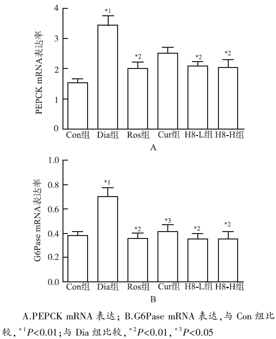
