LI Lisheng1,, LUO Yunmei1, LIU Juan2, FU Xiaoxia1, YANG Danli1, XIE Xiaolong1
1.Department of Pharmacology & Key Lab of Basic Pharmacology of Education Ministry,Joint International Research Lab of Ethnomedicin of Ministry of Education, Zunyi Medical College,Zunyi 563000,China
2.Institute of Clinical Medcine,Affiliated Hospital of Zunyi Medical College,Zunyi 563000,China
Objective To investigate the effects of icariin (ICA) on partial vasoactive substances in monocrotaline (MCT)-induced pulmonary arterial hypertension (PAH) rat model. Methods Sixty male SD rats were randomly divided into five groups:normal control group,model control group,ICA low-,middle- and high-dose(20,40,80 mg·kg-1·d-1) group,12 rats in each group.Except for normal control group, the rats were injected with MCT (50 mg·kg-1·d-1) to establish PAH model.After 1 week MCT-injection,ICA was given by intragastric administration for 3 weeks according to different groups.Mean pulmonary artery pressure (mPAP) was recorded through catheter connected with Power Lab system.Except for normal control group, the right ventricular hypertrophy index (RVHI) was calculated using formula:right ventricle weight/the weight of left ventricle with septum×100%.The morphology of lung artery was assessed by HE staining.Concentration of angiotensinⅡ(AngⅡ),endothelin (ET),prostaglandine F2α (PGF2α),thromboxane A2(TXA2) and prostacyclin (PGI2) in serum was measured by ELISA kit assay.The protein levels of angiotensin converting enzyme (ACE),cyclooxygenase-2 (COX-2) and thromboxane A2 synthetase (TXAS) were analyzed by Western blotting,expression of ACE,COX-2 and TXAS mRNA was measured by real time RT-PCR. Results Compared with the normal control group,mPAP [(48.5±5.2) mmHg] and RVHI (33.3±3.8)%in model control group were significantly increased (P<0.05),the morphology revealed there was obvious artery remodeling at distal artery,the contents of AngⅡ,PGF2α,TXA2 in serum were elevated,and ACE,COX-2 and TXAS gene expression was up-regulated in rats treated with MCT.ICA (40,80 mg·kg-1·d-1) treatment significantly attenuated mPAP,RVHI and pulmonary artery remodeling (P<0.05),and decreased the contents of serum AngⅡ,ET,PGF2α,TXA2,and PGI2,and inhibited the gene expression of ACE,COX-2 and TXAS. Conclusion ICA decreases the contents of AngII,ET,PGI2,PGF2α and TXA2 in the serum of MCT-induced PAH rats,which may be one of the mechanisms underlying ICA inhibiting PAH.
Fig.1
Histomorphology of pulmonary arterioles among five groups of rats(HE staining,×200) A.normal control group; B.model control group;C.low-dose ICA group;D.medium-dose ICA group;E.high-dose ICA group
Fig.3
Comparison of the protein content of ACE, COX-2 and TXAS in lung tissue among five groups of rats(x¯±s,n=5) Compared with normal control group, *1P<0.05;compared with model control group,*2P<0.05
表4
Tab.4
表4
表4
5组大鼠肺组织COX-2、TXAS和ACE mRNA 表达的比较
Tab.4
Comparison of the mRNA expression of ACE, COX-2 and TXAS in lung tissue among five groups of rats x¯±s
组别
剂量/ (mg·kg-1·d-1)
小鼠/ 只
COX-2/ β-actin
TXAS/ β-actin
ACE/ β-actin
正常对照组
—
12
458.9±86.7
202.8±9.7
222.9±14.1
模型对照组
—
5
743.6±70.6*1
423.3±89.2*1
522.1±89.8*1
ICA
小剂量组
20
7
490.9±129.0
230.1±32.7*2
454.8±99.5
中剂量组
40
8
338.5±82.3*2
135.8±9.4*2
199.4±26.2*2
大剂量组
80
8
237.6±75.0*2
83.1±24.1*2
248.1±45.0*2
F
2.162
8.653
2.722
Compared with normal control group, *1P<0.05;compared with model control group,*2P<0.05
与正常对照组比较,*1P<0.05;与模型对照组比较,*2P<0.05
表4
5组大鼠肺组织COX-2、TXAS和ACE mRNA 表达的比较
Tab.4
Comparison of the mRNA expression of ACE, COX-2 and TXAS in lung tissue among five groups of rats x¯±s
Abstract PURPOSE OF REVIEW: Pulmonary arterial hypertension (PAH) was previously considered a uniformly fatal disease, with patients succumbing to right heart failure and death at an average of 3 years after diagnosis. The past 20 years, however, have seen the development of numerous targeted therapies that have changed the natural history of PAH. As more pharmacologic agents have been approved and utilized, further advances in the design of and endpoints for clinical trials. This study will review some of these notable developments. RECENT FINDINGS: The successful design and completion of long-term, event-driven trials is exemplified in three recent studies: SERAPHIN, GRIPHON, and AMBITION. SERAPHIN and GRIPHON evaluated the newer agents, macitentan, an endothelin receptor antagonist, and selexipag, a prostacyclin receptor agonist, respectively. Both trials were large-scale studies that, in addition to showing marked effect on the primary endpoint of morbidity/mortality, clearly demonstrated that assessment of long-term effects of PAH therapies is feasible for new compounds. The AMBITION study evaluated a treatment strategy, namely up-front combination therapy with tadalafil and ambrisentan compared with monotherapy and showed the combination approach to be superior at decreasing the likelihood of clinical failure. SUMMARY: The evolution of clinical trials in PAH has direct implications for care of these patients. The short and long-term benefits of combination regimens suggest that the multidrug approach to PAH should, in fact, be standard of care for this disease.
ARCHER SL,WEIR EK,WILKINS MR.Basic science of pulmonary arterial hypertension for clinicians:new concepts and experimental therapies[J].Circulation,2010,121(18):2045-2066.
University of Chicago, IL 60637, USA. sarcher@medicine.bsd.uchicago.edu
AKAGIS,NAKAMURAK,MATSUBARAH,et al.Prostag-landin I2 induces apoptosis via upregulation of Fas ligand in pulmonary artery smooth muscle cells from patients with idiopathic pulmonary arterial hypertension[J].Int J Card,2013,165(3):499-505.
Pulmonary vascular remodeling with idiopathic pulmonary arterial hypertension (IPAH) is associated with impaired apoptosis of pulmonary artery smooth muscle cells (PASMCs). We have reported that high-dose prostaglandin I2 (PGI2) therapy markedly improved hemodynamics in IPAH patients. The therapy is thought to reverse vascular remodeling, though the mechanism is unclear. The aim of this study is to assess proapoptotic effects of PGI2 on PASMCs obtained from IPAH patients.We investigated proapoptotic effects of PGI2 in PAH-PASMCs by TUNEL assays, caspase-3,-7 assays and transmission electron microscopy. We examined the expression of Fas ligand (FasL), an apoptosis-inducing member of the TNF cytokine family, in PAH-PASMCs. We measured the serum FasL levels in IPAH patients treated with PGI2.TUNEL-positive, caspase-3, 7-active cells and fragmentation of the nucleus were detected in PAH-PASMCs treated with PGI2. The percentage of apoptotic cells induced by PGI2 at a high concentration was higher than that induced by PGI2 at a low concentration. PCR-array analysis revealed that PGI2 upregulated the FasL gene in PAH-PASMCs, and we measured the FasL expression by quantitative RT-PCR and Western blotting. PGI2 significantly increased the mRNA level of FasL by 3.98 fold and the protein level of FasL by 1.70 fold. An IP receptor antagonist inhibited the induction of apoptosis, elevation of cyclic AMP and upregulation of FasL by PGI2. Serum FasL level had a significant positive correlation with PGI2 dose in IPAH patients treated with PGI2.PGI2 has proapoptotic effects on PAH-PASMCs via the IP receptor and upregulation of FasL.
LIL,SUNJ,XUC,et al.Icariin ameliorates cigarette smoke induced inflammatory responses via suppression of NF-κB and modulation of GR in vivo and in vitro[J].PLoS One,2014,9(8):e102345.
Purpose To investigate the effects of icariin, a major constituent of flavonoids isolated from the herb Epimedium, on cigarette smoke (CS) induced inflammatory responses in vivo and in vitro. Methods In vivo, BALB/c mice were exposed to smoke of 15 cigarettes for 1 h/day, 6 days/week for 3 months and dosed with icariin (25, 50 and 100 mg/kg) or dexamethasone (1 mg/kg). In vitro, A549 cells were incubated with icariin (10, 50 and 100 08M) followed by treatments with CSE (2.5%). Results We found that icariin significantly protected pulmonary function and attenuated CS-induced inflammatory response by decreasing inflammatory cells and production of TNF-α, IL-8 and MMP-9 in both the serum and BALF of CS-exposed mice and decreasing production of TNF-α and IL-8 in the supernatant of CSE-exposed A549 cells. Icariin also showed properties in inhibiting the phosphorylation of NF-κB p65 protein and blocking the degradation of IΚB-α protein. Further studies revealed that icariin administration markedly restore CS-reduced GR protein and mRNA expression, which might subsequently contribute to the attenuation of CS-induced respiratory inflammatory response. Conclusion Together these results suggest that icariin has anti-inflammatory effects in cigarette smoke induced inflammatory models in vivo and in vitro, possibly achieved by suppressing NF-κB activation and modulating GR protein expression.
KOIZUMIH,YUJ,HASHIMOTOR,et al.Involvement of androgen receptor in nitric oxide production induced by icariin in human umbilical vein endothelial cells[J].FEBS Lett,2010,584(11):2440-2444.
Icariin, a flavonoid isolated from Epimedii herba, stimulated phosphorylation of endothelial nitric oxide synthase (eNOS) at Ser1177, Akt (Ser473) and ERK1/2 (Thr202/Tyr204). The icariin-induced eNOS phosphorylation was abolished by an androgen receptor (AR) antagonist, nilutamide in human umbilical vein endothelial cells (HUVECs). Furthermore, it was also reduced in the cells transfected with small interfering RNA in which the expression of AR was broken down. The icariin-induced eNOS phosphorylation was inhibited by wortmannin, a phosphatidylinositol 3-kinase (PI3K) inhibitor and partially attenuated by PD98059, an upstream inhibitor for ERK1/2. These data suggest that icariin stimulates release of NO by AR-dependent activation of eNOS in HUVECs. PI3K/Akt and MAPK-ERK kinase (MEK)/ERK1/2 pathways were involved in the phosphorylation of eNOS by icariin.
LI LS,LUO YM,LIUJ,et al.Icariin inhibits pulmonary hypertension induced by monocrotaline through enhance-ment of NO/cGMP signaling pathway in rats[J].Evid Based Compl Alt Med,2016,4:1-10.
It has been reported that icariin (ICA) increased contents of nitric oxide (NO) and cyclic guanosine monophosphate (cGMP) by improving expression of endothelial nitric oxide synthase (eNOS) and inhibition of phosphodiesterase type 5 (PDE5). In addition, dysfunction of the NO/cGMP pathway may play a crucial role in the pathogenesis of pulmonary hypertension (PH). In this study, the potential protective effects of ICA on PH induced by monocrotaline (MCT, 50???mg/kg) singly subcutaneous injection were investigated and the possible mechanisms involved in NO/cGMP pathway were explored in male Sprague Dawley rats. The results showed that ICA (20, 40, and 80???mg/kg/d) treatment by intragastric administration could significantly ameliorate PH and upregulate the expression of eNOS gene and downregulate the expression of PDE5 gene in MCT-treated rats. Both ICA (40???mg/kg/d) and L-arginine (200???mg/kg/d), a precursor of NO as positive control, notably increased the contents of NO and cGMP in lung tissue homogenate, which were inversed by treatment with G N -nitro-L-arginine-methyl ester (L-NAME), a NOS inhibitor, and L-NAME-treatment could also inhibit the protective effects of ICA (40???mg/kg/d) on mean pulmonary artery pressure and artery remodeling and tends to inhibit right ventricle hypertrophy index. In summary, ICA is effective in protecting against MCT-induced PH in rats through enhancement of NO/cGMP signaling pathway in rats.
LIUG,HITOMIH,RAHMANA,et al.High sodium aug-ments angiotensin Ⅱ-induced vascular smooth muscle cell proliferation through the ERK 1/2-dependent pathway[J].Hypert Res,2014,37(1):13-18.
Angiotensin II (Ang II)-induced vascular injury is exacerbated by high-salt diets. This study examined the effects of high-sodium level on Ang II-induced cell proliferation in rat vascular smooth muscle cells (VSMCs). The cells were cultured in a standard medium containing 137.565mmol65l(-1) of sodium. The high-sodium medium (14065mmol65l(-1)) contained additional sodium chloride. Extracellular signal-regulated kinase (ERK) 1/2 phosphorylation was determined by western blot analysis. Cell proliferation was evaluated by [(3)H]-thymidine incorporation. Ang II (10065nmol65l(-1)) significantly increased ERK 1/2 phosphorylation and cell proliferation in the both medium containing standard sodium and high sodium. High-sodium level augmented Ang II-induced ERK 1/2 phosphorylation and cell proliferation compared with standard sodium. Pre-treatment with candesartan (165μmol65l(-1), Ang II type 1 receptor blocker) or PD98095 (1065μmol65l(-1), ERK kinase iinhibitor) abolished the proliferative effect induced by high sodium/Ang II. Pre-treatment with 5-N,N-hexamethylene amiloride (3065μmol65l(-1), Na(+)/H(+) exchanger type 1 (NHE-1) inhibitor), but not SN-6 (1065μmol65l(-1), Na(+)/Ca(2+) exchanger inhibitor) or ouabain (165mmol65l(-1), Na(+)/K(+)-ATPase inhibitor) attenuated ERK 1/2 phosphorylation or cell proliferation. Osmotic pressure or chloride had no effect on Ang II-induced proliferative changes. High-sodium level did not affect Ang II receptor expression. Ang II increased intracellular pH via NHE-1 activation, and high-sodium level augmented the pH increase induced by Ang II. These data suggest that high-sodium level directly augments Ang II-induced VSMC proliferation through NHE-1- and ERK 1/2-dependent pathways and may offer new insights into the mechanisms of vascular remodeling by high-sodium/Ang II.
CHESTER AH,YACOUB MH.The role of endothelin-1 in pulmonary arterial hypertension[J].Global Cardiol Sci Pract,2014,2:62-78.
Abstract Pulmonary arterial hypertension (PAH) is a rare but debilitating disease, which if left untreated rapidly progresses to right ventricular failure and eventually death. In the quest to understand the pathogenesis of this disease differences in the profile, expression and action of vasoactive substances released by the endothelium have been identified in patients with PAH. Of these, endothelin-1 (ET-1) is of particular interest since it is known to be an extremely powerful vasoconstrictor and also involved in vascular remodelling. Identification of ET-1 as a target for pharmacological intervention has lead to the discovery of a number of compounds that can block the receptors via which ET-1 mediates its effects. This review sets out the evidence in support of a role for ET-1 in the onset and progression of the disease and reviews the data from the various clinical trials of ET-1 receptor antagonists for the treatment of PAH.
ESKILDSEN MP,HANSEN PB,STUBBEJ,et al.Prostaglandin I2 and prostaglandin E2 modulate human intrarenal artery contractility through prostaglandin E2-EP4,prostacyclin-IP,and thromboxane A2-TP receptors[J].Hypertension,2014,64(3):551-556.
Cyclooxygenase inhibitors decrease renal blood flow in settings with decreased effective circulating volume. The present study examined the hypothesis that prostaglandins, prostaglandin E2 (PGE2) and prostacyclin (PGI2), induce relaxation of human intrarenal arteries through PGE2-EP and PGI2-IP receptors. Intrarenal arteries were microdissected from human nephrectomy samples (n=53, median diameter ≈362 μm, 88% viable, 76% relaxed in response to acetylcholine). Rings were suspended in myographs to record force development. In vessels with K(+)-induced tension (EC70: -log [mol/L]=1.36±0.03), PGE2 and PGI2 induced concentration-dependent relaxation (-log EC50: PGE2=7.1±0.3 and PGI2=7.7). The response to PGE2 displayed endothelium dependence and desensitization. Relaxation by PGE2 was mimicked by an EP4 receptor agonist (CAY10598, EC50=6.7±0.2). The relaxation after PGI2 was abolished by an IP receptor antagonist (BR5064, 10(-8) mol/L). Pretreatment of quiescent arteries with PGE2 for 5 minutes (10(-6) mol/L) led to a significant right shift of the concentration-response to norepinephrine (EC50 from 6.6±0.1-5.9±0.1). In intrarenal arteries with K(+)-induced tone, PGE2 and PGI2 at 10(-5) mol/L elicited increased tension. This was abolished by thromboxane receptor (TP) antagonist (S18886, 10(-6) mol/L). A TP agonist (U46619, n=6) evoked tension (EC50=8.1±0.2) that was inhibited by S18886. Polymerase chain reaction and immunoblotting showed EP4, IP, and TP receptors in intrarenal arteries. In conclusion, PGE2 and PGI2 may protect renal perfusion by activating cognate IP and EP4 receptors associated with smooth muscle cells and endothelium in human intrarenal arteries and contribute to increased renal vascular resistance at high pathological concentrations mediated by noncognate TP receptor.
Prostag-landin I induces apoptosis via upregulation of Fas ligand in pulmonary artery smooth muscle cells from patients with idiopathic pulmonary arterial hypertension
Prostaglandin I and prostaglandin E modulate human intrarenal artery contractility through prostaglandin E2-EP4,prostacyclin-IP,and thromboxane A-TP receptors

 , 罗云梅
, 罗云梅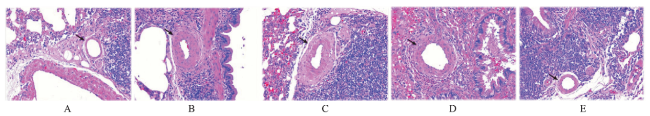

