中国科技论文统计源期刊 中文核心期刊  
美国《化学文摘》《国际药学文摘》
《乌利希期刊指南》
WHO《西太平洋地区医学索引》来源期刊  
日本科学技术振兴机构数据库(JST)
第七届湖北十大名刊提名奖  
医药导报, 2017, 36(9): 967-970
doi: 10.3870/j.issn.1004-0781.2017.09.004
阿帕替尼对非小细胞肺癌细胞侵袭能力的影响*
Affects of Apatinib in the Invasion Ability of Non-small Cell Lung Cancer Cells
李维娜1,, 邓豫2,

摘要:

目的 观察阿帕替尼对非小细胞肺癌细胞株A549及H460侵袭能力及上皮-间质转化(EMT)的影响。方法 通过细胞存活实验筛选阿帕替尼适宜剂量,通过Transwell检测其对细胞侵袭能力的影响,通过光学显微镜观察细胞的形态学变化,通过Western blotting检测EMT相关蛋白的变化。结果 在A549及H460细胞中加入不同浓度的阿帕替尼,当阿帕替尼的浓度≤120 nmol·L-1时,细胞的存活能力没有明显改变。在60及120 nmol·L-1阿帕替尼作用下,A549细胞侵袭能力降低,并发生间质-上皮转化的形态学变化,EMT相关的E-cadherin表达升高,Vimentin、N-cadherin及Snail的表达降低。结论 阿帕替尼可诱导非小细胞肺癌细胞株A549及H460由间质表型向上皮表型转化,并抑制细胞的侵袭能力。

关键词: 阿帕替尼 ; 癌,肺,非小细胞 ; 侵袭 ; 上皮-间质转化

Abstract:

Objective To observe the effect of apatinib on the invasion ability and epithelial-mesenchymal transition (EMT) of non-small cell lung cancer ( NSCLC) cell line A549 and H460. Methods Different doses of apatinib were incubated with the A549 and H460 cells, then the effect of apatinib on the invasion of A549 cells was detected by Transwell assay, and the change of cell morphology was observed by optical microscope, and the expression of EMT related proteins were detected by Western blotting. Results The cell viability of A549 and H460 have no change when the concentration of apatinib was under 120 nmol·L-1. Therefore, 60 nmol·L-1 and 120 nmol·L-1 apatinib were used in the further experiments. The invasion of A549 cells was reduced after apatinib incubation for 48 h, with cell morphology changes, and the expression of E-cadherin was enhanced, with the depresion of N-cadherin,Vimentin and Snail, which were related to the progress of EMT. Conclusion Apatinib could induce the MET, and reduce the invasion ability of A549 and H460 cells.

Key words: Apatinib ; Cancer, lung, non-small cell ; Invasion ; Epithelial-mesenchymal transition

阿帕替尼(apatinib)是近年来新上市的小分子抗血管生成靶向药物,目前主要用于治疗一线化疗药物无效或复发的进展期胃癌[1]。血管内皮生长因子(vascular endothelial growth factor,VEGF)及血管内皮生长因子受体(vascular endothelial growth factor receptor,VEGFR)家族在肿瘤生长、血管生成等多种病理生理学进程中发挥重要作用。阿帕替尼是主要针对VEGFR2的小分子抑制药,而后者是调控肿瘤血管生成的主要受体亚型,所以阿帕替尼能够抑制肿瘤新生血管,从而抑制肿瘤的生长和转移。目前阿帕替尼在国内主要应用于进展期胃癌患者,针对其他实体肿瘤的研究也处于临床试验阶段[1-3]。上皮-间质转换(epithelial-mesenchymal transition,EMT)作为一种复杂而且重要的生物学效应,广泛参与到多种病理生理进程,其中包括恶性肿瘤的侵袭转移[4-5]。笔者以肺癌细胞A549及H460作为研究工具,旨在研究阿帕替尼对于非小细胞肺癌(non-small cell lung cancer,NSCLC)细胞侵袭能力及EMT的作用。

1 材料与方法
1.1 细胞与试剂

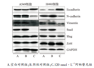
A549及H460人肺癌细胞购自武汉博士德生物工程有限公司。实验用阿帕替尼由江苏恒瑞医药股份有限公司赠予。胎牛血清、达尔伯克必需基本培养液(DMEM)购自武汉祥云博生物工程有限公司。抗E-cadherin抗体(CST,No.3195),抗N-cadherin抗体(CST,No.13116),抗Vimentin抗体(CST,No.5741),抗Snail抗体(CST,No.3879),抗Slug抗体(CST,No.9585),抗Zeb1抗体(CST,No.3396)及抗内参GAPDH抗体(CST,No.5174),预铺好Matrigel的Transwell(孔径8 μm)等其他耗材均购自武汉博士德生物有限公司。Transwell实验应用美国康宁公司耗材,并通过奥林巴斯显微镜观察。免疫印迹(Western blotting)实验应用Bio-rad电泳仪。

1.2 实验方法

1.2.1 细胞培养 A549及H460细胞培养于含10%胎牛血清的RPMI-1640培养液中,置于37 ℃、5% 二氧化碳(CO2)温箱内培养,以0.25%胰蛋白酶消化传代。

1.2.2 细胞存活能力实验 不同浓度阿帕替尼处理后的A549及H460细胞,分别于0,48 h按照CCK-8试剂盒说明书,每孔加入10% CCK8溶液,继续孵育4 h后,利用酶标仪在波长492 nm测定吸光度并计算抑制率,每组设5个复孔,实验重复3次。

1.2.3 细胞侵袭能力实验 将培养至对数生长的A549细胞用无血清RPMI-1640培养液重悬,以每孔5×10⁴的密度加入到预铺好Matrigel的Transwell小室上室内,下室加入完全培养液600 mL,分别加入阿帕替尼使其终浓度为60,120 nmol·L-1,阴性对照组不加入阿帕替尼,空白对照组不做任何处理。培养箱中常规培养48 h取出上室,用4%多聚甲醛固定后用0.01%结晶紫染色,200倍光学显微镜下计数穿膜细胞,随即取5个视野,取平均值,每组实验设3个复孔,实验重复3次。

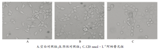
1.2.4 细胞形态学观察 普通光学显微镜观察A549细胞在120 nmol·L-1阿帕替尼处理48 h后的形态学变化。

1.2.5 免疫印迹检测EMT相关蛋白水平变化 采用免疫印迹检测120 nmol·L-1阿帕替尼处理后A549及H460细胞中E-cadherin、N-cadherin、Vimentin、Snail、Slug及Zeb1等EMT相关蛋白水平的变化。收集细胞,加入适量NP-40裂解获取细胞总蛋白液,加入适量十二烷基硫酸钠上样缓冲液100 ℃水浴解交联。蛋白样品行聚丙烯酰胺凝胶电泳,聚偏二氟乙烯膜转膜并用脱脂奶粉封闭非特异性结合,然后依次孵育一抗及二抗后用电化学发光显色液显影。

1.3 统计学方法 采用SPSS12.0版统计学软件进行统计分析。实验数据以均数±标准差( x ¯ ±s)表示,组间均数比较采用t检验,以P<0.05为差异有统计学意义。

2 结果
2.1 阿帕替尼对A549及H460细胞存活能力的影响

在A549及H460细胞中加入不同浓度(60,120,180,240 nmol·L-1)阿帕替尼,观察其对细胞活性的影响,通过CCK8试剂盒检测细胞在492 nm波长下的吸光度,结果提示:当阿帕替尼的浓度≤120 nmol·L-1时对A549及H460细胞的存活没有显著影响,而当阿帕替尼浓度≥180 nmol·L-1时,对A549及H460细胞的存活有明显的抑制作用。因笔者着重研究阿帕替尼对肿瘤细胞侵袭能力的影响,故主要选择60及120 nmol·L-1阿帕替尼,以排除阿帕替尼对细胞增殖、生长的影响(
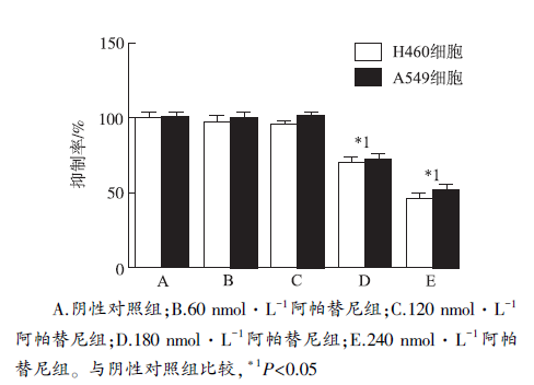
图1)。

图1 5组肺癌细胞存活能力比较(n=5)
A.negative control group;B.60 nmol·L-1 apatinib group;C.120 nmol·L-1 apatinib group;D.180 nmol·L-1 apatinib group;E.240 nmol·L-1 apatinib group .Compared with negative control group, *1P<0.05

Fig.1 Comparison of the viability among five groups of lung cancer cells(n=5)

2.2 阿帕替尼对A549侵袭能力的影响

观察60及120 nmol·L-1阿帕替尼组及阴性对照组处理48 h后的A549细胞,计数穿过Matrigel的细胞数目以代表细胞的侵袭能力。结果提示,120 nmol·L-1阿帕替尼处理后的穿膜细胞数为每视野(32±7)个,60 nmol·L-1阿帕替尼处理后的穿膜细胞数为每视野(45±5)个,相比于阴性对照组的每视野(110±6)个及空白对照组的每视野(115±5)个显著减少(P<0.05)(图2)。

2.3 阿帕替尼对A549细胞形态学的影响

普通光学显微镜观察120 nmol·L-1阿帕替尼处理A549细胞48 h后细胞形态学变化。结果提示A549细胞在未处理及被阴性对照处理后表现为细胞呈长梭形,伪足较多较长,而经120 nmol·L-1阿帕替尼处理后,细胞的形状变圆,细胞伪足变少变短(
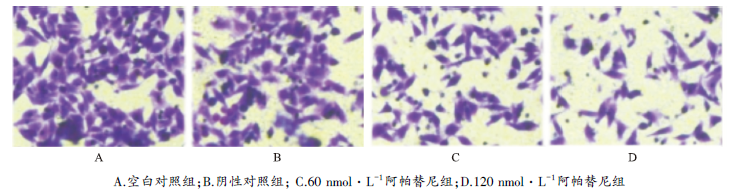
图3)。将上述细胞经胰蛋白酶消化收集后裂解总蛋白供后续实验使用。

图2 阿帕替尼对A549细胞侵袭能力的影响(×200)
A. blank control group;B.negative control group;C.60 nmol·L-1 apatinib group;D.120 nmol·L-1 apatinib group

Fig.2 Effect of apatinib on invasion activity of A549 cells(×200)

图3 阿帕替尼对A549细胞形态改变的影响(×200)
A.blank control group ; B.negative control group;C.120 nmol·L-1 apatinib group

Fig.3 Effect of apatinib on morphological change of A549 cells(×200)

2.4 阿帕替尼对EMT相关蛋白表达的影响

将上述细胞裂解提取总蛋白后进行免疫印迹检测发现,120 nmol·L-1阿帕替尼处理后,A549及H460细胞中上皮性标志E-cadherin的蛋白表达明显升高,相对应的间质性标志N-cadherin及Vimentin的表达明显减少;同时检测了与EMT相关的转录因子的表达,其中Snail的表达明显降低,而Slug及Zeb1的表达无明显变化(图4)。

图4 阿帕替尼对肺癌细胞中EMT相关蛋白的影响
A.blank control group;B.negative control group;C.120 nmol·L-1 apatinib group

Fig.4 Effect of apatinib on EMT related proteins of lung cancer cells

3 讨论

NSCLC是严重威胁人类健康的恶性肿瘤,发病率呈上升趋势[6]。目前手术切除仍是NSCLC的首要治疗方法,但对于某些复发或转移的患者已经丧失手术机会,依赖于化疗等辅助治疗方案。以铂剂为基础的双药联合治疗方案可使得晚期NSCLC患者生存期延长、生活质量改善,并使肿瘤相关症状减轻,一线药物不敏感时也有多西他赛、培美曲赛、吉非替尼等二线药物可供选择,但总体来说,晚期NSCLC患者生存期仍有限,临床亟待新的、更为有效的治疗药物及方法[6]

VEGF是一种重要的细胞因子,可由肿瘤细胞或肿瘤间质细胞所分泌,其作用于肿瘤细胞表面的VEGFR,可直接促进肿瘤细胞的增殖和生长,而作用于肿瘤中的间质细胞如血管内皮细胞,则可促进后者的增殖,而增强肿瘤的血管生成[7]。因此VEGF及VEGFR是目前公认的肿瘤分子靶向治疗的重要靶点。目前针对VEGF及VEGFR的靶向药物包括VEGF的单克隆抗体如贝伐单抗、VEGFR的单克隆抗体如雷莫芦单抗,以及针对VEGFR的酪氨酸激酶小分子抑制药如阿帕替尼[7]。阿帕替尼目前已批准用于二线化疗药物无效及伴有远处转移的进展期胃癌的临床治疗,而在NSCLC、乳腺癌等其他上皮来源肿瘤中的研究正处于临床试验阶段。阿帕替尼主要作用于VEGFR2,后者是介导肿瘤血管生成的关键受体亚型,因此阿帕替尼延长晚期肿瘤患者生存期、改善生活质量的分子机制在于抑制肿瘤的血管生成[1]。而EMT是肿瘤转移的重要分子机制之一,阿帕替尼是否能够影响肿瘤的EMT或间质-上皮转换(mesenchymal-epithelial transition,MET)过程,进而抑制肿瘤的侵袭转移尚不十分清楚,因此笔者着重研究小剂量阿帕替尼对于NSCLC细胞A549侵袭能力的影响。

在NSCLC细胞株A549及H460中,加入不同浓度的阿帕替尼,观察到当阿帕替尼浓度≤120 nmol·L-1时,阿帕替尼对A549及H460细胞的生长存活能力没有显著影响,因此笔者选取60及120 nmol·L-1这两种药物浓度进行后续实验。通过预铺好Matrigel的Transwell小室模拟肿瘤转移时需要穿透的组织细胞基底膜,观察到阿帕替尼可减少A549细胞的穿膜个数,即阿帕替尼抑制了A549细胞的侵袭能力。本研究结果显示,A549细胞经阿帕替尼作用后细胞从长梭形变成椭圆形,伪足减少变短,细胞间隙也相应变小,而这些形态学改变是MET的特征表现。EMT及与其反向的MET作为一种复杂而且重要的生物学效应,广泛参与到多种病理生理进程,其中包括恶性肿瘤的侵袭转移[4-5,8]。阿帕替尼使细胞从转移能力较强的间质表型向转移能力较弱的上皮表型转化,从侧面解释阿帕替尼对A549及H460细胞侵袭能力的抑制作用。为了证实阿帕替尼通过使细胞发生MET从而抑制细胞的侵袭能力,笔者采用Western blotting 检测阿帕替尼对细胞中EMT相关蛋白如E-cadherin、Vimentin等蛋白水平的影响,结果发现上皮标志E-cadherin表达升高,

间质标志N-cadherin及Vimentin表达降低,而作为调控EMT的重要转录因子之一的Snail的表达也明显下降,提示阿帕替尼可能通过调控Snail进而抑制NSCLC细胞EMT及侵袭转移。

综上所述,阿帕替尼可抑制NSCLC细胞株A549及H460发生EMT,从而降低其侵袭转移的能力,阿帕替尼处理后A549及H460细胞中EMT相关转录因子Snail的表达明显下降,提示Snail可能参与阿帕替尼介导的MET变化过程。但阿帕替尼调控Snail的具体分子学机制尚待阐明,也是笔者进一步的研究内容。

The authors have declared that no competing interests exist.

参考文献

[1] GENG R,LI J.Apatinib for the treatment of gastric cancer[J].Expert Opin Pharmacother,2015,16(1):117-122.
Abstract INTRODUCTION: Antiangiogenesis therapy plays an important role in cancer treatment. Apatinib mesylate, a small molecule tyrosine kinase inhibitor targeting vascular endothelial growth factor receptor-2, has been recommended as third-line treatment for metastatic gastric cancer patients. AREAS COVERED: The current review summarizes the publications and conference reports relating to apatinib from preclinical and clinical research in gastric cancer. Apatinib showed good safety, tolerance and treatment efficacy in Phase I/II studies. In a Phase III study, apatinib prolonged the median overall survival of patients with chemotherapy-refractory metastatic gastric cancer by 55 days and the median progression-free survival by 25 days compared with placebo. EXPERT OPINION: Apatinib is a new treatment option for advanced gastric cancer. Apatinib is expected to have a broader application when it has been evaluated worldwide. The key issues are to find biomarkers and overcome drug resistance.
DOI:10.1517/14656566.2015.981526      PMID:25420417      URL    
[本文引用:3]
[2] LI J,QIN S,XU J,et al.Apatinib for chemotherapy-refractory advanced metastatic gastric cancer:results from a randomized,placebo-controlled,parallel-arm,phase Ⅱ trial[J].J Clin Oncol,2013,31(26):3219-3225.
Purpose;Patients with metastatic gastric cancer (mGC) who do not respond to or who experience progression with second-line chemotherapy have no treatment options that clearly confer a survival benefit. This trial investigated the safety and efficacy of apatinib, an inhibitor of vascular endothelial growth factor receptor, as a treatment option for heavily pretreated patients with mGC.Patients and Methods;Patients who experienced treatment failure with at least two chemotherapeutic regimens were randomly assigned to receive placebo (group A), apatinib 850 mg once daily (group B), or apatinib 425 mg twice daily (group C).Results;We enrolled 144 patients onto this study. In groups A, B, and C, the median overall survival (OS) times were 2.50 months (95% CI, 1.87 to 3.70 months), 4.83 months (95% CI, 4.03 to 5.97 months), and 4.27 months (95% CI, 3.83 to 4.77 months), respectively, and the median progression-free survival (PFS) times were 1.40 months (95% CI, 1.20 to 1.83 months), 3.67 months (95% CI, 2.17 to 6.80 months), and 3.20 months (95% CI, 2.37 to 4.53 months), respectively. There were statistically significant differences between the apatinib and placebo groups for both PFS (P Conclusion;Apatinib showed improved PFS and OS in heavily pretreated patients with mGC who had experienced treatment failure with two or more chemotherapy regimens. (C) 2013 by American Society of Clinical Oncology
DOI:10.1200/JCO.2013.48.8585      PMID:23918952      URL    
[本文引用:0]
[3] QIN S,LI J,XU J,et al.Phase Ⅲ study of apatinib in advanced gastric cancer:a randomized,double-blind,placebo-controlled trial[J].J Clin Oncol,2014,32(Suppl):4003.
O-0029PHASE III STUDY OF APATINIB IN ADVANCED GASTRIC CANCER: A RANDOMIZED, DOUBLE-BLIND, PLACEBO-CONTROLLED TRIAL
DOI:10.1093/annonc/mdu193.29      URL    
[本文引用:1]
[4] KIM H Y,JACKSON T R,DAVIDSON L A.On the role of mechanics in driving mesenchymal-to-epithelial transitions[J].Semin Cell Dev Biol,2016,24(16):30137-30139.
The mesenchymal-to-epithelial transition (MET) is an intrinsically mechanical process describing a multi-step progression where autonomous mesenchymal cells gradually become tightly linked, polarized epithelial cells. METs are fundamental to a wide range of biological processes, including the evolution of multicellular organisms, generation of primary and secondary epithelia during development and organogenesis, and the progression of diseases including cancer. In these cases, there is an interplay between the establishment of cell polarity and the mechanics of neighboring cells and microenvironment. In this review, we highlight a spectrum of METs found in normal development as well as in pathological lesions, and provide insight into the critical role mechanics play at each step. We define MET as an independent process, distinct from a reverse-EMT, and propose questions to further explore the cellular and physical mechanisms of MET.
DOI:10.1016/j.semcdb.2016.05.011      PMID:27208723      URL    
[本文引用:2]
[5] JAKOBSEN K R,DEMUTH C,SORENSEN B S,et al.The role of epithelial to mesenchymal transition in resistance to epidermal growth factor receptor tyrosine kinase inhibitors in non-small cell lung cancer[J].Transl Lung Cancer Res,2016,5(2):172-182.
Abstract Inhibition of the epidermal growth factor receptor (EGFR) is an important strategy when treating non-small cell lung cancer (NSCLC) patients. However, intrinsic resistance or development of resistance during the course of treatment constitutes a major challenge. The knowledge on EGFR-directed tyrosine kinase inhibitors (TKIs) and their biological effect keeps increasing. Within the group of patients with EGFR mutations some benefit to a much higher degree than others, and for patients lacking EGFR mutations a subset experience an effect. Up to 70% of patients with EGFR mutations and 10-20% of patients without EGFR mutations initially respond to the EGFR-TKI erlotinib, but there is a severe absence of good prognostic markers. Despite initial effect, all patients acquire resistance to EGFR-TKIs. Multiple mechanisms have implications in resistance development, but much is still to be explored. Epithelial to mesenchymal transition (EMT) is a transcriptionally regulated phenotypic shift rendering cells more invasive and migratory. Within the EMT process lays a need for external or internal stimuli to give rise to changes in central signaling pathways. Expression of mesenchymal markers correlates to a bad prognosis and an inferior response to EGFR-TKIs in NSCLC due to the contribution to a resistant phenotype. A deeper understanding of the role of EMT in NSCLC and especially in EGFR-TKI resistance-development constitute one opportunity to improve the benefit of TKI treatment for the individual patient. Many scientific studies have linked the EMT process to EGFR-TKI resistance in NSCLC and our aim is to review the role of EMT in both intrinsic and acquired resistance to EGFR-TKIs.
DOI:10.21037/tlcr.2016.04.07      PMID:27186512      URL    
[本文引用:2]
[6] BANSAL P,OSMAN D,GAN G N,et al.Recent advances in targetable therapeutics in metastatic non-squamous NSCLC[J].Front Oncol,2016,6(1):112.
Lung adenocarcinoma is the most common subtype of non-small cell lung cancer (NSCLC). With the discovery of epidermal growth factor receptor (EGFR) mutations, anaplastic lymphoma kinase (ALK) rearrangements, and effective targeted therapies, therapeutic options are expanding for patients with lung adenocarcinoma. Here, we review novel therapies in non-squamous NSCLC, which are directed against oncogenic targets, including EGFR, ALK, ROS1, BRAF, MET, human epidermal growth factor receptor 2 (HER2), vascular endothelial growth factor receptor 2 (VEGFR2), RET, and NTRK. With the rapidly evolving molecular testing and development of new targeted agents, our ability to further personalize therapy in non-squamous NSCLC is rapidly expanding.
DOI:10.3389/fonc.2016.00112      PMID:48548691      URL    
[本文引用:2]
[7] RIBATTI D.Tumor refractoriness to anti-VEGF therapy[J].Oncotarget,2016,doi:10.18632/oncotarget.8694.
Vascular endothelial growth factor (VEGF) has been identified as the most potent cytokine involved in tumor angiogenesis and metastasis formation. Clinical results of anti-angiogenic therapies targeting VEGF and its receptors are very modest, resulting in a moderate improvement of overall survival. The clinical outcome is associated with the development of resistance and the increased risk of invasion and metastasis. In this article, I have analyzed the principal mechanisms of resistance to VEGF pathway inhibitors, including normalization of tumor blood vessels, hypoxia, recruitment of inflammatory cells and immature myeloid cells, alternative mechanisms of tumor vessel formation, genomic instability of tumor endothelial cells. In this context, the concept and strategies of anti-angiogenic therapies should be extensively re-considered and re-evaluated. In particular, rational combinations of anti-angiogenic agents based on pharmacokinetic and pharmacodynamics data are needed to overcome resistance and it is extremely important to determine the optimal duration and scheduling of anti-VEGF agents.
DOI:10.18632/oncotarget.8694      PMID:5216828      URL    
[本文引用:2]
[8] 石永利,吴鸿雁,樊祥山,.上皮-间质转变相关蛋白在非小细胞肺癌中的表达及其预后意义[J].中国癌症杂志,2011,21(5):342-347.
背景与目的:非小细胞肺癌(non-small cell lung cancer,NSCLC)患者最主要的死亡原因是手术后肿瘤转移,而上皮-间叶转变(epithelial-mesenchymal transitions,EMT)是肿瘤发生侵袭、转移的重要原因。本研究探讨EMT相关蛋白Snail、Vimentin、E-cadherin和β-catenin在NSCLC患者中的表达及其预后意义。方法:采用组织芯片技术和免疫组织化学EnVision两步法检测Snail、Vimentin、E-cadherin和β-catenin蛋白在165例NSCLC患者中的表达情况。根据免疫组织化学结果对NSCLC患者分组,分析Snail、Vimentin、E-cadherin和β-catenin蛋白在判断患者预后和生存情况中的作用。结果:165例NSCLC患者中,Snail、Vimentin、E-cadherin和β-catenin的阳性表达率分别为45.5%、33.9%、66.7%和50.9%。E-cadherin的表达与Snail呈负相关(P<0.001),而与β-catenin的表达呈正相关(P=0.003)。Snail、Vi...
[本文引用:1]
资源
PDF下载数    
RichHTML 浏览数    
摘要点击数    

分享
导出

相关文章:
关键词(key words)
阿帕替尼
癌,肺,非小细胞
侵袭
上皮-间质转化

Apatinib
Cancer, lung, non-small c...
Invasion
Epithelial-mesenchymal tr...

作者
李维娜
邓豫

LI Weina
DENG Yu