中国科技论文统计源期刊 中文核心期刊
美国《化学文摘》《国际药学文摘》
《乌利希期刊指南》
WHO《西太平洋地区医学索引》来源期刊
日本科学技术振兴机构数据库(JST)
第七届湖北十大名刊提名奖
美国《化学文摘》《国际药学文摘》
《乌利希期刊指南》
WHO《西太平洋地区医学索引》来源期刊
日本科学技术振兴机构数据库(JST)
第七届湖北十大名刊提名奖

 , 丁佳圣
, DING Jiasheng
, 丁佳圣
, DING Jiasheng
目的 探讨4种磷脂囊泡对人肺腺癌细胞(A549)的细胞毒性及促细胞内胰岛素转运的影响。方法 4种胰岛素磷脂囊泡处理A549细胞,采用噻唑蓝比色法(MTT)检测磷脂囊泡细胞毒性,电化学发光法(ECLIA)检测细胞内胰岛素含量。结果 MTT实验结果显示胰岛素浓度≤0.2 U·mL-1时细胞存活率>95%,50%胰岛素磷脂囊泡浓度对细胞存活率的影响以胰岛素脂质体组最高(62.48±2.46)%,乙醇脂质体组最低(16.85±5.05)%,相对安全性顺序依次为:脂质体>脂质微泡>丙二醇脂质体>乙醇脂质体。相同浓度下4种磷脂囊泡促细胞内胰岛素转运的含量排序依次为:脂质微泡>丙二醇脂质体>乙醇脂质体>脂质体。结论 以磷脂类膜材构建的脂质体、脂质微泡具备较高安全性,脂质微泡促进胰岛素细胞内转运能力最强。
Objective To study the cytotoxicity and insulin intracellular delivery of four kinds of phospholipid vesicles as liposome, ethanol liposome, propylene glycol liposome, and lipid microbubble in human lung adenocarcinoma cell line (A549). Methods A549 cells were treated with four kinds of insulin phospholipid vesicles. Methyl thiazolyl tetrazolium (MTT) chromatometry method was employed to determine the cytotoxicity of phospholipid vesicles. The content of intracellular insulin was detected by electrochemical luminescence method (ECLIA). Results MTT experimental results showed that when the insulin concentration was less than 0.2 U·mL-1, the cell survival rate was more than 95%. When concentration of insulin phospholipid vesicles was 50%, the highest cell survival rate was in insulin liposome group (62.48±2.46)%; the lowest cell survival rate in ethanol liposome group (16.85±5.05)%. The relative safety order was liposomes, microbubbles, propylene glycol liposomes, and ethanol liposomes. At the same concentration of phospholipid vesicles, the order of insulin intracellular contents was: lipid microbubbles>propylene glycol liposomes>ethanol liposomes>liposomes. Conclusion Liposomes and lipid microbubbles constructed with phospholipid membrane are of high safety. Lipid microbubbles have the strongest ability in insulin intracellular delivery.
肺部给药具有使药物免受肝脏首关效应、胃肠道内酶降解等优势,是胰岛素等生物大分子药物理想的给药方式。然而胰岛素由于分子量大、水溶性强、肺黏膜通透性较差的原因,经肺部给药时通常需要加入吸收促进剂来增强肺黏膜通透性。磷脂是一种表面活性剂类的黏膜吸收促进剂,将磷脂分散在水中可自然形成多层囊泡状结构,采用一定的制备工艺可形成磷脂囊泡体系,药物可包埋或溶解在囊泡内部,也可吸附或耦合在其外表面,增强药物的透黏膜效果。同时生物膜是由磷脂双分子层组成,内嵌蛋白,肺泡的表面活性物质也主要是由磷脂组成,主要成分是二棕榈酰磷脂胆碱,因此,以磷脂为膜材构建脂质囊泡与肺部内源性物质相同,有较好的生物相容性。已有多项研究报道将药物包裹入脂质体后肺部给药能有效地减少药物对呼吸道和肺部的刺激性和毒性,并增加药物疗效[1,2]。A549细胞为人肺腺癌细胞,属于传代细胞系,起源于肺支气管上皮、支气管腺或肺泡上皮细胞等肺组织正常细胞,具有可稳定地传代培养、相对原代细胞易获取和易培养等优点。因此,本研究选取A549细胞,探讨4种基于磷脂构建的脂质囊泡,即脂质体、乙醇脂质体、丙二醇脂质体、脂质微泡的细胞毒性及促进药物进入细胞内的能力。
人肺癌细胞系A549细胞购自中国科学院上海细胞生物学研究所。
胰岛素注射液(江苏万邦生化医药股份有限公司,批号:8021450);poloxamer188(上海协泰化工有限公司,批号:081006);聚山梨酯-80(江苏海安石油化工厂,批号:121201);氢化蛋黄卵磷脂[上海艾韦特(A.V.T.)医药科技有限公司,批号:84928172];胆固醇(上海君创生物科技有限公司,批号:140715);丙二醇(美国Sigma 公司,批号:STBF373V);无水乙醇(杭州萧山化学试剂厂,批号:20140203)。DMEM 培养液(吉诺生物医药技术有限公司,批号:709791);0.25%胰酶(美国Sigma公司,批号:YWNA0551B);胎牛血清(天津市灏洋生物制品科技有限责任公司,批号:20140925);青-链霉素溶液(100 x,吉诺生物医药技术有限公司,批号:20160110);磷酸盐缓冲液(PBS,吉诺生物医药技术有限公司,批号:20150914);噻唑蓝(MTT,上海华舜生物工程有限公司,批号:20160330);其他化学试剂均为分析纯。
IKA D-7921型磁力加热搅拌器(广州仪科实验有限公司);RE52-AA型旋转蒸发仪(上海亚荣生化仪器厂);YS100型光学显微镜(日本Nikon 公司);H-600型透射电镜(日本Hitachi公司);Nicomp 380型激光粒度分析仪(美国Particle Sizing Systems 公司);BS110S型电子分析天平(北京赛多利斯天平有限公司,感量:0.1 mg);FD-1A-50型冷冻干燥机(北京博医康实验仪器有限公司);96孔细胞培养板(美国Corning Costar公司);HDL Apparatus型超净工作台(苏州安泰空气技术有限公司);CO2培养箱(美国Thermo 公司);BiofugeStratos型超速低温离心机(美国Thermo 公司);E170型自动电化学发光免疫分析仪(罗氏公司)。
薄膜分散法制备胰岛素脂质体,称取氢化蛋黄卵磷脂10 mg、胆固醇5 mg、聚山梨酯80为1~2 mg于西林瓶中,加入无水乙醇2 mL,置65~70 ℃水浴中,搅拌使溶解,于旋转蒸发仪上旋转,使磷脂的乙醇液在壁上成膜,减压除乙醇。加入胰岛素溶液6 mL,室温搅拌30 min,制得胰岛素脂质体悬液。
注入法制备胰岛素乙醇脂质体,称取氢化蛋黄卵磷脂10 mg、胆固醇5 mg、聚山梨酯80为1~2 mg于西林瓶中,用乙醇2 mL溶解,置于磁力搅拌器上,在密闭条件下边搅拌边缓慢细流注入胰岛素溶液4 mL,分别经孔径0.45,0.22 μm微孔滤膜滤过各3遍,得到胰岛素乙醇脂质体悬液。
注入法制备胰岛素丙二醇脂质体,称取氢化蛋黄卵磷脂10 mg、胆固醇5 mg、聚山梨酯80 1~2 mg于西林瓶中,用丙二醇4 mL溶解,置于磁力搅拌器上,在密闭条件下边搅拌边缓慢细流注入胰岛素溶液2 mL,分别经孔径0.45,0.22 μm微孔滤膜滤过各3次,得到胰岛素丙二醇脂质体悬液。
冷冻干燥法制备脂质微泡冻干粉,称取Poloxamer 188 100 mg、聚山梨酯80 10 mg、氢化蛋黄卵磷脂 4 mg于西林瓶中,加入叔丁醇,加热至65~70 ℃,溶解,4 ℃冷置到凝固,冻干(冷冻槽温度-48 ℃,真空度为<100 mBar)20 h制得脂质微泡冻干粉。使用时,加入胰岛素溶液并经手摇振荡后制成胰岛素脂质微泡悬浮液。
取生长状态良好A549细胞,以每孔5×10⁴个接种于96孔板中,DMEM培养液为每孔200 μL。37 ℃、5%CO2,饱和湿度条件下培养。当细胞长至贴壁汇合时,PBS洗2次,分别用细胞培养基稀释胰岛素药物、胰岛素脂质体、胰岛素乙醇脂质体、胰岛素丙二醇脂质体、胰岛素脂质微泡处理A549细胞,药物浓度和各药物载体浓度的设置见
表1 药物浓度和4种胰岛素磷脂囊泡浓度的设置
Tab.1 Concentration design of test drug and four insulin-phospholipid vesicles
取生长状态良好A549细胞分别接种于24 孔平底细胞培养板,每孔8×105个·(500 μL)-1。37 ℃,5%CO2培养24 h。弃培养液,分别用细胞培养基稀释胰岛素药物、胰岛素脂质体、胰岛素乙醇脂质体、胰岛素丙二醇脂质体、胰岛素脂质微泡每孔200 μL处理A549细胞,每种体系设高(50%)、中(25%)、低(6%)3个浓度,每组设3复孔,同时设空白培养液对照组和单纯胰岛素(2 U·mL-1)对照组。在各采样点前37 ℃、5%CO2继续培养。于0,30,60,120,180,240 min 各时间点移除上清液。加入PBS (4 ℃)300 μL 洗涤细胞4次。加入0.1%聚乙二醇辛基苯基醚(Triton X-100,4 ℃)500 μL将细胞重新悬浮,过夜,使细胞裂解。将上述各孔溶液分别过孔径0.45 μm滤膜,通过电化学发光法测定过滤液中胰岛素的含量,即胰岛素进入细胞内的量。
采用Excel 软件对各组数据进行处理,计量资料以均数±标准差(
药物胰岛素对细胞生存率的影响如
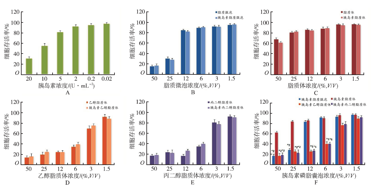
图1
不同浓度的胰岛素、磷脂囊泡及胰岛素磷脂囊泡对A549细胞存活率的影响 (
Fig.1
Effect of different concentration of insulin, phospholipid vesicles and insulin phospholipid vesicles on the viability of A549 cells(
4种磷脂囊泡都按低(6%)、中(25%)、高(50%)3种浓度和同一胰岛素药物浓度(2 U·mL-1)设置,促胰岛素细胞内转运结果如

图2
4种磷脂囊泡促胰岛素细胞内转运结果 (
Fig.2
Insulin intracellular delivery promoted by four kinds of phospholipid vesicles(
脂质体、乙醇脂质体、丙二醇脂质体、脂质微泡都为球形中空结构,由磷脂分子的双亲性作用形成分子膜包围而成密闭球形囊泡,除脂质微泡的外壁为单层磷脂分子膜外,其他的外壁都是一层或多层磷脂双分子膜。因囊泡的外膜结构与细胞膜相似,所以磷脂囊泡的安全性相对较高。从MTT实验的研究结果上看,脂质体与脂质微泡的安全性高于乙醇脂质体和丙二醇脂质体。这与乙醇脂质体和丙二醇脂质体为含醇体系相关,浓度越低细胞存活率越高。药物胰岛素安全性高,实验结果表明胰岛素浓度在10,20 U·mL-1时会抑制细胞生长,浓度≤2 U·mL-1时细胞存活率为>92.37%。因此,在同一磷脂囊泡浓度下,脂质微泡组与胰岛素脂质微泡组对细胞存活率的影响结果相似,脂质体组与胰岛素脂质体组、乙醇脂质体组与胰岛素乙醇脂质体组以及丙二醇脂质体组与胰岛素丙二醇脂质体组的结果亦相似。这表明药物对细胞存活率的影响不大。同一浓度下4种胰岛素磷脂囊泡对细胞存活率的影响,可见胰岛素脂质体组对细胞生长抑制作用最小,其次是胰岛素脂质微泡、胰岛素丙二醇脂质体和胰岛素乙醇脂质体。脂质微泡的储存形式为干粉,临用前加入适当浓度的药物水溶液,也较好地解决脂质微泡长期贮存不稳定的问题。但脂质微泡处方中因含有大量表面活性剂,其安全性较脂质体低。
脂质囊泡有较强的两亲性,使其具有生物膜特性和药物传输能力,可以显著提高药物的跨膜吸收速率及体内生物利用度[3,4]。基于以上4种磷脂囊泡构建胰岛素给药体系研究其促胰岛素细胞内转运的能力,结果显示磷脂囊泡浓度对促进细胞内胰岛素转运影响较大,脂质微泡、丙二醇脂质体、乙醇脂质体以中浓度体积比25%时促胰岛素细胞内转运的效果最好,而脂质体组在高浓度体积比50%时细胞内胰岛素浓度最高。这可能与脂质体细胞安全性最高,而高浓度脂质微泡、丙二醇脂质体与乙醇脂质体对细胞毒性较大致细胞内胰岛素浓度低相关。丙二醇脂质体的促细胞内药物转运能力较脂质体强,其原因可能在于丙二醇脂质体纳米级粒径具有穿过靶组织内皮细胞的能力,能实现在细胞水平和(或)亚细胞水平供药。脂质微泡的平均粒径为3.08 μm,虽然其粒径较脂质体(1.018 μm)等微粒大,但是脂质微泡的含气中空结构,通过微泡爆破时产生的空化效应能在细胞膜上产生可逆性通道,暂时扩大的细胞膜孔径促进药物进入细胞,这可能就是脂质微泡组促细胞药物摄取能力最强的原因[5,6]。据此,提示以脂质微泡为载体构建胰岛素给药体系有望成为一种新型多肽蛋白类药物肺部给药系统。
The authors have declared that no competing interests exist.
