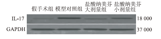
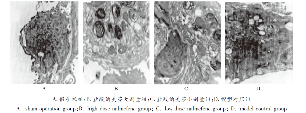
Objective To study the protective effect of nalmefene hydrochloride on lung ischemia-reperfusion injury and its mechanism. Methods 40 rats were randomly divided into model group, high dose of nalmefene group, low dose nalmefene group and sham operation group equally(n=10). The lung ischemia-reperfusion model was established by occlusion of the left pulmonary hilum. The intravenous injection of nalmefene (20,10 μg·kg-1) was applied at 10 minutes before occlusion of the left pulmonary hilum in the high dose of nalmefene group and the low dose of nalmefene group, respectively. The sham operation group without occlusion of the left pulmonary hilum was not given any treatment. At 2 h after reperfusion, all rats were detected arterial blood gas value and then sacrificed. The specimens from the upper lobe of the left lung tissue were preserved to observe pulmonary lesions, detect the ratio of wet / dry weight and the expressions of β-endorphin and interleukin(IL)-17. Results Compared with the model group, the value of PCO2, the degree of pulmonary lesions, the ratio of wet / dry weight and the expressions of β-endorphin and IL-17 in lung tissue were significantly decreased (P<0.01), while the value of PO2 was significantly increased (P<0.01) in the low dose of nalmefene group. Compared with the low dose of nalmefene group, the value of PCO2, the degree of pulmonary lesions, the ratio of wet/dry weight and the expressions of β-endorphin and IL-17 in lung tissue were significantly decreased (P<0.01), while the value of PO2 was significantly increased (P<0.01) in the high dose of nalmefene group.Conclusion Nalmefene hydrochloride may prevent lung ischemia-reperfusion injury in a dose dependent manner by reducing the production of β-endorphin and inhibiting the expression of IL-17 in lung tissue.
表1
4组大鼠动脉血气分析及肺组织W/D
Tab.1
Analysis of arterial blood gas and the ratio of wet/dry weight of lung tissue in four groups of ratsx¯±s,n=10,kPa
组别
PO2
PCO2
W/D
假手术组
13.52±0.11
3.02±0.19
3.04±0.17
模型对照组
6.32±0.15*1
8.29±0.22*1
8.91±0.28*1
纳美芬大剂量组
9.96±0.28*2*3
4.06±0.26*2*3
4.15±0.24*2*3
纳美芬小剂量组
8.59±0.20*2
5.37±0.27*2
5.21±0.25*2
Compared with sham operation group,*1P<0.01; Compared with model control group, *2P<0.01;compared with low-dose nalmefene hydrochloride group, *3P<0.01
Lung reperfusion injury (LIRI) is a pathologic process occurring when oxygen supply to the lung has been compromised followed by a period of reperfusion. The disruption of oxygen supply can occur either via limited blood flow or decreased ventilation termed anoxic and ventilated , respectively. When reperfusion occurs, blood flow and oxygen are reintroduced to the ischemic lung parenchyma, facilitating a toxic environment through the creation of reactive oxygen species, activation of the immune and systems, , and apoptotic . This review will focus on the mechanisms of LIRI, the current supportive treatments used, and the many therapies currently under research for prevention and treatment of LIRI.
ZHENG ZK,WANG JJ,HUH,et al.Short-term inhalation of nitric oxide inhibits activations of toll-like receptor 2 and 4 in the lung after ischemia-reperfusion injury in mice[J].华中科技大学学报(医学版),2013,33(2):219-223.
In order to investigate the effects of different terms of inhaled nitric oxide (NO) preconditioning with low concentration on the activations of Toll -like receptor 2 and 4 (TLR2/4) in the lung ischemia-reperfusion (IR) injury in mice, we divided the male C57BL mice into five groups: sham (S) group, IR group, NO 1-min preconditioning group (15 ppm NO inhalation for 1 min before ischemia, NO 1-min), NO 10-min preconditioning group (15 ppm NO inhalation for 10 min before ischemia, NO 10-min), NO 60-min preconditioning group (15 ppm NO inhalation for 60 min before ischemia, NO 60-min). The changes of partial pressure of oxygen in artery (PaO 2 ), left lung wet-to-dry weight ratio (W/D), and myeloperoxidase (MPO) in the injured lung were measured in every group at 6th h of reperfusion after 60 min of left lung ischemia. The changes of TLR2/4 activations and plasma were measured in this procedure in additional mice. As compared with IR group, PaO2 increased, MPO and W/D decreased evidently after reperfusion in NO 10-min group. The changes in NO 60-min group were similar to those in NO 10-min group. There was no difference between NO 1-min and IR group. In NO inhalation group, the expressions levels of TLR2/4 mRNA and proteins were diminished, concentrations were decreased, and the lung injuries were ameliorated effectively. We concluded that short term inhalation of NO protected lung IR injury. But the protective effect of NO was not increased with extension of inhaled NO. Inhaled NO could inhibit the activations of TLR2/4 in the lung after IR injury. TLR signal pathway might contribute to the effect of protection with NO in this model.
SALIMIV,HENNUS MP,MOKHTARI-AZADT,et al.Opioid receptors control viral replication in the airways[J]. Crit Care Med,2013,41(1):205-214.
Objective: Opioids are frequently used during mechanical ventilation for severe viral infection in infancy. Opioid receptors have immunomodulatory properties, but nothing is known about their antiviral effects. We therefore aimed to investigate the role of opioid receptors in virus-induced airway inflammation.<br/>Patients and Interventions: Two single nucleotide polymorphisms in OPRM1 and OPRD1 were genotyped in 465 infants with severe respiratory syncytial virus infection and 930 control subjects. Subsequently, the mechanism by which opioid receptors affect clinical outcome in respiratory syncytial virus bronchiolitis was studied in BALB/c mice. Animals were injected daily with nalmefene, a nonselective opioid receptor antagonist, and infected by intranasal inoculation of respiratory syncytial virus 24 hrs after the first dose of nalmefene. The potential therapeutic effect of pharmaceutical opioids was studied using mu (DAMGO), kappa (U50488), and delta (DPDPE) opioid receptor agonists 48 hrs after infection.<br/>Measurements and Main Results: In our human study, the A118G single nucleotide polymorphism rs1799971 was associated with respiratory syncytial virus disease severity (p = 0.015). In mice, nalmefene treatment increased viral titers and was associated with more pronounced weight loss. Increased viral replication was associated with increased levels of cytokines and chemokines in the bronchoalveolar lavage fluid, enhanced bronchoalveolar cellular influx, and exaggerated lung pathology. Pharmaceutical opioids, in particular DPDPE, did not affect viral replication. They did induce a decreased influx of neutrophils, but an increased influx of lymphocytes and monocytes into the bronchoalveolar lumen during respiratory syncytial virus infection.<br/>Conclusions: Using a human study and an experimental model, we show that opioid receptor signaling has a potential beneficial role in the outcome of respiratory viral disease. We show that opioid receptor signaling is required to control respiratory syncytial virus replication and thereby to control disease severity. However, we also show that caution is required before using pharmaceutical opioids as anti-inflammatory or antiviral treatment of patients with viral respiratory infection. (Crit Care Med 2013; 41:205-214)
TSOULFASG,SVORONOS C.Lung ischemia-reperfusion injury: when NO(nitric oxide) does not always mean no[J].J Surg Res, 2013,S0022-4804(13):309-310.
[本文引用:1]
[8]
BODNAR RJ.Endogenous opiates and behavior: 2011[J]. Peptides,2012,38(2): 463-522.
This paper is the thirty-fourth consecutive installment of the annual review of research concerning the endogenous opioid system. It summarizes papers published during 2011 that studied the behavioral effects of molecular, pharmacological and genetic manipulation of opioid peptides, opioid receptors, opioid agonists and opioid antagonists. The particular topics that continue to be covered include the molecular-biochemical effects and neurochemical localization studies of endogenous opioids and their receptors related to behavior (Section 2), and the roles of these opioid peptides and receptors in pain and analgesia (Section 3); stress and social status (Section 4); tolerance and dependence (Section 5); learning and memory (Section 6); eating and drinking (Section 7); alcohol and drugs of abuse (Section 8); sexual activity and hormones, pregnancy, development and endocrinology (Section 9); mental illness and mood (Section 10); seizures and neurologic disorders (Section 11); electrical-related activity and neurophysiology (Section 12); general activity and locomotion (Section 13); gastrointestinal, renal and hepatic functions (Section 14); cardiovascular responses (Section 15); respiration (Section 16); and immunological responses (Section 17). (C) 2012 Elsevier Inc. All rights reserved.
GONZALEZ-NUNEZV,JIMENE Z G A,BARRETO-VALER K,/et al.In vivo regulation of the μ opioid receptor: role of the endogenous opioid agents[J]. Mol Med,2013,19(1):7-17.
It is well known that genotypic differences can account for the subject-specific responses to opiate administration. In this regard, the basal activity of the endogenous system (either at the receptor or ligand level) can modulate the effects of exogenous agonists as morphine and vice versa. The 渭 opioid receptor from zebrafish, dre-oprm1, binds endogenous peptides and morphine with similar affinities. Morphine administration during development altered the expression of the endogenous opioid propeptides proenkephalins and proopiomelanocortin. Treatment with opioid peptides (Met-enkephalin [Met-ENK], Met-enkephalin-Gly-Tyr [MEGY] and 尾-endorphin [尾-END]) modulated dre-oprm1 expression during development. Knocking down the dre-oprm1 gene significantly modified the mRNA expression of the penk and pomc genes, thus indicating that oprm1 is involved in shaping penk and pomc expression. In addition, the absence of a functional oprm1 clearly disrupted the embryonic development, since proliferation was disorganized in the central nervous system of oprm1-morphant embryos: mitotic cells were found widespread through the optic tectum and were not restricted to the proliferative areas of the mid- and hindbrain. Transferase-mediated dUTP nick-end labeling (TUNEL) staining revealed that the number of apoptotic cells in the central nervous system (CNS) of morphants was clearly increased at 24-h postfertilization. These findings clarify the role of the endogenous opioid system in CNS development. Our results will also help unravel the complex feedback loops that modulate opioid activity and that may be involved in establishing a coordinated expression of both receptors and endogenous ligands. Further knowledge of the complex interactions between the opioid system and analgesic drugs will provide insights that may be relevant for analgesic therapy.
PENGJ,SARKARS,CHANG SL,et al.Opioid receptor expression in human brain and peripheral tissues using absolute quantitative real-time RT-PCR[J]. Drug Alcohol Depend,2012,124(3):223-228.
Abstract The actions of endogenous opioid peptides are mediated by 3 main classes of opioid receptors; mu (MOR), kappa (KOR), and delta (DOR). We developed an absolute quantitative real-time reverse transcriptase PCR (AQ-rt-RT-PCR) assay to quantify MOR, DOR, and KOR mRNA in 22 human tissues. MOR mRNA was greatly enriched (12-20×10(6)copies/μg) in the cerebellum, nucleus accumbens, and caudate nucleus; moderate (6×10(6)copies/μg) in the dorsal root ganglion, spinal cord, and adrenal gland; low (2×10(4)copies/μg) in the pancreas and small intestine; and absent in the lung, spleen, kidney, heart, skeletal muscle, liver, and thymus. High levels (>8.8×10(6)copies/μg) of DOR mRNA were expressed in the brain and dorsal root ganglion; moderate (1.5×10(6)copies/μg) in the adrenal gland and pancreas; low (2×10(4)-6.5×10(5)copies/μg in the cerebellum, spinal cord, small intestine, skeletal muscle, thymus, lung, and kidney); and very low (3.8×10(3)copies/μg) in the heart. DOR mRNA was not detected in the spleen or liver. KOR mRNA was moderate (1×10(6)copies/μg) in brain regions and dorsal root ganglion, but low (1.6-7×10(5)copies/μg) in the cerebellum, temporal lobe and all other peripheral tissues. Our data demonstrate that the AQ-rt-RT-PCR is a highly reproducible and precise method to study the expression of opioid receptors in various tissues and under different disease conditions.
LAN CC,PENG CK,HUANG SF,et al.Activated protein C attenuates ischemia-reperfusion-induced acute lung injury[J]. Exp Lung Res,2015,41(5):241-250.
Ischemia-reperfusion (IR)-induced acute lung injury (ALI) is implicated in several clinical conditions, such as lung transplantation, acute pulmonary embolism after thrombolytic therapy, re-expansion of collapsed lung from pneumothorax, or pleural effusion, cardiopulmonary bypass, etc. Because mortality remains high despite advanced medical care, prevention and treatment are important clinical issues. Activated protein C (APC) manifests multiple activities with antithrombotic, profibrinolytic, and anti-inflammatory effects. We therefore conducted this study to determine the beneficial effects of APC in IR-induced ALI. IR-induced ALI was conducted in a rat model of isolated-perfused lung in situ. The animals were divided into the control group, IR group, and IR+APC group. There were six adult male Sprague-Dawley rats in each group. The IR caused significant pulmonary microvascular hyperpermeability, pulmonary edema and dysfuction, increased cytokines (tumor necrosis factor (TNF)-伪, IL-17, CXCL-1), and neutrophils infiltration in lung tissues. Administration of APC significantly attenuated IR-induced ALI with improving microvascular permeability, pulmonary edema, pulmonary dysfunction, and suppression inflammatory response. The current study demonstrates the beneficial effects of APC in IR-induced ALI. This protective effect is possibly associated with the inhibition of TNF-伪, IL-17A, CXCL1, and neutrophils infiltration in lung tissues. However, the current results were obtained in an animal model and it is still necessary to confirm these findings in human subjects. If we can demonstrate the benefits of APC to protect IR lung injury, we can postulate that APC is a potential therapeutic drug for lung preservation.
OSBORN MD,LOWERY JJ,SKORPUT AG,et al.In vivo characterization of the opioid antagonist nalmefene in mice[J].Life Sci,2010,86(15/16):624-630.
AIMS: The current study assessed the in vivo antagonist properties of nalmefene using procedures previously used to characterize the opioid antagonists naloxone, naltrexone, 6beta-naltrexol and nalbuphine. MAIN METHODS: ICR mice were used to generate antagonist dose-response curves with intraperitoneal (i.p.) nalmefene against fixed A(90) doses of morphine in models of morphine-stimulated hyperlocomotion and antinociception. Additional dose-response curves for antagonist precipitated opioid withdrawal were run in mice treated acutely (100mg/kg, s.c., -4h) or chronically (75mg pellet, s.c., -72h) with morphine. Comparisons were made between antagonist potency and degree of precipitated withdrawal. KEY FINDINGS: Nalmefene produced dose- and time-related antagonism of morphine-induced increases in locomotor activity with a calculated ID(50) (and 95% confidence interval) of 0.014 (0.007-0.027)mg/kg. Nalmefene produced rapid reversal of morphine-induced locomotor activity (5.1min for 50% reduction in morphine effect). A 0.32mg/kg dose of nalmefene produced blockade of morphine-induced antinociception in the 55 degrees C tail-flick test that lasted approximately 2h. Nalmefene was able to potently precipitate withdrawal in mice treated acutely or chronically with morphine. SIGNIFICANCE: These results demonstrate that nalmefene is similar to naloxone and naltrexone with respect to its in vivo pharmacology in mice. Specifically, nalmefene produces potent antagonism of morphine agonist effects while precipitating severe withdrawal. The compound has a slower onset and longer duration of action compared to naloxone and naltrexone. The data allows for a more complete preclinical comparison of nalmefene against other opioid antagonists including the putative opioid neutral antagonist 6beta-naltrexol.

 , 曾昆
, 曾昆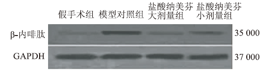

