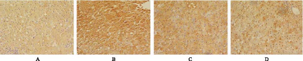
Objective To investigate the effect and mechanism of Baihuadan Xiaogu decoction (BHD) in liver fibrosis. Methods Macaca fascicularis were divided into four groups: the normal control group, the model control group, the BHD-L group and BHD-H group.Hepatic fibrosis models establish by CCl4 (0.5 mL·kg-1) twice a week for 6 month, at the same time, high fat diet was feed.After six months, BHD-L and BHD-H (1.0,4.0 g·kg-1) were administered orally to groups respectively, whereas the normal control and model control groups were orally administered with pure water.Two months later, the blood sample was collected for the examination of alanine aminotransferase (ALT), glutamic oxalacetic transaminase (AST), and total bilirubin (T-BiL).The contents of transforming growth factor-β (TGF-β), alpha-smooth muscle actin (α-SMA), matrix metalloprotein (MMP-2) and tissue inhibitor of metalloproteinase-1 (TIMP-1) in serum were analyzed by ELISA assay.The expression of TGF-β1 in liver biopsy specimens was detected by immunohistochemical staining. Results Compared with model control group, both BHD groups can reduce the levels of Macaca fascicularis serum ALT, AST, T-BiL.It can reduce the levels of TGF-β1, α-SMA and related matrix metalloprotein, regulatory effects on the expression of TGF-β1. Conclusion BHD can prevent liver fibrosis in Macaca fascicularis by reducing the level of TGF-β1, α-SMA and regulate the liver extracellular matrix metabolism.
Key words:
decoction
;
Traditional Zhuang medicine
;
Liver fibrosis/macaca fascicularis
表2
4组食蟹猴血清 TGF-β1、α-SMA、MMP-2、TIMP-1 含量的比较
Tab.2
Comparion of the serum content of TGF-β1,α-SMA,MMP-2 and TIMP-1 among four groups of Macaca fascicularis \(\overline{x}\)±s,n=3
组别
TGF-β1
α-SMA
空白对照组
12.22±0.22
24.37±0.30
模型对照组
17.99±5.40*1
25.68±1.94*1
BHD-L组
13.94±0.97*3
27.08±1.49
BHD-H组
12.36±0.38*3
24.09±0.77*3
组别
MMP-2
TIMP-1
空白对照组
12.54±0.25
123.02±4.07
模型对照组
17.55±3.69*1
159.42±30.53*2
BHD-L组
14.60±10.58*3
131.37±2.05*3
BHD-H组
13.07±0.44*3
121.26±4.58*3
Compared with blank control group,*1P<0.05,*2P<0.01;compared with model control group,*3P<0.05
与空白对照组比较,*1P<0.05,*2P<0.01;与模型对照组比较,*3P<0.05
表2
4组食蟹猴血清 TGF-β1、α-SMA、MMP-2、TIMP-1 含量的比较
Tab.2
Comparion of the serum content of TGF-β1,α-SMA,MMP-2 and TIMP-1 among four groups of Macaca fascicularis \(\overline{x}\)±s,n=3
GRESSNER AM,WEISKIRCHENR.Modem pathogenetic concepts of liver fibrosis suggest stellate cells and TGF-beta as major players and the rapeutic targets[J].Cell Mol Med,2006,10(1):76-99.
Abstract Hepatic fibrosis is a scarring process that is associated with an increased and altered deposition of extracellular matrix in liver. At the cellular and molecular level, this progressive process is mainly characterized by cellular activation of hepatic stellate cells and aberrant activity of transforming growth factor-尾1 and its downstream cellular mediators. Although the cellular responses to this cytokine are complex, the signalling pathways of this pivotal cytokine during the fibrogenic response and its connection to other signal cascades are now understood in some detail. Based on the current advances in understanding the pleiotropic reactions during fibrogenesis, various inhibitors of transforming growth factor-尾 were developed and are now being investigated as potential drug candidates in experimental models of hepatic injury. Although it is too early to favour one of these antagonists for the treatment of hepatic fibrogenesis in human, the experimental results obtained yet provide stimulatory impulses for the development of an effective treatment of choice in the not too distant future. The present review summarises the actual knowledge on the pathogenesis of hepatic fibrogenesis, the role of transforming growth factor-尾 and its signalling pathways in promoting the fibrogenic response, and the therapeutic modalities that are presently in the spotlight of many investigations and are already on the way to take the plunge into clinical studies.
Objective : Guizhi Fuling Wan which inhibited hepatic fibrosis in rats was observed and its possible mechanism of action was explored. Method : According to the random-number table, wistar rats of 60 were divided into control group of 8 (group A) and model group of 52.The control group was injected subcutaneously with normal saline by a dose of 3 mL·kg, model group was injected subcutaneously with 40% CCl to make rat liver fibrosis model by a dose of 3 mL·kg for the consecutive 6 weeks. And according to the random-number table again, model group rats were divided into the model group(group B) and the treatment group which comprised low dose (group C), middle dose (group D) and high dose (group E). Group A and group B were given normal saline by a dose of 9.0 mL·kg. Group C, group D and group E were given Guizhi Fuling Wan by the dose of 0.45, 0.9, 1.8 g·kg·drespectively, and CCl would be maintained subcutaneously during period of 4 weeks. After 4 weeks of treatment, general condition, body weight, liver and spleen weight and their coefficient of rats were compared in each group;rats of pathological changes in each group were determined by naked eye,light microscopy and electron microscopy,rspectively. The protein expression of α-SMA, TGF-, CTGF, Co-Ⅰ and Co-Ⅲ was detected by using immunohistochemical stains in each group. The mRNA genetic expression of α-SMA, TGF-,CTGF,Co-Ⅰ and Co-Ⅲ was detected by using Real-time quantitative PCR in each group. Result : Immunohistochemical stains and real-time quantitative PCR: In model group, protein and mRNA genetic expression of α-SMA,TGF-,CTGF, Co-Ⅰ and Co-Ⅲ was significantly increased in liver tissue (,CTGF,Co-Ⅰ and Co-Ⅲ in liver tissue of rats. Among the treatment group,group D and group E decreased mRNA genetic expression of α-SMA,Co-Ⅰ and Co-Ⅲ significantly (, CTGF obviously(, CTGF significantly(, CTGF, Co-Ⅰ and Co-Ⅲ gene and protein expression, have a good anti-liver fibrosis.

 , 陈少锋
, 陈少锋