中国科技论文统计源期刊 中文核心期刊  
美国《化学文摘》《国际药学文摘》
《乌利希期刊指南》
WHO《西太平洋地区医学索引》来源期刊  
日本科学技术振兴机构数据库(JST)
第七届湖北十大名刊提名奖  
HERALD OF MEDICINE, 2018, 37(6): 662-666
doi: 10.3870/j.issn.1004-0781.2018.06.005
叶酸壳寡糖修饰的聚乳酸-羟基乙酸共聚物纳米粒对耐紫杉醇人卵巢癌细胞增殖的影响
Effect of Poly (lactic-co-glycolic acid) Nanoparticles Surface Modified with Folic Acid Conjuncted Chitosan Oligosaccharide on Proliferation of SKOV-3/TAX Cells
邓艾平1,, 王奕1, 刘珏1, 胡振夏2, 符旭东2

摘要:

目的 制备由叶酸壳寡糖修饰的聚乳酸-羟基乙酸共聚物(PLGA)纳米粒(F-CS-PLGA-NPs),考察其体外对人卵巢癌细胞(SKOV-3)和耐紫杉醇卵巢癌细胞(SKOV-3/TAX)的抑制作用。方法 采用碳二亚胺法制备叶酸耦联的壳寡糖,以此作为普通纳米粒(PLGA-NPs)的向导材料,采用界面沉积法制备F-CS-PLGA-NPs,噻唑蓝(MTT)法测定对SKOV-3和SKOV-3/TAX体外增殖的抑制作用,流式细胞仪检测细胞凋亡率。结果 体外实验结果显示PLGA-NPs和F-CS-PLGA-NPs对SKOV-3/TAX的增殖抑制作用比SKOV-3更不敏感。F-CS-PLGA-NPs对SKOV-3和SKOV-3/TAX的半数抑制浓度(IC50)分别为(23.17±2.45)和(88.81±10.69) nmol·L-1结论 叶酸壳寡糖对PLGA-NPs的修饰可增加其对耐药肿瘤细胞的靶向性,为耐药肿瘤的治疗提供新的思。

关键词: 聚乳酸-羟基乙酸共聚物 ; 纳米粒 ; 叶酸壳寡糖修饰 ; 增殖 ; 人卵巢癌细胞/耐紫杉醇

Abstract:

Objective To encapsulate PLGA nanoparticles modified with folic acid conjuncted chitosan (F-CS-PLGA-NPs) and to study its in vitro inhibitory effect on SKOV-3 and SKOV-3/TAX. Methods Folic acid conjuncted chitosan oligosaccharide was synthesized using carbodiimide as the catalys, which was used as nanoparticles materials to obtain the paclitaxel loaded nanoparticles. MTT experiment was adopted to evaluate the inhibitory effect of F-CS-PLGA-NPs on SKOV-3 and SKOV-3/TAX cells. Results The in vitro experiments showed that the inhibitory effect of PLGA-NPs and F-CS-PLGA-NPs on proliferation of SKOV-3/TAX was less sensitive than that on the proliferation of SKOV-3. The IC50 of F-CS-PLGA-NPs on SKOV-3 and SKOV-3/TAX cells were (23.17±2.45) nmol·L-1 and (88.81±10.69) nmol·L-1. Conclusion Folic acid conjuncted chitosan oligosaccharide modification could increase the targeting efficiency of nanoparticles on drug-resistant tumor cell, which might be a potential formulation for treating drug-resistant cancer cells.

Key words: Poly (lactic-co-glycolic acid) ; Nanoparticles ; Folic acid conjuncted chitosan oligosaccharide modify ; Proliferation ; SKOV-3/TAX

紫杉醇(paclitaxel,PTX)通过促进微管蛋白聚合抑制解聚,保持微管蛋白稳定,抑制细胞有丝分裂[1],是卵巢癌治疗的一线药物之一。目前临床缺乏其靶向制剂,且卵巢癌通过细胞内化疗药物外排、细胞内化疗药物代谢解毒增加、DNA损伤修复能力增加、信号传导通障碍、细胞凋亡异常等多种机制产生耐药[2], 其中药物外排所致耐药最为常见。叶酸受体是一种糖基化磷脂酰肌醇连接的膜糖蛋白,在90%以上的卵巢癌细胞上呈现高表达[3],其通过内化方式将叶酸衍生物带入细胞,可成功避免药物外排而将药物导入细胞内,增强了对耐药肿瘤细胞的增殖抑制作用。笔者在本实验拟构建一种叶酸壳寡糖修饰的聚乳酸-羟基乙酸共聚物[poly(lactic-co-glycolic acid),PLGA]纳米粒(F-CS-PLGA-NPs),评价其体外对人卵巢癌细胞(SKOV-3)和耐紫杉醇卵巢癌细胞(SKOV-3/TAX)增殖的抑制作用。

1 材料与方法
1.1 仪器

UV-2550紫外-可见分光光度计(日本岛津公司),NICOLET 5700 FTIR Spectrometer红外光谱仪(美国热电公司),Nano-ZS90激光粒度仪(英国马尔文仪器有限公司),H-300型透射电镜(日本日立集团),FEI Quanta 200扫描电子显微镜(荷兰FEI公司),JEOL JFC-1600 AUTO FINE COATER 离子溅射仪(日本电子株式会社),流式细胞仪(FACSCalibur,美国Becton Dickinson),LGJ冷冻干燥机(军事医学科学院实验仪器厂),PC-2000型高效液相色谱(天津市兰博实验仪器设备有限公司),JY92-2D超声细胞粉碎机(宁波新芝生物科技股份有限公司),RT-6500酶标仪(深圳雷杜生命科学股份有限公司),BP211D电子分析天平(德国赛多利斯集团,感量:0.01/0.1 mg)。

1.2 试药

紫杉醇(武汉远城科技发展有限公司,批号:20100925,含量99.8%),叶酸对照品(中国食品药品检定研究院,批号:100074-200412,含量≥98%),叶酸(上海如吉生物科技发展有限公司,批号:20110125,含量≥97%),壳寡糖(分子量2 100,脱乙酰度95%,山东奥康生物科技有限公司,批号:20100809),端羧基聚乳酸羟基乙酸共聚物(PLGA-COOH,二单体比例75:25,分子量10 000,山东医疗器械研究所,批号:20100612),1-乙基-(3-二甲基氨基丙基)碳酰二亚胺盐酸盐(EDC·HCl,北京中生瑞泰科技有限公司,批号:20100625),聚乙烯醇AH-26(PVA AH-26,国药集团化学试剂有限公司,批号:20100116),紫杉醇注射液(TAXOL,北京华素制药股份有限公司,批号:1102291),达尔伯克改良伊格尔培养基(DMEM,美国Hyclone公司,批号:NAD1569),RPMI-1640培养基(美国Invitrogen 公司,批号:NAB1581),细胞周期凋亡试剂盒(江苏碧云天生物技术研究所),其他试剂为分析纯。

1.3 细胞

人卵巢癌上皮细胞(SKOV-3,武汉大学细胞典藏中心)。

1.4 耐紫杉醇卵巢癌细胞(SKOV-3/TAX)的诱导

取对数生长期SKOV-3细胞,0.25%胰蛋白酶消化后调整至浓度为1×10⁵个·mL-1的细胞悬浮液,接种于75 cm2的培养瓶中培养,使用含紫杉醇终浓度为300 μg·mL-1的RPMI-1640培养基培养2 h后,换用不含药液培养基继续培养,待细胞生长至对数生长期时重复剂量诱导,每隔4周测定一次紫杉醇对细胞的半数抑制浓度(IC50)值[4,5]

1.5 激光共聚焦显微观察细胞形态

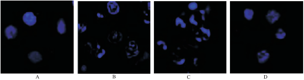
分别取对数生长期的SKOV-3/TAX细胞,稀释成1×105个·mL-1,接种于底部置有盖玻片的6孔板,待细胞生长铺满底部50%时,加入紫杉醇终浓度为100 nmol·L-1的聚乳酸-羟基乙酸共聚物纳米粒(PLGA-NPs)、叶酸壳寡糖修饰的PLGA-NPs(F-CS-PLGA-NPs)、F-CS-PLGA-NPs+叶酸干预,培养24 h后取出盖玻片,用磷酸盐缓冲液(PBS)反复清洗3次,滴加5.0 μg·mL-1DAPI溶液,避光孵育30 min后于激光共聚焦显微镜下观察细胞形态。

1.6 噻唑蓝(MTT)法测定纳米粒体外对SKOV-3和SKOV-3/TAX的抑制作用

取处于对数生长期的SKOV-3和SKOV-3/TAX细胞接种于96孔板,待细胞生长铺满底部后加入紫杉醇终浓度为10,25,50,100,250,500,1 000 nmol·L-1的F-CS-PLGA-NPs,并设PLGA-NPs、TAXOL液组,培养48 h后;加入MTT和二甲亚砜(DMSO)后用酶联免疫吸附测定(ELISA)检测仪于波长492 nm处测定吸光度(A)值;细胞存活率:存活率(%)=(实验组A值-空白组A值)/(正常组A值-空白组A值)×100%。耐药指数(resistance index,RI)计算公式:RI=耐药细胞的IC50/敏感细胞的IC50

1.7 流式细胞检测细胞凋亡率

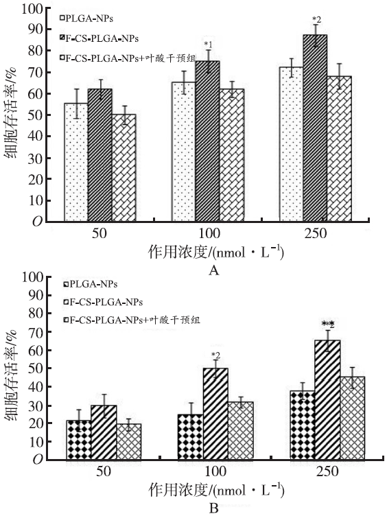
分别取对数生长期的SKOV-3和SKOV-3/TAX细胞稀释成1×105个·mL-1后接种于6孔板中,加入紫杉醇终浓度为50,100,250 nmol·L-1的PLGA-NPs、F-CS-PLGA-NPs、F-CS-PLGA-NPs+叶酸干预组,培养12 h后弃去旧培养基,每孔加入4 ℃ PBS 2 mL反复清洗3次,加入胰酶200 μL消化,去除消化液后加入含有20%胎牛血清的PBS终止消化,离心后用预冷的结合缓冲液490 μL重悬得到细胞悬液,加入Annexin V-FITC 5 μL和碘化丙啶(propidium iodide,PI)5 μL,避光孵育15 min,上机检测。

1.8 统计学方法

采用SPSS16.0版统计软件进行分析计,数据以均数±标准差( x ¯ ±s)表示,采用独立样本t检验分析,以P<0.05为差异有统计学意义。

2 结果
2.1 RI测定结果

经历约30周的诱导期后,SKOV-3和SKOV-3/TAX的IC50结果见表1。

表1 不同紫杉醇制剂对SKOV-3和SKOV-3/TAX的IC50和RI
Tab.1 IC50 and RI of various paclitaxel formulation on SKOV-3 and SKOV-3/TAX
制剂 IC50/(nmol·L-1) RI
SKOV-3 SKOV-3/TAX
TAXOL 60.58±5.12*1 535.65±53.54*1 8.8
PLGA-NPs 46.72±5.18*1 290.87±43.86*1 6.2
F-CS-PLGA-NPs 23.17±2.45 88.81±10.69 3.8

Compared with F-CS-PLGA-NPs,F=13.84,*1P<0.01

与F-CS-PLGA-NPs比较,F=13.84,*1P<0.01

表1 不同紫杉醇制剂对SKOV-3和SKOV-3/TAX的IC50和RI

Tab.1 IC50 and RI of various paclitaxel formulation on SKOV-3 and SKOV-3/TAX

2.2 细胞形态的观察

在倒置显微镜下观察SKOV-3和SKOV-3/TAX的细胞形态,可见SKOV-3呈椭圆型、细胞质居中;SKOV-3/TAX细胞体积明显增大、形态不规则、胞质内颗粒增多,见图1。

用荧光显微镜观察SKOV-3/TAX细胞对纳米粒的摄取,可见给予PLGA-NPs后,细胞核的染色质高度凝聚、边缘化,而给予F-CS-PLGA-NPs后,细胞核裂解为碎块,产生许多凋亡小体,说明细胞对F-CS-PLGA-NPs摄取的作用显著增强,并且这一作用可被游离叶酸阻断,说明叶酸的表面修饰可以增强SKOV-3/TAX对纳米粒的摄取。结果见图2。

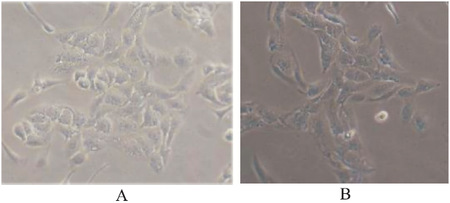
图1 倒置显微镜下SKOV-3(A)和SKOV-3/TAX(B)的细胞形态(×1 000)

Fig.1 Cell morphology of SKOV-3 (A)and SKOV-3/TAX(B)observed by inverted microscop(×1 000)

图2 SKOV-3/TAX在激光共聚焦显微镜下细胞形态
A.正常细胞;B.PLGA-NPs组;C.F-CS-PLGA-NPs组;D.F-CS-PLGA-NPs+叶酸干预组

Fig.2 Cell morphology of SKOV-3/TAX observed by laser scanning confocal microscope
A.normal cell;B.PLGA-NPs group;C.F-CS-PLGA-NPs group;D.F-CS-PLGA-NPs and folic acid group

2.3 体外浓度抑制情况

将不同浓度紫杉醇制剂作用于SKOV-3细胞48 h后,F-CS-PLGA-NPs显示出较TAXOL和PLGA-NPs更强的增殖抑制作用,随着紫杉醇浓度增加,对细胞的抑制作用增强,结果见
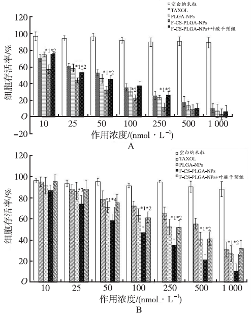
图3A。将不同浓度的紫杉醇制剂作用于SKOV-3/TAX 细胞48 h后,当紫杉醇浓度≥25 nmol·L-1时,F-CS-PLGA-NPs对SKOV-3/TAX的杀伤作用强于TAXOL和PLGA-NPs,可能是由于SKOV-3/TAX细胞表面转运蛋白介导的药物外排使TAXOL和PLGA-NPs不能有效进入细胞体内,而F-CS-PLGA-NPs则还可以通过其表面的叶酸受体与SKOV-3/TAX细胞表面的叶酸及其类似物特异性结合,介导细胞內吞将F-CS-PLGA-NPs摄入细胞内;在紫杉醇浓度较低(<25 nmol·L-1)时,F-CS-PLGA-NPs对SKOV-3/TAX的增殖抑制作用与PLGA-NPs和TAXOL组相当,这说明对耐药细胞的增殖作用可能还与纳米粒表面叶酸浓度有关,结果见图3B。细胞培养基中加游离叶酸封闭叶酸受体,可显著降低F-CS-PLGA-NPs对细胞的杀伤作用,进一步证明了药物对细胞的作用是由细胞表面的叶酸受体介导的。

图3 不同浓度紫杉醇对SKOV-3(A)和SKOV-3/TAX (B)增殖抑制作用(x¯±s,n=6)
与TAXOL 组比较,*1P<0.01,*3P<0.05;与PLGA-NPs组比较,*2P<0.01,*4P<0.05

Fig.3 Inhibitory effect of different concentrations of PTX on the proliferation of SKOV-3 (A)and SKOV-3/TAX (B)(x¯±s,n=6)
Compared with TAXOL group;*1P<0.01,*3P<0.05;Compared with PLGA-NPs group, *2P<0.01,*4P<0.05

2.4 流式细胞仪检测细胞凋亡率

PLGA-NPs和F-CS-PLGA-NPs均能诱导SKOV-3和SKOV-3/TAX的凋亡,同一浓度下,PLGA-NPs和F-CS-PLGA-NPs对SKOV-3/TAX的凋亡率比SKOV-3更低,说明SKOV-3/TAX对紫杉醇的耐药可能与肿瘤细胞表面转运蛋白介导的药物外排机制有关,结果见图4。

图4 不同浓度紫杉醇诱导SKOV-3(A)和SKOV-3/TAX(B)凋亡作用(x¯±s,n=6)
与PLGA-NPs组比较,*1P<0.05,*2P<0.01

Fig.4 Apoptosis induction of different concentration of PTX on SKOV-3(A)and SKOV-3/ TAX (B)(x¯±s,n=6)
Compared with PLGA-NPs group, *1P<0.05,*2P<0.01

3 讨论

本实验依据卵巢癌肿瘤细胞表面叶酸受体的高表达,F-CS-PLGA-NPs与其结合时能特异性地将纳米粒包裹的药物导入细胞内,杀伤肿瘤细胞;紫杉醇在临床上主要用于卵巢癌和乳腺癌的治疗,故选择卵巢癌上皮细胞SKOV-3和SKOV-3/TAX细胞作为体外评价细胞。采用大剂量冲击诱导耐药细胞的形成,与临床治疗模式更相似。结果表明,SKOV-3/TAX的细胞体积较SKOV-3明显增大,且胞质颗粒增加,这与紫杉醇通过促进微管蛋白聚合抑制解聚,抑制细胞有丝分裂的作用有关。

实验结果显示,F-CS-PLGA-NPs与TAXOL和PLGA-NPs比较,对SKOV-3/TAX的IC50均差异有统计学意义,说明SKOV-3/TAX可能耐药机制与膜转运蛋白过表达所致的药物外排有关,这与文献[6]报道一致。膜转运蛋白主要有P-糖蛋白(P-gp)、多药耐药相关蛋白(Mrps)、乳腺癌耐药蛋白(Bcrp)等。P-gp作为一种依赖ATP酶供能的跨膜药泵,可把细胞内的药物泵出或使之溢流到细胞外;Mrp1是结合转运蛋白盒,可降低药物在细胞内的积累[7]。研究显示,紫杉醇所致肿瘤细胞耐药与P-gp和Mrps过量表达有关[8]。F-CS-PLGA-NPs对SKOV-3和SKOV-3/TAX的增殖抑制作用强于TAXOL和PLGA-NPs,且这种作用能被游离的叶酸所阻断;当紫杉醇浓度≥25 nmol·L-1时,F-CS-PLGA-NPs对SKOV-3/TAX的杀伤作用显著强于TAXOL和PLGA-NPs,可能是其表面的叶酸受体与细胞表面的叶酸特异性结合后将药物导入细胞内,F-CS-PLGA-NPs进入细胞并不能逆转细胞对紫杉醇的耐药性,但能将更多的紫杉醇靶向入肿瘤细胞;同时F-CS-PLGA-NPs表面的正电荷基团促使其与带负电荷的肿瘤细胞接近,从而加强抗肿瘤效果。

对紫杉醇耐药的卵巢癌细胞最常见的耐药机制包括细胞内微管蛋白浓度降低、微管蛋白亚型表达改变、膜转运蛋白过表达等,本实验在纳米粒表面进行叶酸修饰后仅能说明F-CS-PLGA-NPs不受细胞膜转运蛋白过表达的影响,不能说明其对所有耐药肿瘤细胞均有较强的增殖抑制作用,有关卵巢癌对紫杉醇的具体耐药机制还需要进一步研究。

The authors have declared that no competing interests exist.

参考文献

[1] PEREZ E A.Microtubule inhibitors:differentiating tubulin-inhibiting agents based on mechanisms of action,clinical activity,and resistance[J].Mol Cancer Ther,2009,8(8):2086-2095.
DOI:10.1158/1535-7163.MCT-09-0366      URL    
[本文引用:1]
[2] 杨晶珍,刘海清,张剑.卵巢癌耐药机制的研究进展[J].中国老年学杂志,2015,35(4):1117-1120.
[本文引用:1]
[3] ZHAO X B,LEE R J.Tumor-selective targeted delivery of genes and antisense oligodeoxyribonucleotides via the folate receptor[J].Adv Drug Deliv Rev,2004,56(8):1193-1215.
Gene therapy is a promising approach for the treatment of cancer. The main obstacle for the clinical application of cancer gene therapy is the lack of gene transfer vectors that are safe, efficacious, and tumor-selective. In recent years, targeted gene delivery through cellular receptors, using either viral or nonviral vectors, is emerging as a novel approach to enhance the efficacy of tumor-selective gene delivery. The folate receptor (FR), which is absent in most normal tissues and elevated in over 90% of ovarian carcinomas and at a high frequency in other human malignancies, is an attractive tumor-selective target. FR-targeted vectors include folate-derivatized adenoviruses, cationic polymers, cationic liposomes, and pH-sensitive liposomes. In addition, FR-targeted liposomes have been evaluated for the targeted delivery of antisense oligodeoxyribonucleotides (ODNs). These vectors have invariably shown impressive FR-selectivity in cell culture assays and, in addition, shown promising tumor-specific gene transfer activity in several in vivo models. There are important theoretical advantages for FR-targeted vectors over traditional non-targeted vectors in therapeutic gene and oligodeoxyribonucleotides delivery in vivo to cancer cells. Further preclinical characterization of these vectors is, therefore, warranted to determine their potential utility in cancer gene therapy.
DOI:10.1016/j.addr.2004.01.005      PMID:15094215      URL    
[本文引用:1]
[4] 樊蓓,李红霞,吴玉梅,.紫杉醇诱导的卵巢癌耐药细胞株的生物学特性及其保存[J].肿瘤防治研究,2015,42(4):334-339.
目的 研究紫杉醇诱导的卵巢癌耐药细胞株的生物学特性,探索耐药细胞的长期保存条件,以建立稳定的体外实验模型。方法 选用卵巢癌化疗敏感细胞株SKOV3,分别采用大剂量冲击和小剂量浓度递增诱导建立耐药的卵巢癌细胞株SKOV3/TAX300、SKOV3/TAX30。四甲基偶氮唑盐(MTT)法检测耐药细胞对化疗药物紫杉醇的IC50;比较耐药细胞和敏感细胞的细胞平板克隆形成、生长曲线倍增时间的差异;荧光定量PCR检测敏感细胞和耐药细胞中耐药相关基因的表达;比较无药冻存和加药冻存的方式对耐药细胞的保存效果。结果 耐药细胞SKOV3/TAX300,耐药指数为4.60,耐药细胞SKOV3/TAX30,耐药指数为51.31;与敏感细胞相比,耐药细胞体积增大,形态不规则,倍增时间延长。多药耐药基因1(MDR1)在两种耐药细胞中高表达,肺耐药相关蛋白(LRP)、蛋白激酶C&alpha;(PRKCA)基因仅在SKOV3/TAX300中的表达增高,BCL-2样1基因(BCL2L1)在SKOV3/TAX30中低表达。耐药细胞冻存在含有药物的条件下,有利于耐药性的保持。结论 大剂量冲击和小剂量浓度递增两种方法诱导的耐药细胞为研究紫杉醇耐药机制建立了良好的体外模型,两种耐药细胞SKOV3/TAX300和SKOV3/TAX30具有不同的生物学特性。
[本文引用:1]
[5] ZHANG J,ZHAO J,ZHANG W,et al.Establishment of paclitaxel-resistant cell line and the underlying mechanism on drug resistance[J].Int J Gynecol Cancer,2011,22(9):1450-1456.
The objective of this study was to establish a taxol (TAX)-resistant human ovarian carcinoma cell line and investigate its drug-resistant mechanism. Using the dose calculated from clinical chemotherapy, we established a TAX-resistant human ovarian carcinoma cell line OC3/TAX300 by intermissive and repeated exposure to TAX of a high concentration at 300 渭g/mL for 2 hours each time. The drug sensitivity was examined by tetrazolium dye (MTT) test. Distribution of cell cycle, DNA content analysis, and P-glycoprotein (P-gp) expression were detected by flow cytometry. We detected the differential gene expression by use of cDNA microarray. The reverse transcription-polymerase chain reaction and Western blot were used to verify the representative mRNA expression and their protein expression. OC3/TAX300 cells were established after 10 months with stable resistance, and the drug resistance index was 6.70. It displayed significant cross-resistance to topotecan. Distribution of cell cycle revealed a higher percentage of G2 + M phase (P < 0.01), a lower percentage of S phase (P < 0.05), and overexpression of P-gp (P < 0.01). The cDNA microarray analysis showed that there were 134 significantly differential expression genes in all, of which up-regulated and down-regulated genes were 17 and 117, respectively. The up-regulated genes JAK2 (Janus kinase 2) and HSPC154 were confirmed by reverse transcription-polymerase chain reaction and Western blot. The OC3/TAX300 cell line is an ideal model to investigate the mechanism of TAX resistance. Taxol resistance in this cell could be related to overexpression of P-gp and the change of cell cycle profiles. The differential expression genes of JAK2 and HSPC154 may be candidate genes associated with TAX resistance in ovarian carcinoma cell lines.
DOI:10.1097/IGC.0b013e31826e2382      PMID:23051955      URL    
[本文引用:1]
[6] YANG X,SHEN J,GAO Y,et al.NSC23925 prevents the development of paclitaxel resistance by inhibiting the introduction of P-glycoprotein (Pgp)and enhancing apoptosis[J].Int J Cancer,2015,137(8):2029-2039.
Strategies to prevent the emergence of drug resistance will increase the effectiveness of chemotherapy treatment and prolong survival of women with ovarian cancer. The aim of our study is to determine the effects of NSC23925 on preventing the development of paclitaxel resistance in ovarian cancer both in cultured cells in vitro and in mouse xenograft models in vivo , and to further elucidate these underlying mechanisms. We first developed a paclitaxel-resistant ovarian cancer cell line, and demonstrated that NSC23925 could prevent the introduction of paclitaxel resistance by specifically inhibiting the overexpression of P-glycoprotein (Pgp) in vitro . The paclitaxel-resistant ovarian cancer cells were then established in a mouse model by continuous paclitaxel treatment in combination with or without NSC23925 administration in the mice. The majority of mice continuously treated with paclitaxel alone eventually developed paclitaxel resistance with overexpression of Pgp and antiapoptotic proteins, whereas mice remained sensitivity to paclitaxel and displayed lower expression levels of Pgp and antiapoptotic proteins after administered continuously with combination of paclitaxel鈥揘SC23925. Paclitaxel鈥揘SC23925-treated mice experienced significantly longer overall survival time than paclitaxel-treated mice. Furthermore, the combination of paclitaxel and NSC23925 therapy did not induce obvious toxicity as measured by mice body weight changes, blood cell counts and histology of internal organs. Collectively, our observations provide evidence that NSC23925 in combination with paclitaxel may prevent the onset of Pgp or antiapoptotic-mediated paclitaxel resistance, and improve the long-term clinical outcome in patients with ovarian cancer.
DOI:10.1002/ijc.29574      PMID:4529776      URL    
[本文引用:1]
[7] MAHADEVAN D,SHIRAHATTI N.Strategies for targeting the multidrug resistance-1 (MDR1)/P-gp transporter in human malignancies[J].Curr Cancer Drug Targets,2005,5(6):445-455.
ATP-binding cassette (ABC) transporters are a super family of channel proteins that include multidrug resistance 1 (MDR1/P-gp) and multi-drug resistance related proteins (MRPs) whose functions include the efflux of ions, nutrients, lipids, amino acids, peptides, proteins and drugs. The three-dimensional structures of bacterial and human ABC transporters demonstrate that these proteins are ATP-dependent molecular machines that scan the inner membrane leaflet for lipids/drugs and flip them to the outer membrane leaflet. In many human cancers, the level of expression of MDR1 is an important independent prognostic factor that determines response to combination chemotherapy. Intrinsic and acquired resistance to chemotherapy exposure are due to a high level of MDR1 expression that enhances drug efflux, with associated poor clinical outcome and lower complete remission (CR) rates. Recent clinical trials in hematological and solid malignancies have shown promise for a prolonged remission and improved overall survival when the MDR1 P-gp is inhibited when combined with chemotherapy. Structure-based homology modeling of these ABC transporters may help design novel drug candidates to both the membrane-spanning domain (MSD) and the nucleotide-binding domain (NBD) located within the cytoplasm. This review will highlight advances in the utilization of homology modeling in the drug discovery process and how this will impact on fundamental insights to the development of novel therapeutics that could alter and/or inhibit their functions.
DOI:10.2174/1568009054863609      PMID:16178818      URL    
[本文引用:1]
[8] SARKADI B,HOMOLYA L,SZAKACS G,et al.Human multidrug resistance ABCB transporters:participation in a chemoimmunity defense ststerm[J].Physiol Rev,2006,86(4):1179-1236.
In this review we give an overview of the physiological functions of a group of ATP binding cassette (ABC) transporter proteins, which were discovered, and still referred to, as multidrug resistance (MDR) transporters. Although they indeed play an important role in cancer drug resistance, their major physiological function is to provide general protection against hydrophobic xenobiotics. With a highly conserved structure, membrane topology, and mechanism of action, these essential transporters are preserved throughout all living systems, from bacteria to human. We describe the general structural and mechanistic features of the human MDR-ABC transporters and introduce some of the basic methods that can be applied for the analysis of their expression, function, regulation, and modulation. We treat in detail the biochemistry, cell biology, and physiology of the ABCB1 (MDR1/P-glycoprotein) and the ABCG2 (MXR/BCRP) proteins and describe emerging information related to additional ABCB- and ABCG-type transporters with a potential role in drug and xenobiotic resistance. Throughout this review we demonstrate and emphasize the general network characteristics of the MDR-ABC transporters, functioning at the cellular and physiological tissue barriers. In addition, we suggest that multidrug transporters are essential parts of an innate defense system, the "chemoimmunity" network, which has a number of features reminiscent of classical immunology.
DOI:10.1152/physrev.00037.2005      PMID:17015488      URL    
[本文引用:1]
资源
PDF下载数    
RichHTML 浏览数    
摘要点击数    

分享
导出

相关文章:
关键词(key words)
聚乳酸-羟基乙酸共聚物
纳米粒
叶酸壳寡糖修饰
增殖
人卵巢癌细胞/耐紫杉醇

Poly (lactic-co-glycolic ...
Nanoparticles
Folic acid conjuncted chi...
Proliferation
SKOV-3/TAX

作者
邓艾平
王奕
刘珏
胡振夏
符旭东

DENG Aiping
WANG Yi
LIU Jue
HU Zhenxia
FU Xudong