中国科技论文统计源期刊 中文核心期刊
美国《化学文摘》《国际药学文摘》
《乌利希期刊指南》
WHO《西太平洋地区医学索引》来源期刊
日本科学技术振兴机构数据库(JST)
第七届湖北十大名刊提名奖
美国《化学文摘》《国际药学文摘》
《乌利希期刊指南》
WHO《西太平洋地区医学索引》来源期刊
日本科学技术振兴机构数据库(JST)
第七届湖北十大名刊提名奖

 , 但卫斌, 谢俊杰, DAN Weibin, XIE Junjie
目的 探讨重楼总皂苷(RPTS)对肝癌HepG2细胞放射敏感性的影响及其机制。方法 HepG2细胞培养后分为4组。空白对照组:用正常培养液继续培养48 h;RPTS组:用低细胞毒性浓度的RPTS(25 μg·mL-1)干预48 h;单纯照射组:先用正常培养液继续培养48 h,再用6 MV的X线进行照射;照射+RPTS组:先用低细胞毒性浓度的RPTS(25 μg·mL-1)干预48 h,再用6 MV的X线进行照射。采用CCK-8法检测重楼总皂苷对HepG2细胞增殖的影响;流式细胞术检测重楼总皂苷对细胞周期及凋亡的影响; Western blotting法检测MUC-1蛋白的表达情况。结果 与空白对照组比较,RPTS能明显抑制HepG2细胞增殖,其抑制率随药物浓度的增加、药物持续时间的延长明显增大。与RPTS组比较,照射+RPTS组早期凋亡率和晚期凋亡率明显增加(
Objective To investigate the effect of total saponins from
原发性肝癌是我国第四常见的恶性肿瘤,在肿瘤相关致死病因中排名第三,严重威胁人民的健康和生命[1,2]。过去考虑全肝耐受放射剂量较低、放射线对于周围正常组织的影响,很难提高肝脏肿瘤的靶区放射剂量,所以放射治疗(放疗)在肝癌治疗中的应用有限。近年来,随着对放射生物学及放射物理学的不断认识、放疗技术及设备的不断发展,使放疗在肝癌治疗中的应用更加广泛。但肿瘤的辐射抗性以及大剂量照射对正常肝组织的损伤仍然是肝癌放疗中难以突破的瓶颈。重楼是我国传统中药,具有清热解毒、凉肝定惊、消肿镇痛等功效,现代药理研究发现它具有抗病毒、抗肿瘤、免疫调节等功效[3]。重楼总皂苷(
RPTS由成都曼思特生物科技有限公司(批号:A0125,含量≥98%)提供,实验前用二甲亚砜(DMSO)配制成一定浓度的保存液,保存于-20 ℃条件下,实验时用小剂量伊格尔培养液(MEM)配制成不同浓度的药液。CCK-8试剂盒(C0038)购于上海碧云天生物技术有限公司;碘化丙啶(PI)染色试剂盒(PAB180014)及Annexin V-FITC/PI凋亡试剂盒(PAB180012)均购自bio-Swamp公司;BCA蛋白浓度测定试剂盒(P0010S)购于上海碧云天生物技术有限公司;鼠抗MUC-1抗体[C595(NCRC48)]购自Abcam公司。
肝癌HepG2细胞购于中国典型培养物保藏中心(武汉大学保藏中心),用MEM Hyclone培养基、10%胎牛血清、青霉素-链霉素溶液配置成的完全培养基培养于37 ℃、5%二氧化碳(CO2)饱和湿度的细胞培养箱中,实验时取对数生长期细胞备用。
采用瑞典医科达直线加速器(规格型号:ELEKTA-Precise)6 MV X线进行照射。设照射野大小为16 cm×16 cm,采用设源皮距(SSD)照射技术,SSD为100 cm,板缘距离射野边缘约2 cm,剂量率为200 cGy·min-1,照射剂量设为单次8 Gy。
取指数生长期细胞,配制成4×10³个·mL-1的单细胞悬液,保持每孔100 μL接种于96孔板中,复孔3个,置于37 ℃、5%CO2饱和湿度的培养箱中培养24 h,待细胞贴壁后,再给予不同浓度的RPTS(0.0,30,60,120,240 μg·mL-1)干预细胞,每个浓度设5个复孔,每个96孔板边缘均加入磷酸盐缓冲液(PBS)100 μL,再分别继续培养24,48,72 h。然后吸出原液,PBS洗涤1次,每孔中加入用MEM稀释的CCK-8 100 μL,再置于培养箱中继续孵育3 h。用酶联免疫吸附测定(ELISA)法测定每孔在波长450 nm处吸光度(
取对数生长期细胞,清洗消化后制成3×10⁵个·mL-1的单细胞悬液,接种于6孔板中,置于培养箱中继续培养24 h,待细胞贴壁后再分别设空白对照组、RPTS组、单纯照射组、照射+RPTS组。空白对照组:用正常培养液继续培养48 h;RPTS组:用低细胞毒性浓度的RPTS(25 μg·mL-1)干预48 h;单纯照射组:先用正常培养液继续培养48 h,再用6 MV的X线进行照射;照射+RPTS组:先用低细胞毒性浓度的RPTS(25 μg·mL-1)干预48 h,再用6 MV的X线进行照射。照射完成后用PBS洗涤2次,胰酶消化,离心收集细胞,并重悬于Binging Buffer 200 μL中,再根据PI染色试剂盒及Annexin V-FITC/PI凋亡检测试剂盒的说明书进行细胞染色,最后用流式细胞仪检测细胞周期分布及凋亡情况。实验重复3次。
细胞培养和分组方法同“1.5”项。照射完成后收集细胞并提取蛋白,再根据BCA蛋白浓度测定试剂盒说明书测定各组蛋白的含量,每组样品各取蛋白30 μg进行上样,根据Western blotting法操作步骤检测各组中MUC-1蛋白的表达情况。实验重复3次。
采用SPSS19.0版统计软件进行统计分析,计量资料以均数±标准差 (
与空白对照组比较,RPTS能明显抑制HepG2细胞增殖,其抑制率随药物浓度的增加、作用时间的延长明显增大。见
RPTS可影响HepG2细胞的周期分布,减少G0/G1期细胞分布率,将细胞阻滞于S期(
表1 4组HepG2细胞作用48 h后细胞周期分布情况
Tab.1 Distribution of cell cycle in four groups of HepG2 cells after 48 h treatment %,$\bar{x}±s$
与空白对照组比较,RPTS组在RPTS(25 μg·mL-1)干预HepG2细胞48 h后,早期凋亡率和晚期凋亡率均显著增加(
表2 4组HepG2细胞48 h后细胞凋亡变化情况
Tab.2 Apoptosis changes in four groups of HepG2 cells after 48 h treatment %,$\bar{x}±s$
RPTS+照射组MUC-1蛋白的表达最低,呈现时间依赖性(
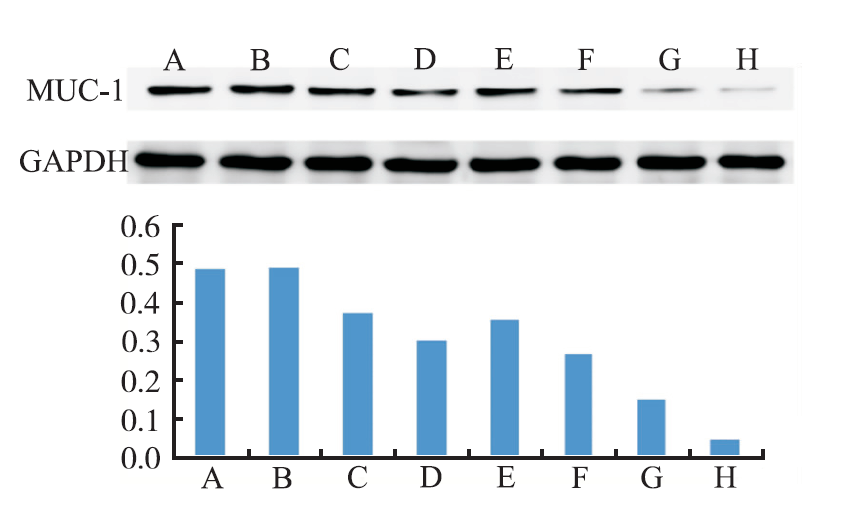
图2
4组HepG2细胞在48和72 h MUC-1蛋白表达
A.空白对照组(48 h);B.空白对照组(72 h);C.RPTS组(48 h);D.RPTS组(72 h);E.单纯照射组(48 h);F.单纯照射组(72 h);G.照射+RPTS组(48 h);H.照射+RPTS组(72 h)
Fig.2
Protein expression of MUC-1 in four groups of HepG2 cells at 48 h and 72 h
A.blank control group(48 h);B.blank control group(72 h);C.RPTS group(48 h);D.RPTS group(72 h);E.irradiation group(48 h);F.irradiation group(72 h);G.irradiation+RPTS group(48 h);H.irradiation+RPTS group(72 h)
根据原发性肝癌诊疗规范,对伴有门静脉/下腔静脉癌栓或肝外转移的Ⅲa期、Ⅲb期肝癌患者,可行姑息性放疗;部分患者术前放疗可使肿瘤体积缩小或降期,而获得手术切除机会[7,8,9];而肝外转移的患者,也可用于等待肝癌肝移植前的治疗,或用于减轻疼痛、梗阻或出血等症状,使肿瘤发展减缓,从而延长生存期[10,11,12];而中央型肝癌切缘距肿瘤≤1 cm的窄切缘术后需要行辅助放疗降低复发概率[13]。
虽然放疗在肝癌治疗中的应用日趋广泛,但是肿瘤的辐射抗性以及大剂量射线照射对正常组织的损伤仍然是肝癌放疗中难以突破的瓶颈。肝细胞对放射线存在显著的剂量-体积效应,其放射耐受量、再生能力与照射体积-剂量、肝脏的功能状态密切相关[14]。肝功能为Child-Pugh A级时,在常规分割放疗下,全肝的耐受剂量为28~30 Gy[15],在非常规低分割放疗(每次分割剂量4~8 Gy)下,全肝的耐受量为23 Gy[16];肝功能为Child-Pugh B级时,肝脏对射线的耐受量明显下降。考虑到亚洲PLA患者常伴有肝硬化和脾功能亢进,导致胃肠道瘀血和凝血功能差,所以放射耐受剂量更低于临床推荐剂量[17]。而肝脏肿瘤的放疗剂量与低分化鳞癌(如鼻咽癌)相近,致死量约为60 Gy/6周,所以正常肝组织的耐受剂量远低于致死量[18]。因此,寻找高效低毒的放射增敏剂是目前肝癌放射治疗的一个研究热点。
中药具有多靶点、多效性的特点,其毒副作用较低,在改善放疗敏感性方面有其自身的特点,作为放疗辅助用药已广泛应用于恶性肿瘤的防治。许多活血化瘀类中药能通过改善血液循环及组织供氧来实现放疗的增敏作用,如莪术油、川芎嗪、川红注射液、通窍活血汤等中药制剂,通过扩张血管、增加血流、改善微循环、破坏肿瘤组织周围和内部纤维蛋白的聚集、降低血液黏稠度、减轻血管闭塞等作用,提高瘤体的氧效值,改善乏氧细胞放射敏感性[19,20]。而部分中药提取物则通过增强对肿瘤细胞DNA的损伤、抑制乏氧细胞损伤的修复来起到增敏作用,如在乏氧条件下,马蔺子甲素可明显降低HeLa细胞中谷胱甘肽含量,抑制DNA的合成和DNA链断裂后的重接修复,从而起到放射增敏作用[21];另外一些中药提取物则可以通过调节肿瘤细胞周期,从而提高放射敏感性,如β-榄香烯可以提高H460癌细胞株G2/M期细胞比例,从而起到放疗增敏作用[22];还有一些补益固本类中药提取物则能通过诱导肿瘤细胞凋亡、促进DNA分子损伤、抑制肿瘤细胞增殖来实现放疗增敏,如人参皂苷Rg3能通过抑制细胞活性、诱导细胞凋亡及DNA分子损伤来实现对食管癌细胞的放疗增敏[23]。
根据实验结果可知,RPTS能抑制HepG2细胞的增殖,且在一定范围内具有时间和浓度依赖性。从细胞周期分布情况可知,低细胞毒性的RPTS能使HepG2细胞周期分布发生改变;而低细胞毒性的RPTS可协同X线进一步诱导细胞发生凋亡,这说明改变细胞周期分布、诱导细胞凋亡可能是RPTS发挥抗肿瘤作用、提高肝癌细胞放射敏感性的一个重要机制。
MUC-1蛋白是一种在大部分肿瘤细胞中都过量表达的糖蛋白,它与肿瘤血管生成、增殖、转移、以及新陈代谢在内的诸多方面都关系密切[24,25,26,27,28]。MUC-1基因作为原发性肝癌的致癌基因,已被发现具有加速肝癌细胞转移和扩散的能力[29],而JAK/STAT信号通路对肝癌细胞的生长增殖起着至关重要的作用[30],抑制STAT3信号通路能增加肝癌细胞的辐射诱导凋亡[31]。笔者前期的研究也发现[32],MUC-1蛋白能通过激活JAK2/STAT3信号通路提高肝癌细胞的放射抵抗性;而MUC-1蛋白通过JAK2/STAT3信号通路以及抗凋亡蛋白Mcl-1和Bcl-xL的诱导作用抑制辐射诱导的细胞凋亡。这意味着降低MUC-1蛋白的表达可能是提高肝癌放疗敏感性的一个潜在靶点。在本实验中,RPTS能单独抑制HepG2细胞中MUC-1蛋白的表达,而当RPTS与X线联合应用,则能进一步抑制MUC-1蛋白的表达,说明RPTS可协同X线进一步抑制MUC-1蛋白的表达
The authors have declared that no competing interests exist.
| [1] |
|
| [2] |
|
| [3] |
|
| [4] |
|
| [5] |
|
| [6] |
|
| [7] |
|
| [8] |
|
| [9] |
|
| [10] |
|
| [11] |
Background Increased collagen deposition provides physical and biochemical signals to support tumor growth and invasion during breast cancer development. Therefore, inhibition of collagen synthesis and deposition has been considered a strategy to suppress breast cancer progression. Collagen prolyl-4-hydroxylase ?? subunit 2 (P4HA2), an enzyme hydroxylating proline residues in -X-Pro-Gly- sequences, is a potential therapeutic target for the disorders associated with increased collagen deposition. However, expression and function of P4HA2 in breast cancer progression are not well investigated. Methods Gene co-expression analysis was performed in the published microarray datasets to identify potential regulators of collagen I, III, and IV in human breast cancer tissue. Expression of P4HA2 was silenced by shRNAs, and its activity was inhibited by 1, 4-DPCA, a prolyl-4-hydroxylase inhibitor. Three-dimensional culture assay was used to analyze roles of P4HA2 in regulating malignant phenotypes of breast cancer cells. Reduced deposition of collagen I and IV was detected by Western blotting and immunofluorescence. Control and P4HA2-silenced breast cancer cells were injected into fat pad and tail vein of SCID mice to examine effect of P4HA2 on tumor growth and lung metastasis. Results Using gene co-expression analysis, we showed that P4HA2 was associated with expression of Col1A1, Col3A1, and Col4A1 during breast cancer development and progression. P4HA2 mRNA levels were significantly upregulated in breast cancer compared to normal mammary tissue. Increased mRNA levels of P4HA2 correlated with poor clinical outcome in breast cancer patients, which is independent of estrogen receptor status. Silencing P4HA2 expression or treatment with the P4HA inhibitor significantly inhibited cell proliferation and suppressed aggressive phenotypes of breast cancer cells in 3D culture, accompanied by reduced deposition of collagen I and IV. We also found that knockdown of P4HA2 inhibited mammary tumor growth and metastasis to lungs in xenograft models. Conclusion These results suggest the critical role of P4HA2 in breast cancer progression and identify P4HA2 as a potential therapeutic target and biomarker for breast cancer progression.
[本文引用:1]
|
| [12] |
|
| [13] |
DOI:10.1111/liv.12857
URL
[本文引用:1]
|
| [14] |
目的:研究不同大剂量分割照射模式对BALB/c-nu裸鼠移植瘤(人原发性肝细胞癌细胞系HepG2)抑制肿瘤作用的差异.方法:将原发性肝细胞癌移植 瘤种鼠的肿瘤组织制备成1 mm3大小的肿瘤组织块,选择实验裸鼠右后肢外侧小腿腓肠肌处接种,建立原发性肝细胞癌细胞系移植瘤裸鼠照射模型.待肿瘤直径达1.0 cm时将40只实验裸鼠分成4个组:未照射空白对照组、5 Gy×6次分割组(5 Gy组)、10 Gy×3次分割组(10 Gy组)、15 Gy * 2次分割组(15 Gy组).各照射组均在2周内完成照射,照射完成后继续观察裸鼠肿瘤体积的变化.结果:实验过程中无出现裸鼠死亡,亦未观察到明显的裸鼠进食、活动减少, 皮疹、腹泻、脱皮等不良反应.三种大剂量分割方案均对裸鼠的移植瘤有明显抑制作用.5 Gy组、10 Gy 组和15 Gy组肿瘤抑制率分别为30.2%,68.4%,73.1%.相对于5Gy和10 Gy照射组,15 Gy组对移植瘤的抑制作用更显著.结论:BALB/c-nu裸鼠移植瘤(人肝细胞癌细胞系HepG2)对大剂量分割照射模式能较好耐受.在相同总剂量情况 下,剂量越高,移植瘤的生长抑制作用越强.15 Gy×2次较10 Gy×3次和5Gy×6次有更强的肿瘤生长抑制作用.
[本文引用:1]
|
| [15] |
Purpose: To describe the dose—volume tolerance for radiation-induced liver disease (RILD) using the Lyman—Kutcher—Burman (LKB) normal tissue complication probability (NTCP) model. Methods and Materials: A total of 203 patients treated with conformal liver radiotherapy and concurrent hepatic arterial chemotherapy were prospectively followed for RILD. Normal liver dose—volume histograms and RILD status for these patients were used as input data for determination of LKB model parameters. A complication was defined as Radiation Therapy Oncology Group Grade 3 or higher RILD ≤4 months after completion of radiotherapy. A maximal likelihood analysis yielded best estimates for the LKB NTCP model parameters for the liver for the entire patient population. A multivariate analysis of the potential factors associated with RILD was also completed, and refined LKB model parameters were obtained for patient subgroups with different risks of RILD. Results: Of 203 patients treated with focal liver irradiation, 19 developed RILD. The LKB NTCP model fit the complication data for the entire group. The “n” parameter was larger than previously described, suggesting a strong volume effect for RILD and a correlation of NTCP with the mean liver dose. No cases of RILD were observed when the mean liver dose was <31 Gy. Multivariate analysis demonstrated that in addition to NTCP and the mean liver dose, a primary hepatobiliary cancer diagnosis (vs. liver metastases), bromodeoxyuridine hepatic artery chemotherapy (vs. fluorodeoxyuridine chemotherapy), and male gender were associated with RILD. For 169 patients treated with fluorodeoxyuridine, the refined LKB model parameters were n = 0.97, m = 0.12, tolerance dose for 50% complication risk for whole organ irradiated uniformly [TD 50(1)] = 45.8 Gy for patients with liver metastases, and TD 50(1) = 39.8 Gy for patients with primary hepatobiliary cancer. Conclusion: These data demonstrate that the liver exhibits a large volume effect for RILD, suggesting that the mean liver dose may be useful in ranking radiation plans. The inclusion of clinical factors, especially the diagnosis of primary hepatobiliary cancer vs. liver metastases, improves the estimation of NTCP over that obtained solely by the use of dose—volume data. These findings should facilitate the application of focal liver irradiation in future clinical trials.
[本文引用:1]
|
| [16] |
|
| [17] |
|
| [18] |
|
| [19] |
|
| [20] |
|
| [21] |
中药马蔺子系鸢尼科植物马蔺[IrisLactea Pall.Var.Chinensis(Fisch)Koidz]的成熟种子,始见于神农本草经,本草纲目称为蠡实。我们从马蔺子中提取的Iq7611,为一新型醌类结构,经药理和临床研究证明具有显著的放射增敏效果。
|
| [22] |
|
| [23] |
目的探讨人参皂苷Rg3(简称 Rg3)对食管癌EC109细胞的放射增敏作用。方法采用四甲基偶氮唑盐比色法观察不同浓度的Rg3(10、20、50、100、200、400、 600mmol/L)作用EC109细胞24、48及72h的细胞存活率,克隆形成实验及单击多靶模型计算10mmol/L Rg3预处理24h经0、1、3、6和9Gy X射线照射后的辐射增敏比(SER)。根据实验设计分为对照组、单纯照射组及Rg3+照射组,流式细胞仪及Foci焦点形成实验分别检测3组经8Gy X射线照射48h的细胞凋亡率和DNA分子损伤情况。结果在10~600mmol/L范围内,Rg3可降低EC109细胞的存活率,呈剂量和时间依赖 性;10mmol/L Rg3处理EC109细胞的SER为1.28。Rg3+照射组的凋亡率为(62.33±4.60)%,高于对照组的(6.46±1.23)%和单纯照射组 的(30.68±3.55)%,差异有统计学意义(P0.05);Rg3+照射组的Foci焦点细胞数为(64±12)个,亦高于对照组的(6±3)个和 单纯照射组的(32±6)个,差异有统计学意义(P0.05)。结论 Rg3对食管癌EC109细胞有放射增敏作用,能够抑制细胞活性并诱导细胞凋亡与DNA分子损伤。
[本文引用:1]
|
| [24] |
|
| [25] |
|
| [26] |
|
| [27] |
|
| [28] |
|
| [29] |
|
| [30] |
MUC1 (CD227), a membrane tethered mucin glycoprotein, is overexpressed in >60% of human pancreatic cancers (PCs), and is associated with poor prognosis, enhanced metastasis and chemoresistance. The objective of this study was to delineate the mechanism by which MUC1 induces drug resistance in human (BxPC3 and Capan-1) and mouse (KCKO, KCM) PC cells. We report that PC cells that express high levels of MUC1 exhibit increased resistance to chemotherapeutic drugs (gemcitabine and etoposide) in comparison with cells that express low levels of MUC1. This chemo resistance was attributed to the enhanced expression of multidrug resistance (MDR) genes includingABCC1, ABCC3, ABCC5andABCB1. In particular, levels of MRP1 protein encoded by theABCC1gene were significantly higher in the MUC1-high PC cells. In BxPC3 and Capan-1 cells MUC1 upregulates MRP1 via an Akt-dependent pathway, whereas in KCM cells MUC1-mediated MRP1 upregulation is via an Akt-independent mechanism. In KCM, BxPC3 and Capan-1 cells, the cytoplasmic tail motif of MUC1 associates directly with the promoter region of theAbcc1/ABCC1gene, indicating a possible role of MUC1 acting as a transcriptional regulator of this gene. This is the first report to show that MUC1 can directly regulate the expression of MDR genes in PC cells, and thus confer drug resistance.
[本文引用:1]
|
| [31] |
|
| [32] |
|
