中国科技论文统计源期刊 中文核心期刊
美国《化学文摘》《国际药学文摘》
《乌利希期刊指南》
WHO《西太平洋地区医学索引》来源期刊
日本科学技术振兴机构数据库(JST)
第七届湖北十大名刊提名奖
美国《化学文摘》《国际药学文摘》
《乌利希期刊指南》
WHO《西太平洋地区医学索引》来源期刊
日本科学技术振兴机构数据库(JST)
第七届湖北十大名刊提名奖

 , 文益民
, WEN Yiming
, 文益民
, WEN Yiming
目的 探讨7-甲氧基-4’-羟基异黄酮(MHIF)促进成骨细胞成熟矿化是否与NO/cGMP/sGC信号通路相关。方法 检测成骨细胞经不同浓度(0,10-4,10-5,10-6,10-7,10-8 mol·L-1) MHIF处理后骨钙素和碱性磷酸酶确定最适药物浓度。使用一氧化合成酶阻断剂
Objective To determine whether 7-methoxy-4’-hydroxyisoflavone promote maturation of osteoblasts. Methods Osteoblasts was treated by different concentrations (0, 10-4, 10-5, 10-6, 10-7, 10-8 mol·L-1) of 7-methoxy-4’-hydroxyisoflavone. The concentration of osteocalcin(OC) and alkaline phosphatase(ALP) were determined.The expression levels of protein sGC and PKG-1 in cells were detected by Western blotting. Results The group of 10-6 mol·L-1 significantly increased ALP activity and OC content in osteoblasts. The effect of L-NMA on ALP activity and OC content in osteoblasts was inhibited, and the expression of NOS, NO, cGMP, sGC and PKG was inhibited Conclusion 7-methoxy-4’-hydroxyisoflavone promoted osteoblasts differentiation through NO/cGMP/sGC signal pathway.
大豆苷元和黄酮可提高骨密度和增加骨胳矿盐的含量,改善骨胳微观结构[1],但其作用机制尚不清楚。有人通过结构类推,黄酮类化合物具有类似于雌激素样的结构,所以异黄酮可能通过雌激素信号机制促进骨形成。系列研究表明,大豆异黄酮能治疗去卵巢大鼠模型的骨质疏松症,减少去卵巢造成的骨丢失,且对生殖器官无雌激素样作用[2,3],因此异黄酮通过雌激素信号途径调节骨形成并不是其主要作用机制。前期研究表明,异黄酮通过提高一氧化氮(NO)含量发挥改善心血管疾病作用[4]。因此,笔者通过体外培养成骨细胞观察研究黄豆苷元杂质7-甲氧基-4’-羟基异黄酮(7-methoxy-4’-hydroxyisoflavone,MHIF)对NO/cGMP/sGC信号通路的作用机制。
无特定病原体(SPF)级大鼠,出生2 d内,购于甘肃中医药大学动物实验中心,实验动物生产许可证号:SCXK(甘) 2016-0006。
7-甲氧基-4’-羟基异黄酮(MHIF,中国食品药品检定研究院,批号:100348-201102,含量:100%),胎牛血清(兰州荣晔生物生物公司,批号:RF-32-01);达尔伯克改良伊格尔培养基(Dulbecco's modified Eagle's medium,DMEM,美国Gibco公司,批号:11054020);β-磷酸甘油钠、维生素C(批号:A4403)、地塞米松(批号:D4902)、青霉素(批号:M7292)、链霉素(批号:V900929),均购于美国Sigma公司;Ⅱ型胶原酶(Invitrogen公司,批号:17100-017);sGC一抗(美国abcam公司,批号:ab155651);PKG-1一抗(cell signaling公司,批号:#3248);辣根过氧化酶标记二抗(北京Bioword有限公司,批号:BS10043);BCA蛋白定量试剂盒(索莱宝生物公司,批号:F8060);骨钙素测定试剂盒(南京建成生物工程公司,批号:H152),一氧化氮(NO,南京建成生物工程公司,批号:A012-1)、碱性磷酸酶(alkaline phosphatase,ALP)测定试剂盒(南京建成生物工程公司,批号:A059-2);一氧化氮合成酶阻断剂
酶标仪(BIoTek公司,型号:EPOCH);离心机(湖南湘仪公司,型号:Primo R)。
大鼠原代颅骨成骨细胞[4],无菌条件下,取出生2 d内SPF级大鼠颅骨,剔除骨膜及软组织,将骨片剪成碎片,用0.25%胰蛋白酶于37 ℃水浴酶解2次,每次10 min,弃消化液。0.1%Ⅱ型胶原酶于37 ℃水浴连续消化4次,第1次10 min消化结束后弃掉消化液,以后每次水浴消化20 min,结束后收集消化液,加入含有10%胎牛血清DMEM终止消化,收集液200×
待成骨细胞传代培养融合生长达到80%~90%时,使用不同浓度 (10-4 ,10-5 ,10-6 ,10-7和10-8 mol·L-1) MHIF对成骨细胞进行处理,其中对照组补加与药物组等体积药物溶剂(DMSO),药物浓度参考前期文献报道黄酮类化合物[4],使用不同浓度MHIF对成骨细胞进行处理后,通过测定ALP活性找到最合适浓度的药物浓度。
待成骨细胞传代培养融合生长达到80%~90%时,加入100 μmoL·L-1的L-NMA预处理成细胞,然后对成骨细胞进行随机分组,使用10-6 mol·L-1MHIF进行处理检测各项结果。
1.6.1 碱性磷酸酶活性的测定 MHIF处理成骨细胞后,使用碱性磷酸酶试剂盒测定其活性。具体操作参见说明书,按照1:1的比例分别加入基质液1和基质液2,然后37 ℃水浴15 min后加入显色剂,避光混匀后在507 nm波长测定吸光度(
1.6.2 骨钙素含量测定 MHIF处理成骨细胞后,测定培养液中骨钙素含量。收集血清,室温放置5 min 后,400×
1.6.3 NO含量检测 MHIF处理后,每组取培养液 500 μL,测定培养液中硝酸盐含量,以硝酸盐含量来反映一氧化氮(NO)生成量。具体操作按照硝酸还原酶法一氧化氮试剂盒说明书。将各样品加至96孔板中,紫外分光光度计上测定波长550 nm处
1.6.4 cGMP含量检测 MHIF处理后,用4 ℃预冷磷酸盐缓冲液(PBS)漂洗培养细胞3次,每皿细胞加入0.25%胰蛋白酶1.5 mL收集细胞,4 ℃、200×
1.6.5 蛋白电泳 MHIF处理成骨细胞后,开始提取总蛋白,取出培养成骨细胞后PBS漂洗2次,每一个60 mm皿中加入蛋白裂解液300 μL,放免沉淀检测法(radio immunoprecipitation assay,RIPA),低温静置裂解30 min,收集裂解液,5000×
采用SPSS16.0版统计软件进行分析,结果以均数±标准差(
结果见
结果见
实验结果显示,MHIF组ALP活性显著高于对照组(
实验结果显示,MHIF组骨钙素含量高于对照组(
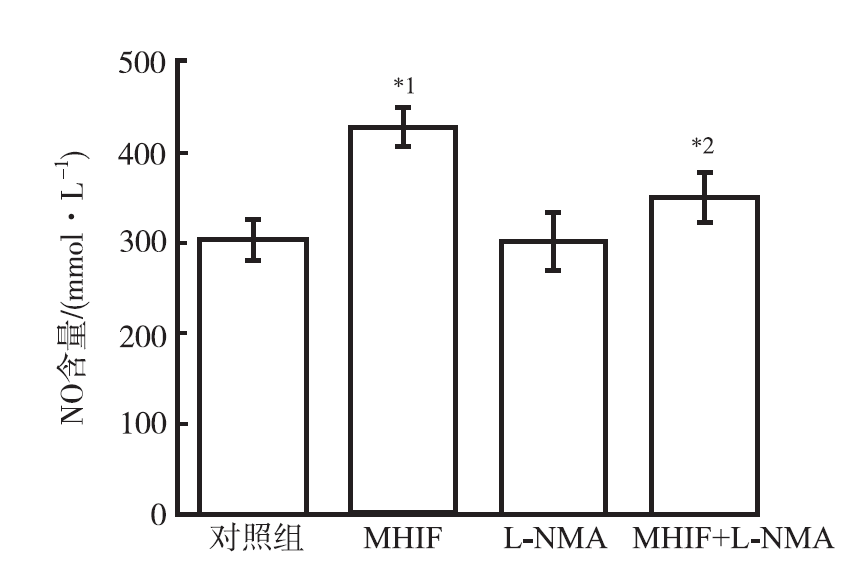
实验结果显示,MHIF组NO含量高于对照组(
实验结果显示,MHIF组cGMP含量高于对照组(
近年来,研究发现黄酮类化合物具有重要的药理活性,尤其是对骨代谢调节作用,但具体机制尚未完全清楚。笔者在本研究首先研究不同浓度MHIF对体外培养大鼠颅骨成骨细胞的骨形成活性的影响,1×10-6 mol·L-1MHIF能显著提高成骨细胞中ALP活性和骨钙素含量,并且存在一定剂量依赖性。目前研究表明,通过提高血管内皮细胞一氧化氮改善心脑血管疾病[5],本实验研究MHIF促进体外培养成骨细胞成骨性分化过程中是否激活NO信号通路。
成骨细胞在骨代谢过程中扮演着骨形成的角色,如果能有效促进成骨细胞成骨性分化,那么就能促进骨形成。因此,本研究通过体外培养大鼠颅骨成骨细胞在不同的药物干预后其成骨性分化的相关指标,碱性磷酸酶和骨钙素是成骨细胞成骨性分化的标志性指标,因此首先从ALP活性和骨钙素含量水平[6],筛选最佳促进成骨细胞成骨性分化的的最适浓度为10-6 mol·L-1,同时实验结果表明促进体外培养成骨细胞分化过程具有剂量依赖性,黄酮类化合物调节骨骼代谢存在剂量的依赖性。
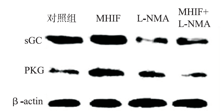
NO参与调控多种生理过程,包括骨重建 [7,8]。NO可结合到可溶性鸟苷酸环化酶(soluble guanylate cy-clase,sGC)的血红素基团上,提高该酶活性[9],由此引起cGMP水平升高,从而导致 cGMP 依赖性蛋白激酶级联激活,以及进一步激活下游信号通路发挥生理活性作用。体外研究表明,由成骨细胞自身产生的少量 NO,可能是成骨细胞内生长因子产生的调节剂[10,11,12]。NO 供体内 NO 的缓慢释放诱导体外成骨细胞的生长和分化[10]。实验结果显示,MHIF促进体外培养成骨细胞成熟与矿化过程中激活NO信号途径。使用L-NMA预处理成骨细胞后发现,MHIF促进成骨细胞中ALP活性和骨钙素含量的能力受到抑制,因此初步证明MHIF促进体外培养成骨细胞分化过程中需要激活一氧化氮合酶参与其中。其次,本研究进一步观察发现,一氧化氮合酶活性被抑制后,MHIF提高成骨细胞NO和cGMP含量均受到抑制,以成骨细胞中sGC和PKG蛋白的表达水平也受到抑制,因此进一步证明MHIF在促进成骨细胞骨形成过程中通过NO/cGMP/sGC信号过程发挥作用。
成骨细胞在骨形成过程中发挥重要作用,因此,本实验应用成骨细胞研究MHIF对体外培养成骨细胞的药理活性,以及其促进成骨性分化的分子机制,实验结果表明MHIF在促进体外培养成骨细胞成骨性分化过程中激活NO信号通路,实验因此推测这个机制为MHIF调节骨代谢可能机制之一,此实验结果为异黄酮类化合物的药物促进活性的机制奠定基础,下一步有待从动物实验水平证明其活性和作用机制。
The authors have declared that no competing interests exist.
| [1] |
|
| [2] |
|
| [3] |
|
| [4] |
DOI:10.1002/jcb.23007
URL
[本文引用:3]
|
| [5] |
|
| [6] |
|
| [7] |
Nitric oxide plays a central role in the regulation of skeletal homeostasis. In cells of the osteoblastic lineage, NO is generated in response to mechanical stimulation and estrogen exposure. Via activation of soluble guanylyl cyclase (sGC) and cGMP-dependent protein kinases (PKGs), NO enhances proliferation, differentiation, and survival of bone-forming cells in the osteoblastic lineage. NO also regulates the differentiation and activity of bone-resorbing osteoclasts; here the effects are largely inhibitory and partly cGMP-independent. We review the skeletal phenotypes of mice deficient in NO synthases and PKGs, and the effects of NO and cGMP on bone formation and resorption. We examine the roles of NO and cGMP in bone adaptation to mechanical stimulation. Finally, we discuss preclinical and clinical data showing that NO donors and NO-independent sGC activators may protect against estrogen deficiency-induced bone loss. sGC represents an attractive target for the treatment of osteoporosis.
[本文引用:1]
|
| [8] |
ABSTRACTChondroadherin (CHAD) is a leucine-rich protein promoting cell attachment through binding to integrin α2β1 and syndecans. We observed that CHAD mRNA and protein were lower in bone biopsies of 50-year-old to 65-year-old osteoporotic women and in bone samples of ovariectomized mice versus gender/age–matched controls, suggesting a role in bone metabolism. By the means of an internal cyclic peptide (cyclicCHAD), we observed that its integrin binding sequence impaired preosteoclast migration through a nitric oxide synthase 2–dependent mechanism, decreasing osteoclastogenesis and bone resorption in a concentration-dependent fashion, whereas it had no effect on osteoblasts. Consistently, cyclicCHAD reduced transcription of two nitric oxide downstream genes, migfilin and vasp, involved in cell motility. Furthermore, the nitric oxide donor, S-nitroso-N-acetyl-D,L-penicillamine, stimulated preosteoclast migration and prevented the inhibitory effect of cyclicCHAD. Conversely, the nitric oxide synthase 2 (NOS2) inhibitor, N5-(1-iminoethyl)-l-ornithine, decreased both preosteoclast migration and differentiation, confirming a role of the nitric oxide pathway in the mechanism of action triggered by cyclicCHAD. In vivo, administration of cyclicCHAD was well tolerated and increased bone volume in healthy mice, with no adverse effect. In ovariectomized mice cyclicCHAD improved bone mass by both a preventive and a curative treatment protocol, with an effect in line with that of the bisphosphonate alendronate, that was mimicked by the NOS2 inhibitor [L-N6-(1-Iminoethyl)-lysine.2 dihydrochloride]. In both mouse models, cyclicCHAD reduced osteoclast and bone resorption without affecting osteoblast parameters and bone formation. In conclusion, CHAD is a novel regulator of bone metabolism that, through its integrin binding domain, inhibits preosteoclast motility and bone resorption, with a potential translational impact for the treatment of osteoporosis. 08 2014 American Society for Bone and Mineral Research.
[本文引用:1]
|
| [9] |
|
| [10] |
|
| [11] |
|
| [12] |
|


