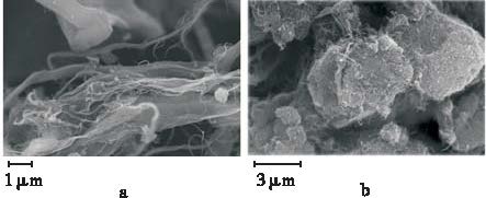
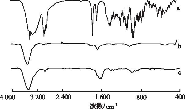
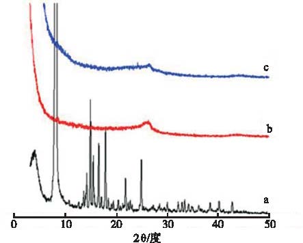
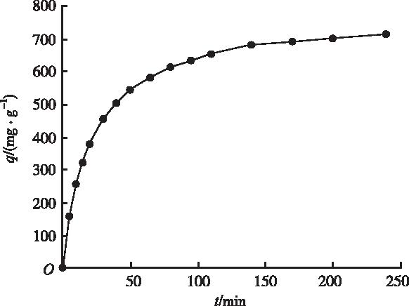
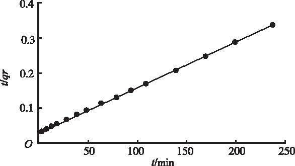
Objective To investigate the method for preparing oridonin-single-walled carbon nanotubes (ORI-SWCNTs) nanocomposite and study its adsorption kinetics. Methods ORI-SWCNTs nanocomposite was prepared by using the method of solution mixing. The synthesized ORI-SWCNTs nanocomposite was characterized by using Laser particle size analyzer, Fourier transform infrared, DSC analysis, powder X-ray diffraction and electron microscopy techniques. Results The encapsulation efficiency and loading capacity of ORI in SWCNTs-COOH nanocarrier was estimated to be about (70.23±2.1) % and (27.29±1.2) %, respectively. The Zeta potential was (-34.29±1.2) mV, partical size was about (458±18) nm. The absorption of ORI on SWCNTs-COOH could be explained by pseudo-second-order model. Conclusion The established preparation process of ORI-SWCNTs nanocomposite by solution mixing is feasible, with higher loading efficiency and encapsulate efficiency..
CHENS,GAOJ,HALICKA HD,et al.The cytostatic and cytotoxic effects of oridonin(Rubescenin),a diterpenoid from Rabdosia rubescens,on tumor cells of different lineage[J]. Int J Oncol,2005,26(3):579-588.
Rabdosia rubescens is a herbal medicine used to treat esophageal cancer in China. In this study, the sesquiterpene oridonin, an isoprenoid, was isolated from Rabdosia rubescens. Mass spectroscopy and carbon 13 NMR spectroscopy were used to identify the structure of the purified compound. It was then evaluated for biological activity against human cell lines derived from prostate (DU-145, LNCaP), breast (MCF-7), and ovarian (A2780 and PTX10) cancers. Oridonin exhibited anti-proliferative activity toward all cancer cell lines tested, with an IC50 estimated by the MTT cell viability assay ranging from 5.8卤2.3 to 11.72卤4.8 碌M. Flow cytometric analysis demonstrated that oridonin induced a G1 phase arrest in androgen receptor-positive LNCaP cells containing wt p53, while it blocked the cell cycle at G2 and M phases in androgen receptor-negative DU-145 cells with mutated p53; the arrest in M was verified by examination of cell morphology and by the increased frequency of cells with Ser-10 phosphorylated histone H3. The increased incidence of apoptosis, identified by characteristic changes in cell morphology, was seen in tumor lines treated with oridonin. Notably, at concentrations that induced apoptosis among tumor cells, oridonin failed to induce apoptosis in cultures of normal human fibroblasts. Western blot analysis was used to determine the protein expression of cancer suppressor genes, p53 (wt) and Bax, and the proto-oncogene, Bcl-2 in LNCaP cells following treatment with oridonin. Oridonin up-regulated p53 and Bax and down-regulated Bcl-2 expression in a dose-dependent manner. To further explore the possible interaction between oridonin and DNA, its absorption spectrum was measured in the presence and absence of double stranded (ds) DNA. Spectral shifts and an increase in absorption band intensity were observed indicating interaction of oridonin with DNA bases. The nature of the binding is not clear at present though no evidence of histone H2AX phosphorylation on Ser-139 was apparent in DU-145 cells treated with oridonin that would indicate the induction of ds DNA breaks. In conclusion, oridonin inhibits cancer cell growth in a cell cycle specific manner and shifts the balance between pro- and anti-apoptotic proteins in favor of apoptosis. The present data suggest that further studies are warranted to assess the potential of oridonin in cancer prevention and/or treatment.
IKEZOET,CHEN SS,TONG XJ,et al.Oridonin induces growth inhibition and apoptosis of a variety of human cancer cells[J].Int J Oncol,2003,23(4):1187-1193.
PC-SPES is an eight herbal mixture that was shown to have activity against prostate cancer. Recently, we purified oridonin from Rabdosia rubescens, one component of PC-SPES, by high performance liquid chromatography (HPLC). The ability of oridonin to inhibit the proliferation of cancer cells was examined by MTT assay. Oridonin effectively inhibited the proliferation of a wide variety of cancer cells including those from prostate (LNCaP, DU145, PC3), breast (MCF-7, MDA-MB231), non-small cell lung (NSCL) (NCI-H520, NCI-H460, NCI-H1299) cancers, acute promyelocytic leukemia (NB4), and glioblastoma multiforme (U118, U138) with ED50s ranging from 1.8 to 7.5 micro g/ml. TUNEL assay and cell cycle analysis showed that oridonin induced apoptosis and G0/G1 cell cycle arrest in LNCaP prostate cancer cells. In addition, expression of p21waf1 was induced in LNCaP and NCI-H520 cells in a p53-dependent manner. Interestingly, when p53 was suppressed by over-expression of E6 from human papilloma virus type 16 (HPV-16), these cells lost their sensitivity to oridonin-induced growth inhibition and apoptosis. Taken together, oridonin inhibited the proliferation of cancer cells via apoptosis and cell cycle arrest with p53 playing a central role in several cancer types which express the wild-type p53 gene. Oridonin may be a novel, adjunctive therapy for a large variety of malignancies and probably represents one of the major, active components of PC-SPES.
LOUH,ZHANGX,GAOL,et al.In vitro and in vivo anti-tumor activity of oridonin nanosuspension[J].Int J Pharm,2009,379(1):181-186.
The aim of the present study was to evaluate the antitumor activity of an oridonin (ORI) nanosuspension relative to ORI solution both in vitro and in vivo. ORI nanosuspension with a particle size of 897.202±0214.202nm was prepared by the high pressure homogenization method (HPH). MTT assay showed that ORI nanosuspension could significantly enhance the in vitro cytotoxicity against K562 cells compared to the ORI solution, the IC 50 value at 3602h was reduced from 12.8502μmol/L for ORI solution to 8.1102μmol/L for ORI nanosuspension. Flow cytometric analysis demonstrated that the ORI nanosuspension also induced a higher apoptotic rate in K562 cells compared to ORI solution. In vivo studies in a mouse model of sarcoma-180 solid tumors demonstrated significantly greater inhibition of tumor growth following treatment with ORI nanosuspension than ORI solution at the same dosage. The mice injected with ORI nanosuspension showed a higher reduction in tumor volume and tumor weight at the dose of 2002mg/kg compared to the ORI solution ( P 02<020.01), with the tumor inhibition rate increased from 42.49% for ORI solution to 60.23% for the ORI nanosuspension. Taken together, these results suggest that the delivery of ORI in nanosuspension is a promising approach for the treatment of the tumor.
CHEN HL,MA XY,LIZ,et al.Functionalization of sing-le-walled carbon nanotubes enables efficient intracellular delivery of siRNA targeting MDM2 to inhibit breast cancer cells growth[J].Biomed Pharmacother,2012,66(5):334-338.
The delivery of DNA or RNA to cells represents the limiting step in the development of cancer gene therapy and RNA interference protocols. Single walled carbon nanotubes (SWNTs) are of interest as carriers of biologically active molecules because of their ability to cross cell membranes. In this study, we developed a novel strategy for chemical functionalization of SWNTs (f-SWCNTs) with DSPE-PEG-Amine to bind small interfering RNA (siRNA) by disulfide bonds applied to siRNA-mediated gene silencing in breast cancer cells. Results indicated the efficiency of f-SWNTs carrying siRNA reached 83.55%, and the new f-SWNTs-siRNA-MDM2 complexes were successfully introduced into the breast carcinoma B-Cap-37 cells at a concentration of 100 nM in mediums, and caused proliferation inhibition of B-Cap-37 cells significantly. The proliferation inhibition ratio of B-Cap-37 cells was detected as 44.53% for 72 h, and the apoptosis ratio was measured as 30.45%. It was obvious that MDM2 can serve as a novel therapeutic target by an effective carrier system of DSPE-PEG-Amine-functionalized SWNTs, which would be very advanced and significant to therapy of breast cancer further.
MENG LJ,ZHANG XK,LU QH,et al.Single walled car-bon nanotubes as drug delivery vehicles:targeting doxorubicin to tumors[J].Biomaterials,2012,33(6):1689-1698.
Single walled carbon nanotubes (SWNTs) are emerging as promising delivery vehicles for cancer diagnostics and chemotherapies due to their unique properties, including, remarkable cell membrane penetrability, high drug-carrying capacities, pH-dependent therapeutic unloading, prolonged circulating times and intrinsic fluorescent, photothermal, photoacoustic and Raman properties. In this leading opinion paper, we systemically discuss and evaluate the relationship of the biological safety of SWNTs with their physicochemical properties such as their length, purity, agglomeration state, concentration and surface functionalization. Other relevant issues, including the cellular uptake mechanism, biodistribution and metabolism of SWNTs are also reviewed. The design and preparation of SWNT-based drug delivery systems (DDSs) and their pharmacokinetic, cancer targeting and therapeutic properties both in聽vitro and in聽vivo are highlighted. Future opportunities and challenges of SWNT-based DDSs are also discussed.
WANGL,ZHANG MY,ZHANGN,et al.Synergistic enh-ancement of cancer therapy using acombination of docetaxel and photothermal ablation induced by single-walled carbon nanotubes[J].Int J Nanomedicine,2011,6:2641-2652.
Single-walled carbon nanotubes (SWNT) are poorly soluble in water, so their applications are limited. Therefore, aqueous solutions of SWNT, designed by noncovalent functionalization and without toxicity, are required for biomedical applications. In this study, we conjugated docetaxel with SWNT via accumulation and used a surfactant to functionalize SWNT noncovalently. The SWNT were then conjugated with docetaxel (DTX-SWNT) and linked with NGR (Asn-Gly-Arg) peptide, which targets tumor angiogenesis, to obtain a water-soluble and tumor-targeting SWNT-NGR-DTX drug delivery system. SWNT-NGR-DTX showed higher efficacy than docetaxel in suppressing tumor growth in a cultured PC3 cell line in vitro and in a murine S180 cancer model. Tumor volumes in the S180 mouse model decreased considerably under near-infrared radiation compared with the control group. The SWNT-NGR-DTX drug delivery system may be promising for high treatment efficacy with minimal side effects in future cancer therapy.
KAWAGUCHIM,YAMAZAKIJ,OHNOJ,et al.Preparation and binding study of a complex made of DNA-treated single walled carbon nanotubes and antibody for specific delivery of a “molecular heater” platform[J].Int J Nanomedicine,2012,7:4363-4372.
Carbon nanotubes have been explored as heat-delivery vehicles for thermal ablation of tumors. To use single-walled carbon nanotubes (SWNT) as a “molecular heater” for hyperthermia therapy in cancer, stable dispersibility and smart-delivery potential will be needed, as well as lack of toxicity. This paper reports the preparation of a model complex comprising DNA-treated SWNT and anti-human IgG antibody and the specific binding ability of this model complex with the targeted protein, ie, human IgG. Treatment with double-stranded DNA enabled stable dispersibility of a complex composed of SWNT and the antibody under physiological conditions. Quartz crystal microbalance results suggest that there was one immobilized IgG molecule to every 21,700 carbon atoms in the complex containing DNA-treated SWNT and the antibody. The DNA-SWNT antibody complex showed good selectivity for binding to the targeted protein. Binding analysis revealed that treatment with DNA did not interfere with binding affinity or capacity between the immobilized antibody and the targeted protein. The results of this study demonstrate that the DNA-SWNT antibody complex is a useful tool for use as a smart “molecular heater” platform applicable to various types of antibodies targeting a specific antigen.
YUAN JF,GAO HC,CHING CB.Comparative protein profile of human hepatoma HepG2 cells treated with grapheme and single walled carbon nanotubes:an iTRAQ-coupled 2D LC-MS/MS proteome analysis[J].Toxicol Lett,2011,207(3) :213-221.
Graphitic nanomaterials are promising candidates for applications in electronics, energy, materials and biomedical areas. Nevertheless, few detailed studies related to the mechanistic understanding of these nanomaterials with the living systems have been performed to date. In the present study, our group applied the iTRAQ-coupled 2D LC鈥揗S/MS approach to analyze the protein profile change of human hepatoma HepG2 cells treated with graphene and single-walled carbon nanotubes (SWCNTs), with the purpose of characterizing the interactions between living system and these nanomaterials at molecular level. Overall 37 differentially expressed proteins involved in metabolic pathway, redox regulation, cytoskeleton formation and cell growth were identified. Based on the protein profile, we found SWCNTs severely interfered the intracellular metabolic routes, protein synthesis and cytoskeletal systems. Moreover, our data suggested that SWCNTs might induce oxidative stress, thereby activating p53-mediated DNA damage checkpoint signals and leading to apoptosis. However, only moderate variation of protein levels for the cells treated with graphene was observed, which indicated graphene was less toxic and might be promising candidate for biomedical applications. We envision that this systematic characterization of cellular response at protein expression level will be of great importance to evaluate biocompatibility of nanomaterials.
LI CC,LIN JL,HUANG SJ,et al.A new and acid-exclu-sive method for dispersing carbon multi-walled nanotubes in aqueous suspensions[J].Colloids Surf A,2007,297(1):275-281.
Carbon multi-walled nanotubes (MWNTs) can be easily dispersed in water using a new and effective method herewith by pretreatment with H 2O 2 followed by dispersing with ammonium polyacrylic acid. The structure morphology and surface chemistry of H 2O 2-treated MWNTs were characterized, respectively, by transmission electron microscopy and Fourier transform-infrared spectroscopy, and the results were compared with MWNTs treated with a mixture of concentrated HNO 3 and H 2SO 4 acids in a 3:1 volume ratio. It was found that the MWNTs with H 2O 2-treatment had fewer microstructure defects than those treated with concentrated acids. The dispersion stability of as-prepared aqueous suspensions was analyzed by rheology and settling experiments. The aqueous suspension prepared by the new method has neutral pH and high dispersion stability, but that prepared from the acid-treated MWNTs was acidic with pH 2鈥3 although their dispersion stability was also good. Besides, the dispersed suspension from the new method stayed highly fluid without becoming coagulated or gelled at solid contents up to 5 wt.%.
TAN JM,KARTHIVASHANG,ARULSELVANP,et al.Characterization and in vitro studies of the anticancer effect of oxidized carbon nanotubes functionalized with betulinic acid[J].Drug Des Devel Ther,2014,8:2333-2343.
Julia M Tan,1 Govindarajan Karthivashan,2 Palanisamy Arulselvan,2 Sharida Fakurazi,2,3 Mohd Zobir Hussein1 1Materials Synthesis and Characterization Laboratory, Institute of Advanced Technology (ITMA), Universiti Putra Malaysia, Serdang, Selangor, Malaysia; 2Laboratory ofnbsp;Vaccine and Immunotherapeutics, Institute of Bioscience (IBS), Universiti Putra Malaysia, Serdang, Selangor, Malaysia; 3Department ofnbsp;Human Anatomy, Faculty of Medicine and Health Sciences, Universiti Putra Malaysia, Serdang, Selangor, Malaysia Abstract: Among the array of nanomaterials, carbon nanotubes have shown great potential as drug carriers in the field of nanomedicine, owing to their attractive physicochemical structure, which facilitates functionalization of therapeutic molecules onto their external walls or being encapsulated inside the tubes. The aim of this preliminary study was to formulate betulinic acid (BA), a poorly water-soluble drug, in oxidized multiwalled carbon nanotubes (MWCNT-COOH) for enhanced delivery efficiency into cancer cells with reduced cytotoxicity. The synthesized MWCNT-BA nanocomposite was characterized using ultraviolet-visible, Fourier transform infrared, thermogravimetric analysis, powder X-ray diffraction, and field emission scanning electron microscopy techniques. The loading of BA in MWCNT-COOH nanocarrier was estimated to be about 14.5%ndash;14.8% (w/w), as determined by ultraviolet-visible and thermogravimetric analysis. Fourier transform infrared study shows that the peaks of the resulting MWCNT-BA nanocomposite correlate to the characteristic functional groups of BA and MWCNT-COOH. The powder X-ray diffraction results confirmed that the tubular structures of MWCNT-COOH were not affected by the drug loading mechanism of BA. The release profiles demonstrated that approximately 98% of BA could be released within 22nbsp;hours by phosphate-buffered saline solution at pH 7.4nbsp;compared with about 22% within 24nbsp;hours at pH 4.8. The biocompatibility studies revealed that MWCNT-BA at concentrations lt;50nbsp;micro;g/mL expressed no cytotoxicity effects for mouse embryo fibroblast cells after 72nbsp;hours of treatment. The anticancer activity of MWCNT-BA was observed to be more sensitive to human lung cancer cell line when compared with human liver cancer cell line, with half maximal inhibitory concentration values of 2.7nbsp;and 11.0nbsp;micro;g/mL, respectively. Our findings form a fundamental platform for further investigation of the MWCNT-BA formulation against different types of cancer cells. Keywords: multiwalled carbon nanotubes (MWCNTs), drug delivery, controlled release, cytotoxicity, A549nbsp;cell line, HepG2nbsp;cell line
LAGERGREN S.About the theory of so-called adsorption of soluble substances [J].Kun Sven Veten Hand,1898,24(4):1-39.
[本文引用:1]
[14]
HO YS.Adsorption of heavy metals from waste streams by peat [D].Birmingham:University of Birmingham,1995.
[本文引用:1]
[15]
HO YS,MCKAYG.Pseudo-second order model for sorp-tion processes[J].Process Biochem,1999,34(5):451-465.
A literature review of the use of sorbents and biosorbents to treat polluted aqueous effluents containing dyes/organics or metal ions has been conducted. Over 70 systems have been reported since 1984 and over 43 of these reported the mechanism as being a pseudo-first order kinetic mechanism. Three sorption kinetic models are presented in this paper and have been used to test 11 of the literature systems previously reported as first order kinetics and one system previously reported as a second order process. In all 12 systems, the highest correlation coefficients were obtained for the pseudo-second order kinetic model.
BOPARAI HK,JOSEPHM,O’CARROLL D M.Kinetics and thermodynamics of cadmium ion removal by adsorption onto nano zerovalent iron particles[J].J Hazard Mater,2011,186(1):458-465.
Nano zerovalent iron (nZVI) is an effective adsorbent for removing various organic and inorganic contaminants. In this study, nZVI particles were used to investigate the removal of Cd(2+) in the concentration range of 25-450 mg L(-1). The effect of temperature on kinetics and equilibrium of cadmium sorption on nZVI particles was thoroughly examined. Consistent with an endothermic reaction, an increase in the temperature resulted in increasing cadmium adsorption rate. The adsorption kinetics well fitted using a pseudo second-order kinetic model. The calculated activation energy for adsorption was 54.8 kJ mol(-1), indicating the adsorption process to be chemisorption. The intraparticle diffusion model described that the intraparticle diffusion was not the only rate-limiting step. The adsorption isotherm data could be well described by the Langmuir as well as Temkin equations. The maximum adsorption capacity of nZVI for Cd(2+) was found to be 769.2 mg g(-1) at 297 K. Thermodynamic parameters (i.e., change in the free energy (G(o)), the enthalpy (H(o)), and the entropy (S(o))) were also evaluated. The overall adsorption process was endothermic and spontaneous in nature. EDX analysis indicated the presence of cadmium ions on the nZVI surface. These results suggest that nZVI could be employed as an efficient adsorbent for the removal of cadmium from contaminated water sources.
Functionalization of sing-le-walled carbon nanotubes enables efficient intracellular delivery of siRNA targeting MDM2 to inhibit breast cancer cells growth
Single walled car-bon nanotubes as drug delivery vehicles:targeting doxorubicin to tumors
2012
Synergistic enh-ancement of cancer therapy using acombination of docetaxel and photothermal ablation induced by single-walled carbon nanotubes
2011
Preparation and binding study of a complex made of DNA-treated single walled carbon nanotubes and antibody for specific delivery of a “molecular heater” platform
2012
Comparative protein profile of human hepatoma HepG2 cells treated with grapheme and single walled carbon nanotubes:an iTRAQ-coupled 2D LC-MS/MS proteome analysis

 , 郭波红
, 郭波红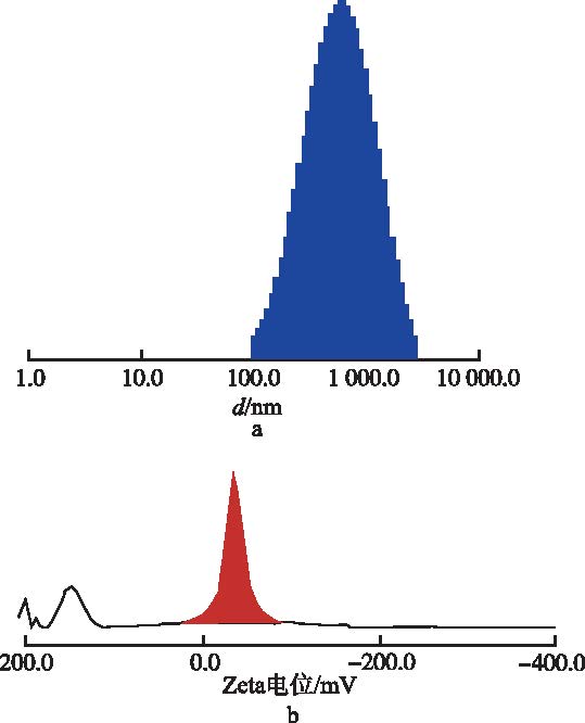


