Objective To explore the molecular mechanism of the interaction between monocytes and renal tubular epithelial cells in the process of renal interstitial fibrosis and the effects of astragaloside IV on the interaction of the two cells. Methods By using in vitro co-culture system,monocyte cells (U973) and human renal proximal tubular epithelial cells (HK-2) were cultured and treated with astragaloside IV.The mRNA expression of Arg,marker gene of M1 monocytes iNOs and M2 monocytes were tested by real-time PCR.The cell surface marker of U973 TLR-4 was detected by FACS,and the change of TBK/IRF3 signaling expression was explored through mRNA and protein level. Results Astragaloside IV treatment inhibited the increase of iNOs and the decrease of Arg-1 induced by M1 transformation in HK-2 cells stimulated monocytes.Further,the surface marker TLR-4 was also decreased and the TBK/IRF3 signaling pathway was blocked by astragaloside IV . Conclusion Astragaloside IV inhibits renal interstitial fibrosis by blocking the TBK/IRF3 signaling pathway,inhibiting M1 differentiations of U973 cell and the expression of TLR4,and then relieves the production of inflammatory factors.
Fig.1
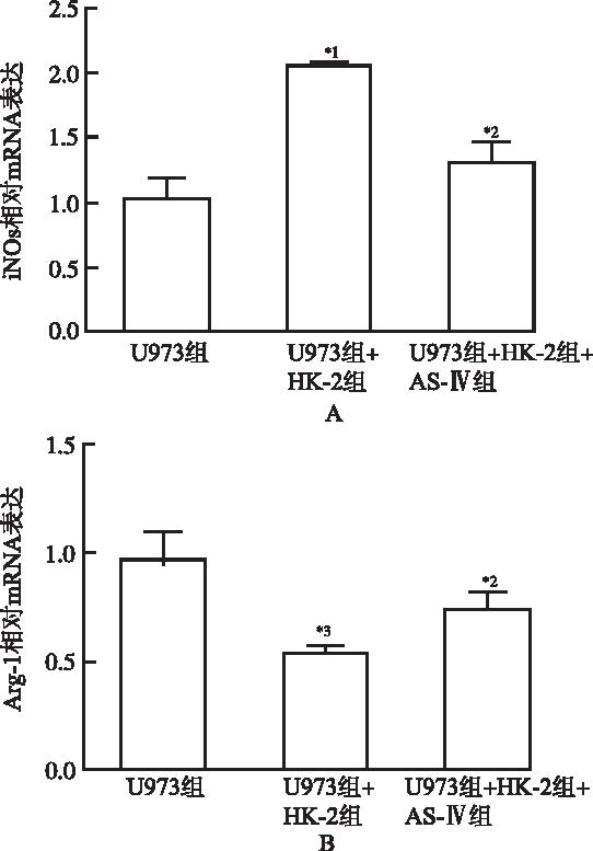
Inhibition of M1 differentiation of U973 cells treated by Astragaloside IV(\(\overline{x}\)±s,n=3) A.mRNA expression of iNOs;B.mRNA expression of Arg-1;Gapdh was used as the internal standard,compared with U973 group,*1P<0.01,*3P<0.05;compared with U973+HK-2 group,*2P<0.05
Fig.2
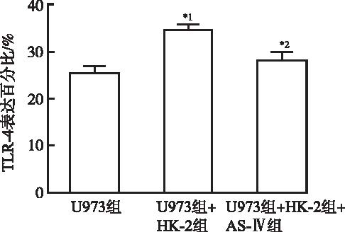
Inhibition of TLR-4 increase of U973 cells by Astragaloside IV(\(\overline{x}\)±s,n=3) Compared with U973 group,*1P<0.05;compared with U973+ HK-2 group,*2P<0.05
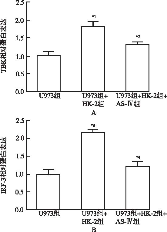
Fig.5
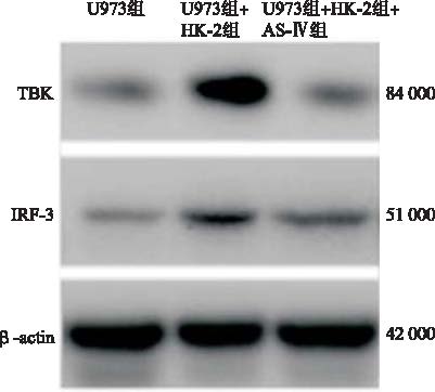
Analysis of gray intensity on the bonds in Fig.4 by Image J saftware(\(\overline{x}\)±s,n=3) Compared with U973 group,*1P<0.05,*3P<0.01;compared with U973+HK-2 group,*2P<0.05,*4P<0.01
LIUF,ZHUANGS.Role of receptor tyrosine kinase signaling in renal fibrosis[J].Intern J Mol Sci,2016,17(6):972-981.
Renal fibrosis can be induced in different renal diseases, but ultimately progresses to end stage renal disease. Although the pathophysiologic process of renal fibrosis have not been fully elucidated, it is characterized by glomerulosclerosis and/or tubular interstitial fibrosis, and is believed to be caused by the proliferation of renal inherent cells, including glomerular epithelial cells, mesangial cells, and endothelial cells, along with defective kidney repair, renal interstitial fibroblasts activation, and extracellular matrix deposition. Receptor tyrosine kinases (RTKs) regulate a variety of cell physiological processes, including metabolism, growth, differentiation, and survival. Many studies fromin vitroand animal models have provided evidence that RTKs play important roles in the pathogenic process of renal fibrosis. It is also showed that tyrosine kinases inhibitors (TKIs) have anti-fibrotic effects in basic research and clinical trials. In this review, we summarize the evidence for involvement of specific RTKs in renal fibrosis process and the employment of TKIs as a therapeutic approach for renal fibrosis.
MENG XM,NIKOLIC-PATERSON D J,LAN H Y.Inflammatory processes in renal fibrosis.[J].Nat Rev Neph,2014,10(9):493-503.
Many types of kidney injury induce inflammation as a protective response. However, unresolved inflammation promotes progressive renal fibrosis, which can culminate in end-stage renal disease. Kidney inflammation involves cells of the immune system as well as activation of intrinsic renal cells, with the consequent production and release of profibrotic cytokines and growth factors that drive the fibrotic process. In glomerular diseases, the development of glomerular inflammation precedes interstitial fibrosis; although the mechanisms linking these events are poorly understood, an important role for tubular epithelial cells in mediating this link is gaining support. Data have implicated macrophages in promoting both glomerular and interstitial fibrosis, whereas limited evidence suggests that CD4(+) T cells and mast cells are involved in interstitial fibrosis. However, macrophages can also promote renal repair when the cause of renal injury can be resolved, highlighting their plasticity. Understanding the mechanisms by which inflammation drives renal fibrosis is necessary to facilitate the development of therapeutics to halt the progression of chronic kidney disease.
SUAREZ-ÁLVAREZB,LIAPISH,ANDERS HJ.Links between coagulation,inflammation,regeneration,and fibrosis in kidney pathology laboratory investigation[J]. J Technic Meth Path,2016,96(4):378-390.
[本文引用:1]
[5]
LIQ,LIU BC,LV LL,et al.Monocytes induce proximal tubular epithelial-mesenchymal transition through NF-kappa B dependent upregulation of ICAM-1[J].J Cell Bioch,2011,112(6):1585-1592.
Inflammatory cell infiltration plays a key role in the pathogenesis of tubulointerstitial damage in chronic renal diseases. In addition to secreting the profibrotic cytokines, monocytes themselves have been demonstrated to be directly associated with renal fibrogenesis. However, how infiltrating monocytes interact with resident cells and the underlying mechanisms remain elusive. In this study we investigated the effects of monocytes on phenotypic changes of human proximal tubular HK-2 cells. The typical epithelial cell morphology of HK-2 cells disappeared after co-culture with monocytes, accompanied by decreased E-cadherin expression, and increased -SMA and fibronectin expression, suggesting that HK-2 cells undergo epithelial090009mesenchymal transition (EMT). Further analysis revealed that the effects were dependent on direct contact of the two types of cells as conditioned medium had no effects. Interestingly, administration of CD18 antibody directly inhibited this process. Furthermore, by microarray and RT-PCR we found that NF-kB signaling may play a role in this process and blockade of this signaling pathway in HK-2 cells could inhibit ICAM-1 expression and EMT phenotypes. Taken together, these findings suggest that monocytes infiltration could directly induce EMT of HK-2 cells via upregulation ICAM-1 through NF-kB signaling pathway. J. Cell. Biochem. 112: 15850900091592, 2011. 0008 2011 Wiley-Liss, Inc.
JIALALI,MAJOR AM,DEVARAJS.Global toll-like receptor 4 knockout results in decreased renal inflammation,fibrosis and podocytopathy[J].J Diab Its Complic,2014,28(6):755-761.
Type 1 diabetes mellitus (T1DM) is a pro-inflammatory state with increased toll-like receptor (TLR) activity. Inflammation is crucial in diabetic nephropathy (DN). We tested the effect of global deficiency of TLR4 on renal inflammation, fibrosis and podocytopathy using control (C) and streptozotocin (STZ) induced diabetic wildtype (WT) and TLR4-knockout (TLR4KO) mice. Following STZ treatment, mice were euthanized at 17weeks and plasma and kidneys collected. Compared to C, STZ-WT mice had significantly increased macrophage and TLR4 immunostaining in kidney, significant increases in MyD88, Interferon Regulatory Factor-3, NFKappaB activity, TNF-Alpha, IL-6, and MCP-1; all these were significantly decreased in the STZ-TLR4KO compared to STZ-WT mice. Compared to C, there were significant increases in fibrosis markers (collagen 4, and transforming growth factor-beta) in STZ-WT which were significantly decreased in the STZ-TLR4KO versus STZ-WT. Podocyte numbers and podocin were decreased in the STZ-WT versus C and increased in the STZ-TLR4KO mice. Global genetic deficiency of TLR4 also ameliorates renal inflammation, fibrosis and podocytopathy and could be important in DN.
GUID,HUANGJ,LIUW,et al.Astragaloside IV prevents acute kidney injury in two rodent models by inhibiting oxidative stress and apoptosis pathways[J].Apoptosis,2013,18(4):409-422.
Oxidative stress and apoptosis play key role in the pathogenesis of acute kidney injury (AKI). We hypothesize that Astragaloside IV(AS-IV) prevents AKI through inhibiting oxidative stress and apoptosis. The rats were divided into sham control, saline-,vehicle-, or AS-IV-treated groups. AS-IV (2002mg/kg) was orally administered once daily to the rats for 7 consecutive days before terminating the experiments. In ischemia-induced AKI model, experimental rats were subjected to bilateral clamping of the renal arteries for 4502min, followed by reperfusion for 2402h. In contrast-induced AKI model, iopamidol (2.902g02iodine/kg) was administered intravenously into the rats. Renal function, histopathology, oxidative stress and apoptosis were evaluated in these models. Pretreatment with AS-IV significantly decreased blood urea nitrogen, serum creatinine, cystatin C and neutrophil gelatinase-associated lipocalin levels, as well as urinary kidney injury molecule-1 level and tubular injury. AS-IV also reduced oxidative stress and tubular cell apoptosis. The p38 mitogen-activated protein kinase phosphorylation and caspase-3 activity were elevated in kidney tissues from AKI rats, accompanied by an increase in Bax expression and a decrease in Bcl-2 expression at mRNA and protein levels. These changes were prevented by AS-IV pretreatment. Therefore, AS-IV can be developed as a novel therapeutic approach to prevent AKI through targeting inhibition of oxidative stress and apoptosis pathways.
WANGQ,SHAOX,XUW,et al.Astragalosides IV inhibits high glucose-induced cell apoptosis through HGF activation in cultured human tubular epithelial cells[J].Renal Failure,2014,36(3):400-406.
Astragaloside IV (ASI) in Radix Astragali is believed to be the active component. The study aims to investigate whether ASI inhibits tubular epithelial cells apoptosis induced by high glucose and its mechanisms. Tubular epithelial cells in this paper were isolated from human kidney. The cells apoptosis was detected by TUNEL and caspase 3 assay. The protein levels of HGF and TGF-β1 were measured by ELISA. The phospho-p38 production, ERK and JNK were determined by Western blot. ASI could inhibit cells apoptosis induced by high glucose (2565mmol/L) in dose-dependent and time-dependent manners. ASI also inhibited high glucose-induced expression of TGF-β1 and activation of p38 MAPK pathway at the protein level. Furthermore, ASI increased HGF production in human tubular epithelial cells. The ASI inhibition of tubular epithelial cells apoptosis and reduction of TGF-β1 expression induced by high glucose may represent a new treatment for diabetic kidney injury. The mechanism underlying this inhibitory effect may be related to the inhibition of p38 MAPK signaling pathway activation and HGF overproduction.
ZHOUX,SUNX,GOGNX,et al.Astragaloside IV from Astragalus membranaceus ameliorates renal interstitial fibrosis by inhibiting inflammation via TLR4/NF-κB in vivo and in vitro.[J].Intern Immunoph,2017,42:18-24.
61UUO-induce renal fibrosis and increase inflammation.61Astragaloside IV ameliorates renal fibrosis through anti-inflammatory effects.61TLR4/NF-кB signal pathways are involved in inhibiting inflammation.
ITALIANIP,BORASCHID.From monocytes to M1/M2 macrophages:phenotypical vs. functional differentiation[J].Frontiers in Immunology,2014,5:512-514.
Studies on monocyte and macrophage biology and differentiation have revealed the pleiotropic activities of these cells. Macrophages are tissue sentinels that maintain tissue integrity by eliminating/repairing damaged cells and matrices. In this M2-like mode, they can also promote tumor growth. Conversely, M1-like macrophages are key effector cells for the elimination of pathogens, virally infected, and cancer cells. Macrophage differentiation from monocytes occurs in the tissue in concomitance with the acquisition of a functional phenotype that depends on microenvironmental signals, thereby accounting for the many and apparently opposed macrophage functions. Many questions arise. When monocytes differentiate into macrophages in a tissue (concomitantly adopting a specific functional program, M1 or M2), do they all die during the inflammatory reaction, or do some of them survive? Do those that survive become quiescent tissue macrophages, able to react as na07ve cells to a new challenge? Or, do monocyte-derived tissue macrophages conserve a “memory” of their past inflammatory activation? This review will address some of these important questions under the general framework of the role of monocytes and macrophages in the initiation, development, resolution, and chronicization of inflammation.
GUITERASR,FLAQUERM,CRUZADOM,et al.Macrophage in chronic kidney disease.[J].Clinical Kidney J,2016,9(6):765-771.
Chronic kidney disease (CKD) has become a major health problem worldwide. This review describes the role of macrophages in CKD and highlights the importance of anti-inflammatory M2 macrophage activation in both renal fibrosis and wound healing processes. Furthermore, the mechanisms by which M2 macrophages induce renal repair and regeneration are still under debate and currently demand more attention. The M1/M2 macrophage balance is related to the renal microenvironment and could influence CKD progression. In fact, an inflammatory renal environment and M2 plasticity can be the major hurdles to establishing macrophage cell-based therapies in CKD. M2 macrophage cell-based therapy is promising if the M2 phenotype remains stable and is ‘fixed’ byin vitromanipulation. However, a greater understanding of phenotype polarization is still required. Moreover, better strategies and targets to induce reparative macrophagesin vivoshould guide future investigations in order to abate kidney diseases.

 , 米秀华, 沈丽萍

