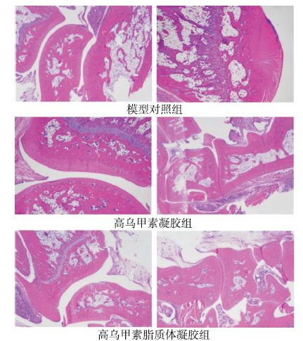
Objective To investigate the efficacy and skin irritation of transdermal formulations of lappaconitine liposome gel. Methods Analgesic effect of lappaconitine liposome gel and lappaconitine gel was investigated by mouse hot plate test and mouse writhing test.Adjuvant arthritis rat model was established to compare the anti-inflammatory effect of the two kinds of transdermal gel by measuring the degree of rat foot swelling, rat serum tumor necrosis factor (TNF-α) and interleukin-2 (IL-2), and observing pathological section of rat ankle joints as observation indexes.Skin irritability of lappaconitine liposome gel was observed by rabbit intact and damaged skin test. Results Compared with the lappaconitine gel group, pain threshold increased and sensitivity to heat decreased with time increase of administration in the lappaconitine liposome gel group, and the difference was statistically significant at 4 h (P<0.01).Frequency of writhing decreased in the lappaconitine liposome gel group at 15 min; the rat’s foot circumference decreased, the expression of TNF-α and IL-2 decreased (P<0.05), and the inflammatory reaction decreased; the lappaconitine liposome gel had nonirritant to the skin. Conclusion Lappaconitine liposome gel has better analgesic and anti-inflammatory effects than lappaconitine gel, and it is not irritant to intact or damaged skin.
GUOT,ZHANG YT,ZHAO JH,et al.Nanostructured lipid carriers for percutaneous administration of alkaloids isolated from Aconitum sinomontanum[J].J Nanobiotechnol,2015,13:47.
Background Lipid-based nanosystems have great potential for transdermal drug delivery. In this study, nanostructured lipid carriers (NLCs) for short-acting alkaloids lappacontine (LA) and ranaconitine (RAN) isolated from Aconitum sinomontanum (AAS) at 69.47 and 9.16% (w/w) yields, respectively, were prepared to enhance percutaneous permeation. Optimized NLC formulations were evaluated using uniform design experiments. Microstructure and in vitro/in vivo transdermal delivery characteristics of AAS-loaded NLCs and solid lipid nanoparticles (SLNs) were compared. Cellular uptake of fluorescence-labeled nanoparticles was probed using laser scanning confocal microscopy and fluorescence-activated cell sorting. Nanoparticle integrity during transdermal delivery and effects on the skin surface were also investigated. Results NLC formulations were less cytotoxic than the AAS solution in HaCaT and CCC-ESF cells. Moreover, coumarin-6-labeled NLCs showed biocompatibility with HaCaT and CCC-ESF cells, and their cellular uptake was strongly affected by cholesterol and lipid rafts. Significantly greater cumulative amounts of NLC-associated LA and RAN than SLN-associated alkaloids penetrated the rat skin in vitro. In vivo microdialysis showed higher area under the concentration???time curve (AUC) 0???t for AAS-NLC-associated LA and RAN than for AAS-SLN-associated alkaloids. Conclusions NLC formulations could be good transdermal systems for increasing biocompatibility and decreasing cytotoxicity of AAS. AAS-NLCs showed higher percutaneous permeation than the other preparations. These findings suggest that NLCs could be promising transdermal delivery vehicles for AAS.
Lappaconitine is a kind of diterpenoid alkaloids extracted from Aconitum sinomontanum Nakai. As a non-opioid analgesic, its hydrobromide is commonly used in the treatment of moderate to severe pain clinically. The clinical application of lappaconitine traditional preparations was limited due to its poor water solubility and sensitivity to light, which made the study of new agents become a trend. In recent years, different formulations of lappaconitine are constantly developed to meet different therapeutic purposes. Research progress on new preparations of lappaconitine was reviewed in recent years. And the information was classified and summarized mainly from three aspects of carrier, sustained-release and immediate-release preparations, to analyse the existing problems and provide some references for further study.
ZHU XC,GE CT,WANGP,et al.Analgesic effects of lappaconitine in leukemia bone pain in a mouse model[J].Peer,2015,3:e936.
Bone pain is a common and severe symptom in cancer patients. The present study employed a mouse model of leukemia bone pain by injection K562 cells into tibia of mouse to evaluate the analgesic effects of lappacontine. Our results showed that the lappaconitine treatment at day 15, 17 and 19 could effectively reduce the spontaneous pain scoring values, restore reduced degree in the inclined-plate test induced by injection of K562 cells, as well as restore paw mechanical withdrawal threshold and paw withdrawal thermal latency induced by injection of K562 cells to the normal levels. Additionally, the molecular mechanisms of lappaconitine analgesic effects may be related to affect the expression levels of endogenous opioid system genes (POMC, PENK and MOR), as well as apoptosis-related genes (Xiap, Smac, Bim, NF-B and p53). Our present results indicated that lappaconitine may become a new analgesic agent for leukemia bone pain management.
ObjectiveTo investigate the analgesic effect of lappaconitine (LA) on burned rats and its supraspinal analgesic mechanism. MethodsMale Sprague-Dawley rats were divided into 6 groups: normal, burn, burn LA,burn LA plus antisense oligodeoxynucleotide (A-ODN),burn LA plus A317491 and burn LA plus ATP. The later 4 groups had LA (6mg/kg) administered intraperitoneally. The pain threshold was characterized by the mechanical withdrawal threshold (MWT) testing; The expression of P2X3 receptor protein in the midbrain periaqueductal gray (PAG) was assessed by Western blot analysis. Observation the intra-PAG injection of P2X3 A-ODN, selective antagonist and agonist of P2X3 receptor on the analgesic effect of LA. ResultsIn burn group, the MWT was decreased while increased in burn LA group. LA induced a significantly up-regulated P2X3 receptor protein expression in PAG. The antinociceptive effect of LA was attenuated significantly by P2X3 A-ODN. The α,β-meATP or A317491 significantly enhanced or attenuated analgesic effect of LA, respectively. Conclusions LA produces an antinociceptive effect on burn pain via increases expression and function of P2X3 receptors in the PAG.
LIUW,SUNY,CHENGZ,et al.Crocin exerts anti-inflammatory and anti-arthritic effects on type II collagen-induced arthritis in rats[J].Pharmaceut Biol,2018,56(1):209-216.
Abstract CONTEXT: Rheumatoid arthritis (RA) is a common systemic auto-immune disease, which is characterized by chronic and symmetry synovial inflammation. Crocin has been reported to exhibit anti-inflammatory effects in animal models. OBJECTIVE: This study investigates the anti-inflammatory and anti-arthritic effects of crocin on type II collagen-induced arthritis (CIA) in Wistar rats. MATERIALS AND METHODS: The CIA rat model was established and randomly divided into five groups with or without crocin treatment (10, 20 or 40090009mg/kg), which was started on day 21 after arthritis induction and persisted for 360002days. The symptoms and molecular mechanisms of CIA and crocin-treated CIA rats were compared and investigated. RESULTS: CIA rats presented severe RA symptoms, including high arthritis score, paw swelling, joint inflammation, bone erosion, chondrocyte death, cartilage destruction, enhanced expressions of matrix metalloproteinase (MMP) and pro-inflammatory cytokines. However, crocin could mitigate these symptoms. Crocin (40090009mg/kg) exhibited the most efficient therapeutic function on CIA rats: the histological scores of joint inflammation, bone erosion, chondrocyte death, cartilage surface erosion, and bone erosion of CIA rats receiving 40090009mg/kg crocin treatment were comparable to the normal rats. MMP-1, -3 and -13 protein expression levels of CIA rats with 40090009mg/kg crocin treatment were decreased to levels similar to normal rats. Moreover, crocin could also inhibit the expression of TNF-02±, IL-17, IL-6 and CXCL8 in serum and ankle tissues of CIA rats. CONCLUSIONS: In summary, crocin exhibits therapeutic potential for RA, by mitigating the symptoms and inhibiting the pro-inflammatory factor expression.

 , 杨爱霞
, 杨爱霞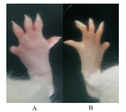
